Tetrahydrobiopterin Inhibits PDGF-stimulated Migration and Pro- liferation in Rat Aortic Smooth Muscle Cells via the Nitric Oxide Synthase-independent Pathway
7
0
0
전체 글
(2) 178. X Jiang, et al. PDGF is increased in atherosclerotic lesions and that PDGF is a principal regulator of mitogenesis in VSMCs [16]. BH4 treatment has been reported to be pharmacologically beneficial for hypertension and atherosclerosis [17]. Moreover, some investigators have shown that BH4 can regulate cell proliferation in various cells, including PC12 and endothelial cells [8,18,19], and migration in kidney cells [9]. It is known that BH4 induces NO production by promoting NOS activity [3] and NO exerts inhibitory effects on migration and proliferation [20,21]. Therefore, it can be assumed that BH4-inhibited cell movements may be associated with NO. However, no studies have investigated whether BH4 inhibits PDGF-induced cell migration and proliferation through NO in VSMCs. In the present study, to clarify the correlation between BH4 and NO in the movements of VSMCs and thus present BH4 as a candidate molecule for inhibiting VSMC responses, we investigated the effects of BH4 and BH2, a BH4 precursor, on cell proliferation and migration in response to PDGF-BB with in vitro and ex vivo analyses.. METHODS Our investigation conformed to the Guide for the Care and Use of Laboratory Animals published by the US National Institutes of Health (NIH Publication No. 85-23, revised 1996). All experiments and animal care were conducted in conformity with the institutional guidelines established by Konkuk University, Korea. Materials BH4 ([6R]-5,6,7,8-tetrahydro-l-biopterin dihydrochoride) and BH2 (7,8-dihydro-l-biopterin) were purchased from Schircks Laboratories (Jona, Switzerland). PDGF-BB was from R&D systems (Minneapolis, MN, USA). Dulbecco's modified eagle medium (DMEM), fetal bovine serum (FBS), penicillin/streptomycin, phosphate buffered saline (PBS), trypsin-ethylenediamine tetraacetic acid (EDTA), and Hank’s balanced salt solution (HBSS) were from Hyclone (Logan, UT, USA) or Invitrogen (Carlsbad, CA, USA). Type I collagen from rat tail tendon, matrigel and collagenase were from BD Bioscience (NJ, USA) and Wako (Richimond, VA, USA). Bovine serum albumin (BSA), NG-nitro-L-arginine methyl ester (L-NAME), and elastase were from Sigma (St. Louis, MO, USA). 48-well microchemotaxis Boyden chamber and polycarbonate membranes were from Neuroprobe (Adderbury, Oxon, UK) and Diff-Quik stain kit was from Sysmex Corp (Kobe, Japan). Other chemical reagents were obtained from Amersham-Pharmacia (Piscataway, NJ, USA) or Roche (Basel, Switzerland). VSMC preparation Rat aortic smooth muscle cells (RASMCs) were isolated from the aortae of male Sprague Dawley (SD) rats (6 weeks old, 160∼180 g, n=5) with collagenase and elastase treatment and were cultured in DMEM containing 10% FBS, 100 U/ml penicillin, 100 μg/ml streptomycin, and 200 mM L-glutamine. For all experiments, cells at passage 3∼8 were grown to 60∼70% confluence and starved in DMEM without FBS for 24 h. After treatment with stimulants and test samples, the cells were lysed with cold extraction buffer (1% Nonidet P-40, 20 mM HEPES [pH 7.5], 150 mM. NaCl, 10% glycerol, 10 mM NaF, 2.5 mM 4-nitrophenylphosphate, 1 mM Na3VO4, 0.5 mM PMSF, 0.7 μg/ml pepstatin, 1 tablet of complete proteinase inhibitor cocktail [Roche, Germany]). Cell migration assay Cell migration assay was performed using the 48-well microchemotaxis Boyden chamber. Polycarbonate membranes with 8-μm pores were coated with 100 μg/ml of type I collagen from rat tail tendons and then dried for 60 min. Cells were harvested with trypsin-EDTA and resuspended in DMEM containing 0.1% BSA. PDGF-BB, BH4 or BH2, and test inhibitor were loaded in the bottom chamber, and the membrane was laid over the cells. After this microchamber o was incubated at 37 C for 90 min, the membranes were fixed and stained using Diff-Quik solution. The number of cells migrating through the membrane was counted in four randomly chosen regions of each well by using a microscope (×200). Cell proliferation assay Cell proliferation was determined using a 5-bromo-2'deoxyuridine (BrdU) incorporation assay (Roche). A microtiter plate was coated with a 100 μg/ml of type I collagen o for 15 min at 37 C. Cells were harvested with trypsinEDTA and resuspended in DMEM (10% FBS, 100 U/ml penicillin, 100 μg/ml streptomycin, and 200 mM L-glutamine). 3 Cells were seeded at a density of 2×10 cells/well. After 12 h, the cells were incubated in FBS-free DMEM for 6 h and treated with PDGF-BB and BH4 or BH2 for 36 h. BrdU-labeling solution was added to the cells and incubated for 12 h. After the denaturation of DNA, peroxidase-labeled anti-BrdU monoclonal antibody was added, and the samples were incubated at room temperature for 90 min. The BrdUantibody complexes were detected with a victor 3 luminometer (PerkinElmer, Boston, MA, USA). Cell viability assay To assess RASMC viability, a 2,3-bis [2-methoxy-4-nitro5-sulfophenyl]-2H-tetrazolium-5-carboxanilide (XTT) assay TM was performed using the WelCount cell viability assay kit (WelGENE, Daegu, Korea). Cells were loaded in 96-well plates at a density of 5×10³ cells/well and incubated for 24 o h at 37 C, and then XTT was added to each well. Plates were incubated in the presence of XTT dye for 4 h to induce the formation of formazan dye, and the optical density value was quantitated with an enzyme-linked immunosorbent assay reader at 450 nm. Aortic sprout assay To examine the migration and proliferation of RASMCs in an ex vivo condition, an aortic ring assay was performed using Matrigel [22]. Aortae from SD rats (5 weeks, n=8), after removing endothelium and adventitium with enzymes, were cut into 1-mm rings. The rings were embedded in 48-well plates coated with Matrigel and simultaneously added with PDGF-BB and BH4 or BH2 in FBS-free DMEM. The rings were stained with Diff-Quik and photographed. The length of the sprouts was analyzed using Scion Image Software on day 5 of incubation..
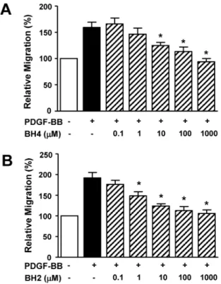
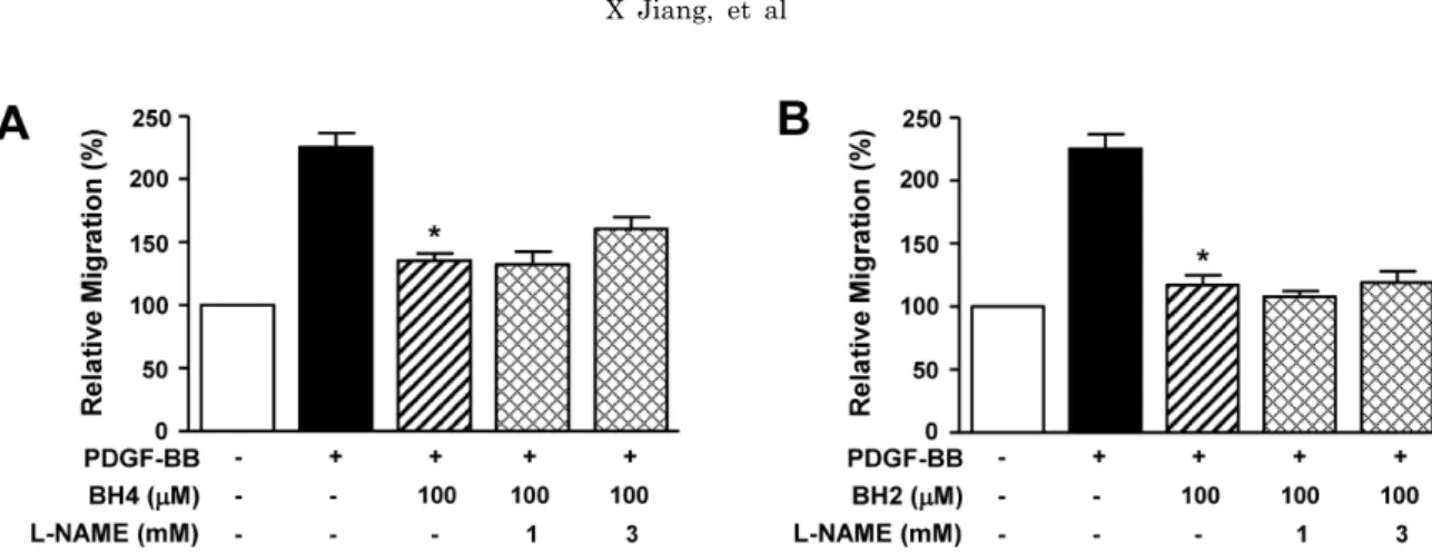
(3) BH4 in Vascular Responses. 179. Statistical analysis. Effect of BH4 on PDGF-BB-induced RASMC proliferation. Data are expressed as the mean±S.E.M. The statistical data evaluation, using GraphPad prism (GraphPad Software, San Diego, CA, USA), was performed by Student’s t-tests for comparisons between pairs of groups and by ANOVA for multiple comparisons. p<0.05 was considered to indicate a significant difference.. The effect of BH4 on the PDGF-induced proliferation of RASMCs was determined with a BrdU incorporation assay. The treatment of RASMCs with BH4 (0.1∼1,000 μM) induced a dose-dependent decrease in PDGF-BB (10 ng/ml)increased proliferation, and this response was maximal with 1,000 μM BH4 (n=8) (Fig. 2A). Furthermore, BH2 (0.1∼ 1,000 μM) treatment inhibited PDGF-BB (10 ng/ml)-increased proliferation in a dose-dependent manner (n=8) (Fig. 2B).. RESULTS Effect of BH4 on PDGF-BB-induced RASMC migration To determine the effects of BH4 on vascular responses, we first examined the migration of RASMCs in response to PDGF by using the Boyden chamber. BH4 (0.1∼1,000 μM) dose-dependently attenuated PDGF-BB (10 ng/ml)-induced RASMC migration and maximally inhibited migration at a concentration of 1,000 μM (n=9) (Fig. 1A). It has been reported that living cells that take up dihydrobiopterin (BH2) reduce it to enzymatically active BH4 [23]. Therefore, after the treatment of RASMCs with BH2, PDGF-BB-induced migration of RASMCs was examined. BH2 (0.1∼1,000 μM) treatment also inhibited PDGF-BB (10 ng/ml)-stimulated RASMC migration in a dose-dependent manner (n=9) (Fig. 1B).. Fig. 1. Effects of BH4 and BH2 on PDGF-BB-induced migration of RASMCs. PDGF (10 ng/ml) and BH4 (0.1∼1,000 μM; A) or BH2 (0.1∼1,000 μM; B) were loaded in the upper chamber and incubated for 90 min (n=9). Migration was examined using a Boyden chamber assay. The migration of RASMCs in the quiescent state was expressed as 100% (n=9). *p<0.05 vs. the PDGF-BB-stimulated response in the absence of BH2 or BH4.. Effects of NOS inhibitor on BH4 inhibition of PDGFinduced migration and proliferation BH4 is commonly known to be an essential cofactor for NOS [1]. To examine whether the effect of BH4 on PDGF-increased migration involved NO, we analyzed the responses to L-NAME, a NOS inhibitor, in RASMCs treated with BH4 (100 μM). As shown in Fig. 3A, L-NAME treatment (1 and 3 mM) did not alter the inhibitory effect of BH4 on PDGF-BB (10 ng/ml)-increased migration (n=8). Moreover, the inhibitory effect of BH2 (100 μM) on PDGFBB (10 ng/ml)-increased migration was also not affected by L-NAME treatment (1 and 3 mM) (n=8; Fig. 3B). Furthermore, L-NAME did not affect BH4-induced inhibition of PDGF-BB (10 ng/ml)-increased proliferation (data not shown).. Fig. 2. Effects of BH4 and BH2 on PDGF-BB-induced proliferation of RASMCs. RASMCs were treated with BH4 (0.1∼1,000 μM; A) or BH2 (0.1∼1,000 μM; B) and then stimulated with PDGF-BB (10 ng/ml) for 36 h. Cell proliferation was quantified using a BrdU incorporation assay. Proliferation in the quiescent state was expressed as 100% (n=8). *p<0.05 vs. the PDGF-BB-induced response in the absence of BH2 or BH4..
(4) 180. X Jiang, et al. Fig. 3. Effects of NOS inhibition on BH4- and BH2-induced inhibition of migration of RASMCs. After treatment with L-NAME (1 and 3 mM) and/or BH4 (100 μM; A) or BH2 (100 μM; B), the cells were loaded into the upper chamber containing PDGF-BB (10 ng/ml) and incubated for 90 min (n=8), as described in the Methods section. Cell migration in the quiescent state was expressed as 100% (n=8). *p<0.05 vs. the PDGF-BB-stimulated response in the absence of BH2 and BH4.. Fig. 4. Effects of BH4 and BH2 on PDGF-BB-induced sprout formation of aortic rings. (A) Aortic rings (1-mm) embedded and cultured in Matrigel were treated with PDGF-BB, vehicle (0.1% DMSO), and BH4 or BH2. The effects were observed on day 5 of the stimulation. Upper panels: effects of vehicle and PDGF-BB (10 ng/ml) on the outgrowth of aortic-ring sprouts. Middle and lower panels: representative photographs showing the effects of BH4 (0.1∼1,000 μM) and BH2 (0.1∼1,000 μM), respectively. (B, C) The statistical data obtained from panel A. The sprout length level in vehicletreated rings was expressed as 100% (n=4). *p<0.05 vs. the PDGF-BB-induced responses in the absence of BH2 and BH4.. Effects of BH4 on the formation of aortic sprouts To confirm the effects of BH4 and BH2 on migration and proliferation, we performed an ex vivo Matrigel assay ex-. amining the sprout formation of aortic rings. As shown in Fig. 4, sprout outgrowth from aortic rings was increased by PDGF-BB (10 ng/ml) treatment, and this response was dose-dependently inhibited by BH4 (0.1∼1,000 μM; n=4). Moreover, BH2 treatment also induced a dose-dependent.
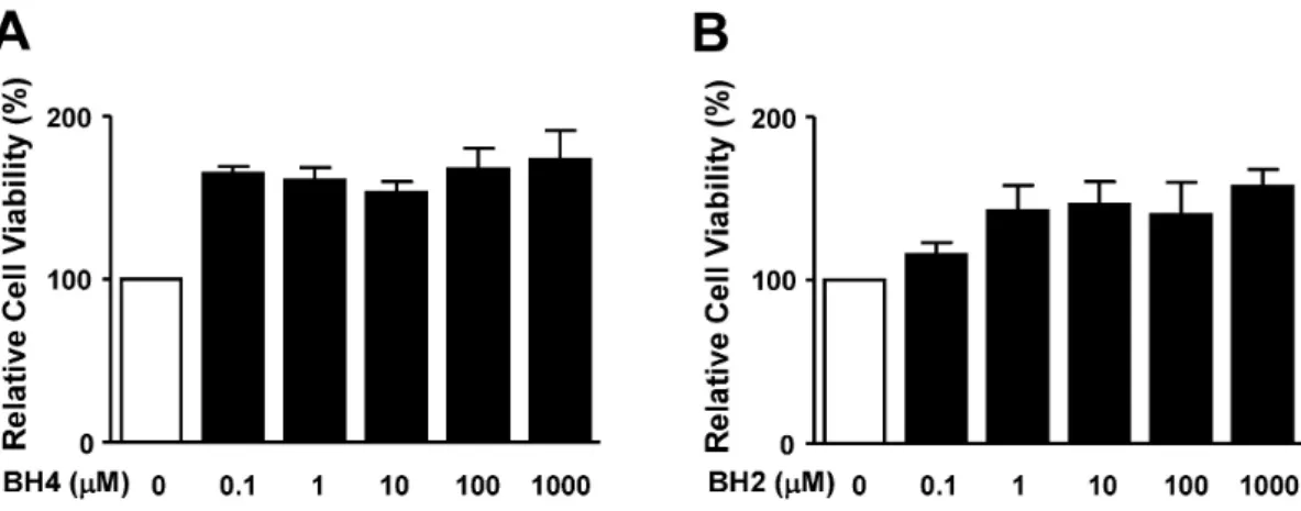
(5) BH4 in Vascular Responses. 181. Fig. 5. Effects of BH4 and BH2 on RASMC viability. Cell viability was evaluated by the XTT assay as described in the Methods section. RASMCs were treated with BH4 (0.1∼1,000 μM; A) or BH2 (0.1∼1,000 μM; B) for 24 h. After addition of the XTT dye, cells were incubated for 4 h. Cell viability was quantified by measuring optical density values with an enzyme-linked immunosorbent assay reader at 450 nm. Cell viability in the quiescent state was considered 100% (n=4).. decrease in PDGF-BB (10 ng/ml)-stimulated aortic ring sprout formation (Fig. 4). Effect of BH4 on RASMC viability To test whether BH4 affects RASMC viability, the cytotoxic effect of BH4 on RASMCs in the quiescent state was analyzed using the XTT assay and a direct cell counting method. The treatment of RASMCs with BH4 (0.1∼ 1,000 μM; n=4) did not produce cytotoxic effects at any treatment concentration in the XTT assay (Fig. 5A). In addition, BH2 (0.1∼1,000 μM; n=4) exerted no cytotoxic effects on RASMCs (Fig. 5B). Effects of BH2 and BH4 on RASMCs measured by the XTT assay were confirmed in a direct cell-count assay (data not shown). From these results, we found that BH4 and BH2 exerted no cytotoxic effects on RASMC viability.. DISCUSSION In this study, we demonstrated for the first time that BH4 attenuated RASMC migration and proliferation in response to PDGF stimulation. Previous investigations reported that BH4 stimulated proliferation of various cells, including vascular endothelial and pheochromocytoma (PC12) cells [18,24]. Moreover, BH4 deficiency is known to promote neointimal formation and cell proliferation in injured vascular walls [8]. It was reported that CCR2-stimulated kidney cell migration was inhibited in plasma or aorta homogenates in mice with elevated BH4 levels due to GTPCH overexpression [9], implying the possibility that BH4 may affect VMSC migration. Our present study results showed that BH4 treatment decreased PDGF-stimulated RASMC migration, indicating that BH4 inhibited the PDGF-induced migration and proliferation of RASMCs. Furthermore, the inhibitory effects of BH4 on PDGF-stimulated proliferation and migration responses in RASMCs were also confirmed by an ex vivo aortic ring assay using Matrigel. Therefore, our present study findings suggest a possible role of BH4 as a therapeutic agent in preventing proliferation and migration, which are pivotal progression. steps of atherosclerosis. BH4 is an essential cofactor of NOS that converts L-arginine to L-citrulline and nitric oxide (NO) [25]. NO plays a key role in preserving vascular homeostasis through modulating vascular activity associated with platelets, endothelium, and smooth muscle cells [26]. Defects in these functions can usually be caused by decreased NO bioavailability and result in the development of diseases such as atherosclerosis and hypertension [27]. NOS enzymatic activity is regulated by BH4 [28]. Low levels of BH4 result in reduced NO production and simultaneously increased superoxide production due to the imbalance of enzymatic coupling between the reduction of oxygen molecules and oxidation of L-arginine [29]. NO and NOS activation are known to inhibit proliferation of VSMCs [30]. Together with proliferation, VSMC migration is an essential process in vascular disorders such as atherosclerosis [11]. Moreover, BH4 induced NO production by promoting NOS activity [3] and NO exerted inhibitory effects on migration of VSMCs [21]. These reports imply the possibility of NO involvement in the effects of BH4 on PDGF-BB-stimulated VSMCs. However, our result in this study revealed that the inhibition of PDGF-induced RASMC responses by BH4 was not affected by a NOS inhibitor. These results indicate that NO does not contribute to the inhibitory effects of BH4 on PDGF-induced VSMC migration and proliferation. Therefore, it is likely that the inhibition of PDGF-induced RASMC migration and proliferation by BH4 treatment is not linked to NO-related effects, although detailed mechanisms of this response were not clarified in this study. Future studies are needed to uncover the NO-independent mechanism of BH4 inhibition of PDGF-BB-induced migration and proliferation. BH4 is biosynthesized by de novo, regeneration, and salvage pathways. De novo synthesis of BH4 is regulated by GTPCH, a rate-limiting enzyme for BH4 biosynthesis [1]. Moreover, BH4 can be reproduced by the regeneration pathway by related enzymes DHFR and DHPR [4,5]. DHPR regenerates BH4 from qBH2, an oxidized and inactive form of biopterin. Another enzyme for BH4 regeneration, DHFR, can regenerate BH4 from BH2. The regeneration pathway of BH4, as well as its de novo synthesis pathway, is involved.
(6) 182. X Jiang, et al. in the regulation of BH4 levels in the circulatory system and deficiency of the enzyme results in functional disorders in rat hypertensive vessels [10]. In this study, we found that BH2 inhibited migration and proliferation of RASMCs in response to PDGF-BB. These findings were also confirmed using an aortic sprout assay. Moreover, BH2 and BH4 did not exert cytotoxic effects in XTT and direct cell-count assays, suggesting that the inhibitory effects of these biopterin derivatives may arise from non-cytotoxic events. However, the data of the XTT assay revealed an enhancement of cell viability because of nonspecific reactions of BH2 and BH4, which may be caused by the increment of the optical density values of these molecules. These results suggest that BH2 has a potent inhibitory role similar to that of BH4 in vascular responses. In summary, we showed that PDGF-BB-increased migration and proliferation in RASMCs were inhibited by BH4 treatment. Ex vivo treatment with BH4 decreased aortic sprout outgrowth in response to PDGF-BB. BH4-induced inhibitory effects on PDGF-induced responses were not affected by NOS inhibition. Moreover, BH2 mimicked the effects of BH4. These results suggest that BH4 inhibits PDGF-BB-induced RASMC migration and proliferation and that these responses may be related to the NOS-independent pathway. Therefore, BH4 and BH2 could also be useful as candidate molecules exerting NO-independent anti-atherosclerotic functions.. ACKNOWLEDGEMENTS This work was supported by Mid-career Researcher Program (2008-0061614) and Basic Science Research Program (2008-313-E00042) through the National Research Foundation of Korea (NRF) funded by the Ministry of Education, Science and Technology (MEST), and by the Korea Research Foundation Grants funded by the Korean Government (Ministry of Education, Science and Technology) (KRF2006-353-E00001).. 9.. 10.. 11.. 12. 13.. 14. 15.. 16. 17. 18.. 19.. REFERENCES 1. Nichol CA, Lee CL, Edelstein MP, Chao JY, Duch DS. Biosynthesis of tetrahydropterin by de novo and savage pathways in adrenal medulla extracts, mammalian cell cultures, and rat brain in vitro. Proc Natl Acad Sci USA. 1983;80:1546-1550. 2. Kwon NS, Nathan CF, Stuehr DJ. Reduced biopterin as a cofactor in the generation of nitrogen oxides by murine macrophages. J Biol Chem. 1989;264:20496-20501. 3. Mayer B, Werner ER. In search of function for tetrahydrobipterin in the biosynthesis of nitric oxide. Arch Pharmacol. 1995;351:453-463. 4. Moens AL, Kass DA. Tetrahydrodiopterin and cardiovascular disease. Aterioscler Thromb Vasc Biol. 2006;26:2439-2444. 5. Zheng J, Yang X, Keith J, Christian H, Fing GD, Gerogory K, Imre K, Chen AF. Gene transfer of human guanosine 5’-triphosphate cyclohydrolase I restores vascular tetrahydrobiopterin level and endothelial function in low rennin hypertension. Circulation. 2003;108:1238-1245. 6 Yang S, Lee YJ, Kim J, Park S, Peris J, Laipis P, Park YS, Chung JHC, Oh SP. A murine model for human sepiapterin reductase deficiency. Am J Hum Genet. 2006;78:575-587. 7. Nicholas JA, Keith M. Regulation of endothelial nitric oxide synthase by tetrahydrobiopterin in vascular disease. Aterioscler Thromb Vas Biol. 2004;24:413-420. 8. Wang CH, Li SH, Weisel RD, Fedak PW, Hung A, Li RK, Rao. 20.. 21. 22.. 23.. 24.. 25. 26.. V, Hyland K, Cherng WJ, Errett L, Leclerc Y, Bonneau D, Latter DA, Verma S. Tetrahydrobiopterin deficiency exaggerates intimal hyperplasia after vascular injury. Am J Physiol Regul Integr Comp Physiol. 2005;289:R299-R304. Ali ZA, Bursill CA, Douglas G, McNeill E, Papaspyridonos M, Tatham AL, Bendall JK, Akhtar AM, Alp NJ, Greaves DR, Channon KM. CCR2-mediated anti-inflammatory effects of endothelial tetrahydrobiopterin inhibit vascular injury-induced accelerated atherosclerosis. Circulation. 2008;118:S71-S77. Lee CK, Han JS, Won KJ, Jung SH, Park HJ, Lee HM, Kim J, Park YS, Kim HJ, Park PJ, Park TK, Kim B. Diminished expression of dihydropteridine reductase is a potent biomarker for hypertensive vessels. Proteomics. 2009;9:4851-4858. Lee HM, Jeon BH, Won KJ, Lee CK, Park TK, Choi WS, Bae YM, Kim HS, Lee SK, Park SH, Irani K, Kim B. Gene transfer of redox factor-1 inhibits neointimal formation: involvement of platelet-derived growth factor-beta receptor signaling via the inhibition of the reactive oxygen species-mediated Syk pathway. Circ Res. 2009;104:219-227 Raines EW. PDGF and cardiovascular disease. Cytokine Growth Factor Rev. 2004;15:237-254. Lee CK, Lee HM, Kim HJ, Park HJ, Won KJ, Roh HY, Choi WS, Jeon BH, Park TK, Kim B. Syk contributes to PDGFBB-mediated migration of rat aortic smooth muscle cells via MAPK pathways. Cardiovasc Res. 2007;74:159-168. Heldin CH, Westermark B. Mechanism of action and in vivo role of platelet-derived growth factor. Physiol Rev. 1999;79: 1283-1316. Won KJ, Lee SC, Lee CK, Lee HM, Lee SH, Fang Z, Choi OB, Jin M, Kim J, Park T, Choi WS, Kim SK, Kim B. Cordycepin attenuates neointimal formation by inhibiting reactive oxygen species-mediated responses in vascular smooth muscle cells in rats. J Pharmacol Sci. 2009;109:403-412 Sachinidis A, Locher R, Vetter W, Tatje D, Hoppe J. Different effects of platelet-derived growth factor isoforms on rat vascular smooth muscle cells. J Biol Chem. 1990;265:10238-10243. Moens AL, Kass DA. Therapeutic potential of tetrahydrobiopterin for treating vascular and cardiac disease. J Cardiovasc Pharmacol. 2007;50:238-246. Anastasiadis PZ, Bezin L, Imerman BA, Kuhn DM, Louie MC, Levine RA. Tetrahydrobiopterin as a mediator of PC12 cell proliferation induced by EGF and NGF. Eur J Neurosci. 1997;9:1831-1837. Shimizu S, Yasuda M, Ishii M, Nagai T, Kiuchi Y, Yamamoto T. Stimulation of in vitro angiogenesis by tetrahydrobiopterin in bovine aortic endothelial cells. Jpn J Pharmacol. 1999;80: 177-180. Stein CS, Fabry Z, Murphy S, Hart MN, Stein CS, Fabry Z, Murphy S, Hart MN. Involvement of nitric oxide in IFN-γmediated reduction of microvessel smooth muscle cell proliferation. Mol Immunol. 1995;32:965-973. Sarkar R, Meinberg EG, Stanley JC, Gordon D, Webb RC. Nitric oxide reversibly inhibits the migration of cultured vascular smooth muscle cells. Circ Res. 1996;78:225-230. Lee HM, Lee CK, Lee SH, Roh HY, Bae YM, Lee KY, Lim J, Park PJ, Park TK, Lee YL, Won KJ, Kim B. p38 mitogenactivated protein kinase contributes to angiotensin II-stimulated migration of rat aortic smooth muscle cells. J Pharmacol Sci. 2007;105:74-81. Hasegawa H, Sawabe K, Nakanishi N, Wakasugi OK. Delivery of exogenous tetrahydrobiopterin (BH4) to cells of target organs: role of salvage pathway and uptake of its precursor in effective elevation of tissue BH4. Mol Genet Metab. 2005;86:S2-S10. Anastasiadis PZ, Jiang H, Bezin L, Kuhn DM, Levine RA. Tetrahydrobiopterin enhances apoptotic PC12 cell death following withdrawal of trophic support. J Biol Chem. 2001;276: 9050-9058. Dawson TM, Snyder SH. Gases as biological messengers: nitric oxide and carbon monoxide in the brain. J Neurosci. 1994;14: 5147-5159. Yetik-Anacak G, Catravas JD. Nitric oxide and the endothelium: history and impact on cardiovascular disease. Vascul.
(7) BH4 in Vascular Responses. Pharmacol. 2006;45:268-276. 27. Puddu GM, Cravero E, Arnone G, Muscari A, Puddu P. Molecular aspects of atherogenesis: new insights and unsolved questions. J Biomed Sci. 2005;12:839-853. 28. Katusic ZS. Vascular endothelial dysfunction: does tetrahydrobiopterin play a role? Am J Physiol Heart Circ Physiol. 2001; 281:H981-H986.. 183. 29. Alp NJ, Channon KM. Regulation of endothelial nitric oxide synthase by tetrahydrobiopterin in vascular disease. Arterioscler Thromb Vasc Biol. 2004;24:413-420. 30. Nakaki T, Nakayama M, Kato R. Nakaki T, Nakayama M, Kato R. Inhibition by nitric oxide and nitric oxide-producing vasodilators of DNA synthesis in vascular smooth muscle cells. Eur J Pharmacol. 1990;189:347-353..
(8)
수치
관련 문서
