大韓放射線홈學會誌 Yol. XX, No. 4, 1984
-Abstract-
限局性 師病變의 經皮的 吸引生檢 f 끼)“GitL τ :,-기 f L」
/ /서울大學校 醫科大學 放射線科學敎室 任 廷 基·林 德·朴 在 亨
서울大學校 醫科大學 病理學敎室
威 훌융 根
%k
Percutaneous Needle Aspiration Biopsy of Localized Pulmonary Lesions
Chung Kie 1m
,
M.D.,
Duk Lim,
M.D.,
Jae Hyung Park,
M.D.Department of Radiology
,
CoIlege of Medicine,
Seoul National UniversityEui Keun Ham
,
M.D.Department of Pathology
,
Col/ege of Medicine,
Seoul National UniversityOver a period of recent two years
,
100 patients who had localized pulmonary lesions and underwent percutaneous needle aspiration and biopsy,
were analized. There were 56 malignancies and 44 benign lesions. The diagnostic accuracy of malignancy including specific cell type and benign disease are 89%and 79% respectively. Differentiation of malignancy vs. benignity was possible in 89% of cases obviating unnecessary exploratory thoracotomy for diagnostic purpose. Five cases were misinterpreted and eight cases were non-diagnostic on cytology. Inadequate material was obtained in two casesi one was due to hardness of the mass
,
which,
later,
confirmed as chondrosarcoma,
and the other was too small (0.8x1.0 cm) to be visible on lateral view.The obtainability of the tissue was 98%. 14 (14%) patients developed pneumothoraxi one of them required treatment and the remainder showed spontaneous resporption. (Transient neglibigle blood tinged sputum was fou nd in 16 (16%) cases.) The method
,
problems and complications are discussed.Authors recommend the percutaneous needle aspiration and biopsy as the initial procedure in diagnostic work-up of pulmonary coin lesions
,
especially when they are smaller,
more peripheral and metastatic neoplasm is suspected.이 論文은 1984 年 8 月 31 日 접수하여 1984 年 11 月 22 日에 채택되었음.
-746 -
1. 서 론
상태에서 뼈骨의 상연을 따라 22 G. 或은 23G. Chiba needle 및 needle bevel 직 상부에 한 개의 구멍 을 뚫어 조직 채취량이 많도록 고안된 22G. Wescott needle 을 限局性 ßr5病했, 즉 ffr5맨揚 내지 ßr5結節의 該斷에 있 사용하여, 수직£로 병소를 穿刺하였다 {Fig. l). 병소가 어서 經皮的 뼈 吸引生檢의 有用性은 이미 알려져 있~ Rl힘部의 後뿔에 더 가까이 위치하더라도됩뼈骨 (scap비a) 며, 실제 閒뼈術을 제외하고는 가장 높은 該斷率을 보 과 겹치연 뼈뿔 前面을 택하였다. 병소에 檢針이 다다르 이고 있다. 저자들은 최근 2 年 간 서울大學校病院 放 연 stylet 을 뽑고 주사기를 연결한 후 피스톤을 장아 射線科에서 시행한 經皮的 뻐 吸引生檢 100 예의 성적과 당겨 음압을 가하면서 짧고 세차게 몇 차례 병변을 관 입상적 의의를 보고하는 바이다.
ll. 대 Ac:>
‘
1982년 10원부터 1984 년 7 월까지 22 개월간 시행 한 113 예의 經皮的 뼈 吸引生檢 종 임상적 추적이 가능 한 100예를 대상으후 했 S며, 이들은 대부분 陽짧細뼈 檢효나 氣管技鏡檢훌 등으로 該斷이 되 지 않아 放射線 科로 위닥된 환자들이다. 대부분은 입원 환자였고 외래 환자는 3 명 이었다.
Ill. 方 法
병소의 위치에 따라 띠없位 (supine) ,或은 趙位 (prone) 로 환자의 자세를 취한 후 $벅f 투시기 Cbiplane f1uoros.
통한 후 檢針과 주사기를 함께 뽑았다. 병소는 평균 2 회 穿刺하였으며, 睡塊가 큰 경우는 睡塊의 변두리에서 조직을 채취하여 塊死組緣만 추출되는 것을 방지하였다.
조직을얻으연 미리 준비된 슬라이드에 쫓妹하여 95 % 알코홀에 고정하고 또한 結核園, 일반細園 빛 힐園의 뚫 妹 염색과 培養檢흉블 의뢰하였으며, 가능하면 細脫群 集切斷法 Cc아 I block cxamination) 도 시행하였다. 시술 직후에는 투시기로 氣뼈의 여부를 관찰하였S며, 시술 3 시간 후에는 반드시 單純뼈部題影을 하여 氣뼈의 발 생 여부를 관찰하였다 3 예의 외래 환자로 시술후 3 시간 동안 관찰 및 單*뺀며部廣影에서 이상 소견이 없음 을 確認하고 귀가 시켰다.
N. 결 과
cope) 로 병소의 위치를 確認하고, 표적부를 소독하며, 性別 분포는 남자가 76 명, 여자가 24 명으로 남자가 2
%
Procaine 으로 局所麻解後 가능한 한 호흡 정지 3 배가량 많았고, 연령군은 20 代에서 70 代까지이며%A
-=:
‘ -'←←←←←←←←←←←←←←「←←←←「←←←←τ二二二二二二二二二二-8Fig. l. A.B. Fine needle is introduced to the mass at left upper lobe through anterior chest wall.
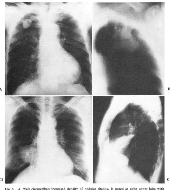
結核이 24 예로 良性 훌愚 중 제일 많았고 CFig.6),
그 외에 아스페르길루스肉穿睡이 4 예 C Fig. 7), 크럽토 코쿠스肉穿睡이 1 예, 題錫이 3 예, 睡題 細뼈나結核園 이 보이지 않아 진단이 不可能했던 예가 8 예, 적절한 조직 채취를 못했던 것이 2 예 있었다 CTable
I ).
Table I. Result of needle aspiration biopsy No.
Tuberculosis Aspergilloma Hamartoma Cryptococcosis Abscess Negative*
Inadequate specimen
4
,3 q) 7
껴/‘
1i
3
l
μ
4 3 1 3 8 2 Squamous cell ca.
Adeno ca.
Small cell ca.
Large cell ca.
‘undifferentiated’ Probable non-small cell ca.
‘poorly differen tiated’
Malignancy of undetermined type Cytologic diagnosis of aspirates 代가 제일 많았다. 병변의 크기는 장경과 단경의 평균치
로 2 αn 이하가 27 예였£며, 제일 작았던 것은 0.8cm 였고, 큰 것은 20 cm 이 넘는 것도 있었다. K훨塊의 평 균 크기는 3.4 cm 이었고 睡塊의 크기와 經皮的 뼈 吸 引生檢의 진단율과는 유의한 상관 관계는 없었다. 睡塊 의 위치는 뼈 中心部기. 6 예였고 나머지는 師 邊Y감部였 다. 經皮的 뼈 吸引生檢術을 도엽한 후, 첫 12 개월 간 은 41 예플 시행했으며, 이 중 37 예에서는 우선적으로 氣管技鏡檢훌를 했으냐 모두 진단을 옷했던 예 들이고,
후반 10 7~ 월 간은 72 예를 시행했£며 이 중 37 예에서
氣管技鏡檢흉를 하여 51 예에서 진단을 내릴 수 있었다.
細6힘病理檢홀의 결과는 惡性 睡揚이 55 예 였으며 이 중 扁平上皮細뼈}훨 Csquamous cell ca.) 이 24 예로 제 일 많았고 CFig.2) 臨찮 Cadeno ca.) 이 15 예 였으며
CFig.3), 大型 未分化 細뼈癡 Cundifferentiated ca. large cell type) 이 7 예, 小型 細뼈홉 Csmall cell ca.) 이 5 예 CFig.4) 未分化 및 非小型 細R빙}폼 Cpoorly di fferentiated ca. non - small cell type) 이 3 예,惡性 睡癡S로만 보고 된 것이 1예 있었다. 良性 睡場으로 는 過誤睡Chamartoma) 이 3 예 있었 A며 αig.5) 이 중 1예는 手術을 하여 확진되었고 2 예는 1 년 이상 추 적하였으나 그 크기의 변화나 증세가 없었다.
* No tumor cell, no A.F.B., necrotic tissue only
Fig. 3. Chest P.A shows round mass with fuzzy border at right upper lobe, medial portion. The mass was punctured and diagnosed as adenocar.
cInoma.
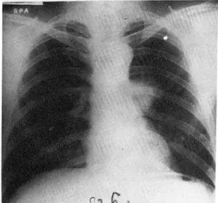
Lobulated mass is noted at right lower lobe without boliterating right cardiac border Right para-tracheal node enalargement is also noted. Percutaneous needle aspiration biopsy was performed and diagnosed as squamous cell carcinoma.
Fig.2.
手術을 하여 該斷이 바뀐 예가 5 예 였는데 2 예의 大型 未分化 細泡훨이 각 각 臨癡 및 轉移f生 精上皮睡
스페 르길루스 肉穿睡 1 예, 類肉睡Csarcoidosis) 1 예 그 ÚTIetastatic seminoma)_으로 판명되었고, 未分化 및 非
리고 5 예의 結核 o 로 확진되었다 CTable 표) • 細뼈病理 小型 細뼈행 2 예는 각 각 過誤睡 및 ~모!치穿睡 tlî
上 談斷을 할 수 없을 정도로 조직 채취가 부적당 했던 것이 2 예 였는데, 1예는 睡塊의 섬한 石짜質 침착으로 檢針이 통과를 못한 예 였고, 手術하여 軟骨肉睡 CCho-
ndrosarcoma) 으로 확진 되 었고 CFig.8) 다은 1 예 는
직경이 O.8cmX 1.0cm 의 결절로서 투시기에서 보이
지 않을 정도로 작았는데, 추적 검사상 결정의 크기의 변화가 없어 結核性 結節로 생각되는 예이다 CTable
ßD-
따라서 經皮的 師 吸引生檢으로 病變 조직의 채취율은 98 %이며 惡性 핏愚을 惡性훌愚으로의 진단율은 95 %, 細뼈型까지 고려한 확진율은 79 %이고, 전체적으로 惡 性과 良性 흉愚의 감별율은 89 %에 이른다CTable 1V).
n 씌 r: ‘
FiS-4. Left hilum is enlarged with adjacent lobulated mass. This was diagnosed as small cell car- cinoma by percutaneous needle aspiration biopsy.
C IymriJomatoid granulomatosis) 으로 꽉진되 었£며, 扁 平上皮細뼈 I예는 眼찮으흐 판명 되었다. 조직 채취는 하였무나 細뼈病理檢흉上 확살한 談斷을 할 수 없었던 예 가 있었는데 이들은 轉移性 扁2JS上皮細脫提 1 예, 아
Table 1I. 13 cases of mismatched histology or negative resu1ts.
Cytologic Dx. No. Final Dx.
. Undifferentiated ca. 2 1: adeno ca.
‘large cell' 1: metastatic
. Poor1y differen tiated ca. 2 1: hamartoma
. Squamous cell ca . . Negative*
1: lymphomatoid granulomatosis 1: adeno ca.
8 1: metastatic squamous cell ca.
m
$
않 m
’
m
비 m냉 ι찌 κ
e x e
째 빼 뼈
’li
’li -‘J
*No tumor cel1, no A.F.B., necrotic tissue only
Fig.5. Chest P-A taken for routine physic외 examma- tion shows well circumscribed round mass with mott1ed calcifications at right mid-lung field.
Chondrocyte and cartilage were demonstrated on the aspirated materia1 and diagnosed as chondromatous hamartoma. So
,
no operation was performed on this patient.Table III. Two cases of inadequate material
No. Mass character Final Dx.
1 Densely calcified mass
at left anterior chest Chondrosarcoma wal1
1 0.8 x 1.0 cm sized nodule
at right upper lung and Tuberculous invisible on lateral granuloma vJew
A
Cl
.""IF""
Fig.6. A. Well circuscribed increased density of nodular shadow is noted at right upper lobe with some emphysematous Langhan’s giant cell were aspirated.
B. Round mass shadow is noted at left upper and pleural invasion is suggested on tomography Fibrostreaky density is also noted at right upper lober. Percutaneous needle aspiration biopsy showed many A.F.B. instead of malignant cell.
Cl,2. Hemorrhagic fluid was aspirated from this loculated effusion at right major fissure con- taining many A.F.B. and histiocyte.
Table IV. Diagnostic accuracy Table V. CompJications
(n=100) Tissue 0 btainability
Benignity vs. malignancy
98%
89% Pneumothorax
. malignancy as malignancy 95% . Spontaneous resorption 13
. type specific accuracy of malignancy 89% . Requiring treatment . type specific accuracy of benign disease ; 79% . Minimal hemoptysis
- 750-
B
C2
14
16
Fig. 7. Numerous hyphae with branching pattern was aspirated from left upper lobe mass and diagno.
sed as aspergilloma. This was confirmed by surgery.
Fig. 8. Densely calcified mass is noted at anterior mediastinum. Percutaneous needle aspiration was tried only to fail due to hardness of the mass. This was confirmed as chondrosarcoma on surgery.
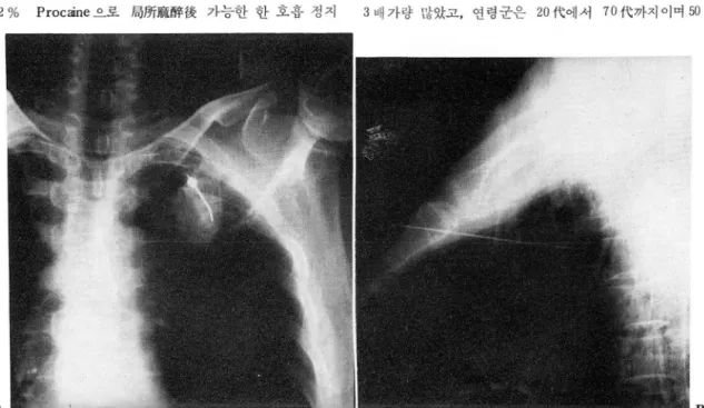
시술로 인한 合佛효으후는 14 예의 氣關이 발생했는데 CFig.9), 13 예에서는 자연 소실됐으며, 1예에서는 氣 뼈 및 臨뼈 Cempyema) 이 생겨 뼈管 CChest - tube) 으 로 치료하였다. 그 외에 시술후 가래에 피가 묻어 나 오거나, 穿刺 부위의 가벼운 통증 등이 있었무냐 일시적 인 것이어서 우시할 정도의 것이었다 CTable V) .
v.
고 찰經皮的 nrlí 吸引生檢은 1909 년 영국의 Holder 등에 의
해 처음 기술된 이래 2> 새로운 檢針의 개발과 2 ,‘, 7) 透視機 해상력의 말전으로 현재는 뼈睡塊의 該斷에 있 어서 보편화된 요긴한 檢훌가되고 있다. 섬세한 針을 사용한 經皮的 細 吸引生檢法은 뼈睡塊에 관한 한 기존 의 氣管技짧生檢法, 氣管技브러쉬生檢法, 經氣管技生檢 法 및 切斷針 Ccuttir몽 needle) 을 사용한 師 生檢法보다 그 확진율이 높으며, 안전하고 쉽게 반복시행 할수 있다 는 장점이 있다 1 J 5 ) 실제로 시울에 要하는 시간은 10 分정도로서, 환자나 시술자에게 부담이 적은 검사임 을 알수 있다. 經皮的 師 吸引生檢의 일반적인 적응증 으로는 소위 말하는 銷殘型 病變 Ccoin Iesion) 이 라 하 겠으며 1
,
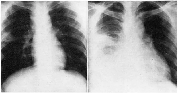
5) 轉移性 뼈癡인 경우 (Fig.l0), 細뼈型을 알 아 등?斷 기간을 단축시킬 수 있겠다. 氣管技內 病變이나 無氣ßr!i, 或은 홉潤性 뼈횟愚인 경우는 圖塊의 위치가 확Fig. 9. Right upper lobe nodule was aspirated and diagnosed as Tbc granuloma. Chest P-A taken 3 hours after aspiration shows pneumothorax.
This was resorbed spontaneously without treatment.
설치 않아 적절한 대상이 되지 않으며, 저자들의 경우 발한 바 있어 CFig.ll), 이런 경우에는 시솔을 피하는 에도 감염에 의한 홈潤性 師훌愚에서 園채취를 위하여 것이 좋으리라 생각된다. 그 외에 유아나 뼈氣睡 Cemph.
經皮的 뼈 吸引生檢을 실시했었으나 該斷을 못했던 경 ysema) 이 섬한 환자, 매우 작은 轉移 結節이 뼈에 퍼져 우가 3 예 있었으며 이 中 1예에서는 뼈의 찢효性 病 있는 경우, 出血性 碩向이 심한 환자는 부적당한 대상 뚫를 助願으후 파급시키는 결과를 招來하여 8郞힘을 유 이 되나 可能한 合佛효과 吸引生檢에 의한 該斷의 必
A
Fig. 10. Multiple variable sized mass are noted at both upper lobes in alleged laryngeal cancer patient.
Lung aspiration material showed squamous cell carcinoma suggesting lung metastasis of laryngeal cancer.
要性을 比較하여 후자의 비중이 클 경우 시술의 대상이 됨은물론이다.
진단율은 96.5 % 3) 에서 86.4 % 5) 까지 보고자마다 다양하며 Lanφnan 6) 이나 Walls 등 )0) 은 뼈睡塊의 진 단에 있어서 氣管技鏡檢훌, 氣管技브리쉬 生檢등 보다는 經皮的 뼈 吸引生檢의 우월성을 주장하고 있다. 특히 病 變이 작을수록, 邊땀部에 있을수록, Pancoast t뻐α 냐 轉移性 뼈}끊등외 경 우에 는 經皮的 ffrli 吸引生檢이 우선적
인 진단법이 된다7.9 , 10>
시슐에 의한 合혐훈으로는 氣뼈이 제일 흔한 것으로,
보고자마다 20 % 내외를 보고하고 있고, 이 증 치료를 要하는 경우는 10 % 미만이라 했£며 1쩔血은 약 8%
정도를 보고하고 있다 3,5 ,11 )
저자들의 경우 1982 年 10 月 처음 經皮的 뼈 吸引生 檢術을 도입한 이래 최근까지 施術 횟수가 증가하는 추 세에 있 a며, 초기의 41 예에서는 무조건 氣管技鏡檢훌 흘 우선적」즈로 시행했£나 )) 후반기에서는 환자를 선 별하여 氣管校鏡檢효를 시행하는 추세로 바뀌고 있으며,
B
Fig. 11. A. Superior mediastinum is widened and triangular shaped pneumonic infiltration is noted at right mid.lung field. Percutaneous needle aspiration biopsy was tried for the organism of right lung infiltration.
B. Lots of pleural effusion developed at right side after lung aspiration. Chest tube was inserted and pus was drained.
經皮的 nrü f!&引生檢에 의한 %뼈病理的 장斷을 팍진의 수단으로 상는 인식의 변화가 두드머지게 나타나고 있 다. 지자들의 경우 외i!!l ‘현者에 대한 施術은 3 예
C
3%) 로서, 49.4 %가 外來 ,월者었던 Lalli 5
,
등의 경 우와 큰 차이릎 보였는데, 이는 앞으로 經皮的 Hrlí I及 引生檢의 談斷的 가치 및 안전성에 대한 임상 의사들의 인식의 폭이 넓어질수록 외래 환자의 비율이 높아지리 라보며, 또는 엽원하더라도 그 기간을 단축시키는 결 과가 되 어 비 용 -효과 Ccost - effectiveness) 변 에 서 도 바람직한 진료가 되리라 믿는다.,
VI. 결 론
저자들은 1982 年 10 月부터, 1984 年 7 月까지 22 개 월간 서울大學校病院 응t~예放져t綠科에서 施行한 113 예 의 經皮的 8市 吸引生檢 中 임상적S로 추적이 가능한
100 예플 대상으로 하여 放射綠學的, 病理學的 및 임상 적 결과를 분석하여 다음과 같은 결론을 얻었다.
1. 經皮的 Hrlí 吸引生檢에 의한 양斷은 惡샘:OC휠 55
예, 良{生 흉愚 35여i , 5?斷이 불확실했던 것이 8 예, 그려고 조직 채취가 불충분 했던 것이 2 예 있었다.
2. 수울을 시행하여 細뼈型, 或은 談斷이 비뀐 것 이 5 예였고, 塊死 組織만 추출됐던 것이 8 예, 조직이 추출되지 않았던 것이 2 예있었다.
3. 經皮的 師 吸引生檢의 組熾 및 組織/IX;‘分 채취 율은 98 %이 며 惡性과 良{生 族愚의 감별 율은 89 %, 惡性 f쫓 愚의 등?斷率은 95 %였고, 細뼈型까지 고러 한 惡性 훗4뿔、
과 良性 훌愚、의 등짧新率은 각각 89 % 및 79 %였다.
4 시솔로 인한 合하효으로는 14예의 氣뼈아 있었 으며 。l 중 13 예는 자연 소실되었고, 1예는 뼈管S로 치료하였다.
5. 시술의 횟수는 초만기의 月 zp:均 3.4 회에서 후반 기에는 7.2 배로 2 배 이상의 증가가 있었£며뽑者를 선별하여 氣管技鏡檢흉를 시행하는 추세로 바뀌었다.
진단율도 초기 41 예의 83 %에서 후반기에는 90 %로
向上되었고, 經皮的 Hr5 吸引生檢을 확진의 수단으로삼
는 인식의 변화가 있었다.
따라서 저 자플은 M에 睡塊가 있는 愚者에서 우선적 으로 간편하고 안전한 經皮的 뻐 吸引生檢을 실시하 여 진단 기간의 단축과 높은 진단율을 기대하는 바 적 극 권장하는 바이다.
REFERENCES
1. 박재형, 엮 덕, 임정기 : 局所的 뼈찢愚의 經皮的師 生檢, 대한 방사선 의학회지 제 20 권 45-50, 1984
2. Zornoza J. 5now J, J r., Lukeman J M Aspiration biopsy of discrete pulmonary lesions using a new thin needle. Radiology 723: 5 79-520
,
7977.3. Westcott J L Direct percutaneous needle aspiration of localized pulmonary lesions; Results in 422 patients. Radiology 737:35
,
7980.4. House AJ5
,
Thomson KR Evaluation of a new transthoracic needle for biopsy of benign and malignant lung lesions. AJ R 729: 2 7 5-22α 7977.5. Lalli AF, McCormack Lj, Zelch M et al : Aspiration biopsies of chest lesions. Radiology 127:35-4'0
,
1978.
6. Landman 5
,
Burgenera FA,
Lim G Comparison of bronchial brushing and percutaneous needle aspira- tion biopsy in the diagnosis of malignant lung lesions. Radiology 175:275-278,
1975.7. J ereb M The usefulness of needle biopsy in chest lesions of different sizes and locations. Radiology
734: 73- 7 5
,
7980.8. Fennessy J J
,
Fry WA,
Manalo-Estrella P : The born- chial brushing technique for obtaining cytologic specimens from peripheral lung lesions. Acta Cytol74:25-3α 7970.
9. 5inner WN Thornbury J R
,
Naylor B : Pulmonary needle aspirations biopsy in the diagnosis of pan- coast tumors,
Radiology 777 :99-7 02,
1974.10. Walls W J, Thornbury J R
,
Naylor B Pulmonary needle aspirations biopsy in the diagnosis of pan- coast tumors,
Radiology 777 :99-102,
7974.11. Heimlich J H Valve drainage of pleural cavity.
Dis Chest 53:282-287
,
7968.12. 5argent EN
,
Turner AF Emergency treatment of pneumotheroax. A simple catheter technique for use in the radiology department. Am J Roent- genol 709:537-535,
1970.13. 5argent EN
,
Turner AF,
Gordouson J : Percutaneous pulmonary needle biopsy. Report of 350 patients Am J Roentgenol 722:758-768,
7974.14. Li ndstrom RR
,
Collins JD,
Byfield JE Obtaining viable tumor ce//s through percutaneous pulmonary needle biopsy. Radiology 94:200-203,
7970.15. Björn Nordenstrõm New instruments for biopsy.
Radiology 777:474-47ε 7975
16. Herman PG