Introduction
Osteological evidence of diseases in archaeologically obtained human skeletal samples is very important to bio- anthropologists and paleopathologists, as it is very helpful for, even essential to, any comprehensive understanding of health and disease patterns in historical populations. This notwithstanding, it is only quite recently that archaeologi- cal skeletons in Korea have been investigated for such pur- poses, given that there have heretofore been very few skele- tal series available from Korean museums, universities or institutes. Joseon Dynasty (1392~1910 AD) tombs typical- ly provide relatively good-preservation-status of cultural and human remains, and have yielded many constructive
osteological traces of human diseases. The relevant papers have reported on diffuse idiopathic skeletal hyperostosis [1], dental caries [2], enamel hypoplasia [3], vertebral de- generative changes [4,5], degenerative joint disease [6,7], and trauma [8]. Although these osteoarchaeological results are impressive, many aspects of paleopathology, for the reason above-mentioned, remain elusive in Korea.
Periostitis, though it is becoming increasingly rare in the present day, is one of the most commonly encountered abnormalities in archaeological samples, prior to the dis- covery of antibiotics and their use as a medical treatment modality [9]. Periostitis is classified as either primary or secondary. Secondary periostitis is caused by specific in- fectious disorders such as syphilis. Of course, we cannot deny that the term primary periostitis, as applied to archae- ological skeletal samples, has perhaps been used only in cases of unknown etiology [9]. The presence of periostitis has been reported worldwide as an important health marker of archaeological skeletons [10-14].
However, as in the case of some other pre-modern dis- eases, there have not yet been any reports on evidence of periostitis in Korean skeletal series. In the present study, in
Evidence of Periostitis in Joseon Dynasty Skeletons
Yi-Suk Kim
1, Deog Kyeom Kim
2, Chang Seok Oh
3,4, Myeung Ju Kim
5, Hye-Ri Kim
6, Dong Hoon Shin
3,4,*
(Received 31 May 2013, revised 24 June 2013, accepted 25 June 2013, Published Online 30 June 2013)
1Department of Anatomy, Ewha Womans University School of Medicine
2Department of Internal Medicine, Seoul National University Boramae Hospital
3Department of Anatomy, Seoul National University College of Medicine
4Institute of Forensic Medicine, Seoul National University College of Medicine
5Department of Anatomy, Dankook University College of Medicine, Chonan
6Department of Rehabilitation Medicine, Seoul National University Bundang Hospital
Abstract : Periostitis is one of the human diseases commonly encountered in archaeological samples. It is known to be an important health indicator for paleopathologists examining skeletal remains. In our recent study on a Joseon skeletal series (n==101), non-specific, primary periostitis was observed only in five individuals (#4,
#29, #137, #175, and #290). Notably, there were no secondary periostitis-suggestive signs (e.g. syphilis), except for those caused by fractures (#33 and #41). As this is the inaugural Korean-skeletal-series report on periostitis, the results presented in these pages should prove significant to interested paleopathologists.
Keywords:Joseon, Periostitis, Skeleton, Syphilis, Paleopathology
*This study was supported by from the SNUH research fund (grant number 04-2010-0140).
The author (s) agree to abide by the good publication practice guideline for medical journals.
The author (s) declare that there are no conflicts of interest.
Correspondence to : Dong Hoon Shin (Bioanthropology and Paleopathology Lab Dept of Anatomy; Institute of Forensic Medicine Seoul National Univer- sity College of Medicine 28 Yongon-Dong, Chongno-Gu, Seoul 110-799, Korea)
E-mail : [email protected]
Korean J Phys Anthropol Vol. 26, No. 2 (2013) pp. 81~90
http://dx.doi.org/10.11637/kjpa.2013.26.2.81 Original Article
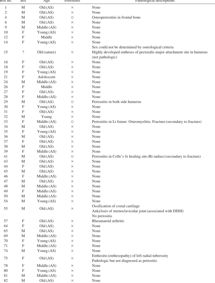
Table 1. Periostitis in the examined cases
Box no. Sex Age Periostitis Pathological descriptions
1 M Old (AS) × None
2 M Old (AS) × None
4 M Old (AS) ○ Osteoperiostitis in frontal bone
6 M Old (AS) × None
9 M Middle (AS) × None
10 F Young (AS) × None
12 F Middle × None
14 F Young (AS) × None
Sex could not be determined by osteological criteria
15 ? Old (suture) × Highly developed entheses of pectoralis major attachment site in humerus (not pathologic)
16 F Old (AS) × None
18 F Old (AS) × None
19 F Young (AS) × None
21 F Adolescent × None
24 M Middle (AS) × None
26 F Middle × None
27 F Old (AS) × None
28 F Middle (AS) × None
29 M Old (AS) ○ Periostitis in both side humerus
30 F Young (AS) × None
31 F Old (AS) × None
32 M Young × None
33 F Middle (AS) ○ Periostitis in Lt femur: Osteomyelitis; Fracture (secondary to fracture)
34 M Old (AS) × None
35 F Young (AS) × None
36 M Old (AS) × None
37 F Old (AS) × None
38 M Old (AS) × None
39 F Middle (AS) × None
41 M Old (AS) ○ Periostitis in Colle’s fx healing site (Rt radius) (secondary to fracture)
43 M Old (AS) × None
44 F Old (AS) × None
45 M Old (AS) × None
46 F Middle (AS) × None
47 M Old (AS) × None
48 M Middle (AS) × None
49 F Middle (AS) × None
50 M Middle (AS) × None
54 M Young (AS) × None
55 M Old (AS) × Ossification of costal cartilage
Ankylosis of sternoclavicular joint (associated with DISH) No periositis
57 F Old (AS) × Rheumatoid arthritis
64 F Old (AS) × None
65 M Old (AS) × None
69 M Middle (AS) × None
70 F Young (AS) × None
71 F Middle (AS) × None
74 M Young (AS) × None
75 F Old (AS) × Enthesitis (enthesopathy) of left radial tuberosity Pathologic but not diagnosed as periositis
78 F Middle (AS) × None
80 F Young (AS) × None
81 M Middle (AS) × None
82 M Old (AS) × None
83 M Adolescent × None
84 M Middle (AS) × None
85 M Old (AS) × None
87 F Young (AS) × None
88 M Middle (AS) × None
89 F Middle (AS) × None
94 M Middle (AS) × None
98 M Middle (AS) × None
99 F Middle (AS) × None
100 M Middle (AS) × None
101 F Middle (AS) × None
102 M Middle (AS) × None
103 F Middle (AS) × None
107 M Middle (AS) × None
110 M Middle (AS) × None
112 M Old (AS) × None
113 F Middle (AS) × None
114 F Middle (AS) × None
115 M Middle (AS) × None
116 M Old (AS) × None
120 F Middle (AS) × None
123 M Middle (AS) × None
129 M Middle (AS) × None
134 F Middle (AS) × None
137 M Old (AS) ○ Periostitis in left femur
140 M Middle (AS) × Lytic enthesopathy of costoclavicular ligament attachment site Pathologic but not diagnosed as periositis
145 M Middle (AS) × Lytic enthesopathy of costoclavicular ligament attachment site Pathologic but not diagnosed as periositis
159 M Old (AS) × None
160 F Middle (AS) × None
162 M Middle (AS) × None
169 M Middle (AS) × None
171 M Middle (AS) × None
172B M Old (AS) × None
173 F Middle (AS) × None
175 F Middle (AS) ○ Periostitis in right tibia
177 M Middle (AS) × None
198 F Young (AS) × None
200 F Old (AS) × None
201 M Middle × None
219 M Middle (AS) × None
220 M Old (AS) × None
221 F Middle (AS) × None
225 M Middle (AS) × None
226 M Old (AS) × None
227 F Middle (AS) × None
228 F Old (AS) × None
229 M Middle × None
230 M Middle (AS) × None
234 M Middle (AS) × None
236 M Old (AS) × None
237 M Middle (AS) × None
249 F Middle × None
290 M Old (AS) ○ Periositis of bilateral iliopubic eminence of hip bones
Highly developed entheses of pectoralis major attachment site (not pathologic) Table 1. Continued
Box no. Sex Age Periostitis Pathological descriptions
order to shed light on the periostitis pattern in a pre-modern Korean population, we examined periostitis-suggestive osteological traces in a Joseon Dynasty skeletal series.
Materials and Methods
Skeletons (n==101) maintained in the Joseon Dynasty Human Sample Collection were examined in this study.
Every such sample initially was confirmed to date from the 16th to 18th centuries, as based on the contextual archae- ological evidence and carbon dating results results [1,4,15].
Sex was determined in each case by reference to the shape of the mastoid process or sciatic notch [16]. Age at death was estimated by the method of Lovejoy et al. [17]. The individuals were classified as adolescent (12~20 years), young adult (20~35 years), middle-aged (35~50 years), and old-aged (over 50 years) groups according to the fol- lowing osteological indicators: the degrees of transverse organization, granularity, apical activity, retro-auricular activity, and articular surface porosity.
We thoroughly examined the collection for the perios- titis when two or more parts of bone elements exhibited active or healed lesions with microprosity and bony spurs as including in the specimen by the presence of at least 50% of individual skeleton. Such signs included many lay- ers of bone parallel to the surface of the underlying bone, Codman’s triangle, a thick bony layer with an irregular sur- face, bony spicules perpendicular to the underlying cortex, and the presence of woven bone on a porous-seeming bony surface, among others [9]. Bones very close to the skin (e.g.
the calvaria or anterior surface of the tibia) were searched especially carefully for evidence of the disease [9]. As for the kinds of traumatic injuries identified in our previous study [4], we also, in the present case, determined whether they were accompanied by any signs of periostitis.
We also looked for osteological-evidentiary traces of periostitis that might have been caused by specific infec- tions [9], for instance syphilis (tertiary syphilis). The pre- sence of caries sicca, a sign of tertiary syphilis in calvaria, was confirmed by central lytic bone destruction with peri- pheral porotic change of periosteal-reactive bone. We were aware that tertiary-syphilitic bone changes are frequently observed in the tibia, the bones surrounding the nasal cav- ity, and the cranial vault [9,18].
Results
Our results are summarized in Table 1. Primary perios- titis was observed in five individuals (#4, #29, #137, #175, and #290). It was found in the frontal bone of the skull (#4), in the humerus (#29), the femur (#137), tibia (#175), and in the hip bones (#290) as well. Generally, as the calvaria and anterior surface of tibia are situated just beneath the skin, they can be damaged by recurrent minor trauma, final- ly becoming the most commonly involved periosteal sites [9,18]. Significantly, in this study, we also observed signs of primary periostitis in the calvaria and tibia (cases #4 and
#175).
Specifically, in case #4, we found periostitis to the mid- dle and left on the frontal bone, about 10.8 mm from the bregma. The overall area was 64.25×37.5 mm. The porous skull surface showed little sign of taphonomic changes (Fig. 1A and 1B). In case #29, the signs, increased microporosity and small body spur, were in the bilateral surgical neck of the humerus, where new bone formation and periosteal-reactive bone changes were observed (Fig.
1C and 1D). Although highly developed entheses at the subscapularis were discovered, it appeared to be degenera- tive change rather than a pathological sign. A porous-seem- ing left-femur surface, which might has been the healing site of a traumatic injury (e.g. green-stick fracture) in youth (Fig. 2A to 2C), was found in case #137. In case #175, we found periosteal signs on the posterior surface of the right tibia (Fig. 3A and 3B). Similar signs were observed in case
#290 as well, where periostitis in the bilateral iliopubic eminence of the hip bones was discovered (Fig. 3C).
Bony changes in two previously reported facture cases (#33 and #41) [19] are likely to be caused by secondary periostitis. In case #33, there were periosteal signs at a femur-fracture site. Periostitis induced by Colles’ fracture could be seen also in case #41, wherein, on the bone sur- face, signs of porosity were apparent. Except for these, we could not find any other bony changes suggestive of secon- dary periostitis in the collection. The osteological signs of tertiary syphilis could not be discovered by us at all.
Discussion
In a previous study on pre-and post-Columbian Pecos Pueblo skeletal remains, periostitis was diagnosed in 13 of
503 (2.6%) skeletons [20]. By sharp contrast, the skeletal samples of the Roman period (3rd~5th centuries) found in Croatia showed periosteal frequencies as high as 66.7% for subadults and 30.4% for adults. In the medieval Polish skeletal samples (10~13th century AD), the prevalence of periostitis was shown 16.7% (8/48) for subadult and 42.2%
(35/83) for adult. In the 12~15th century skeletal sample of the southern Croatian, 20% (4/20) for subadult exhibit the periostitis and 12.1% (7/58) was for adult [21,22].
The authors speculated that these higher incidences re-
flect the relatively low quality of life of those historical people, which condition in that particular walled city, they further surmised, was the result of overcrowding, poor sanitary conditions, and consequent, rampant infectious diseases [14]. In general, periostitis in archaeological ske- letons is known to be an important indicator of response to various stresses. Its prevalence increased as economic and/or societal conditions worsened [9].
In the present study, we did find five cases of primary periostitis (4.9%). This prevalence, just slightly higher than Fig. 1. (A) and (B) Periostitis (asterisk) in frontal bone in case #4. (B) is a magnified image of (A). The surface of the skull was porous.
(C) and (D) Periostitis in case #29. (C) Right humerus. (D) Left humerus. In the surgical neck of the humerus, periosteal changes could be observed (areas marked by broken lines).
A B
C D
that of the above-mentioned pre- and post-Columbian Pecos Pueblo people, is, perhaps surprisingly, far lower than that of the just-noted Roman-period Croatians. How can this be explained? Actually, it was well known to us that the samples we examined in the current study are the remains of individuals buried in Hoegwakmyo tombs, which is to say, the remains of Joseon Dynasty elites [23]. The rela- tively tranquil lives they enjoyed might, in fact, be all the explanation that is needed. A final, definitive answer, how- ever, must await the examination of additional samples.
In this study, we also paid particular attention to secon- dary periostitis. In the trauma cases previously reported [19], we found osteological signs of secondary periostitis. How-
ever in case of periostitis caused by specific infections (e.g.
syphilis), we could not find any osteological evidences in our collection. Syphilis was the major venereal pathogen prevalent among urban adult populations prior to the dis- covery and application of antibiotics. Many countries in those earlier years and centuries suffered estimated infec- tion rates as high as 5% [18,23]. With respect to the origin of syphilis, a number of theories have been debated among paleopathologists around the world. Some researchers have posited “the Columbian Origin of Treponematoses”, according to which syphilis was originally indigenous to America, and then spread to other parts of the world, espe- cially the Old World, after Columbus’ discovery [18,24-27].
Fig. 2. (A) Porous-seeming surface in left femur, possibly caused by periostitis, in case #137. (B) and (C) are magnified images in (A).
A
B
C
Fig. 3. (A) and (B) Case #175. (A) Periostitis in right tibia (indicated by arrow). (B) is a magnified image of the periostitis in (A). (C) Periosteal trace observed in hip bone in case #290 (indicated by arrow in inset).
A B
C
Since osteological investigations have turned up only very rare instances of syphilis in pre-1492 Old World samples, the Columbian origin of treponematoses does seem reason- able [9,28]. However, more studies on Old World samples are still needed for confirmation of hypothesis because another research group still insisted that syphilis was esta- blished in the Old World even before the discovery of the Americas [18,29]. Certainly, if Old World skeletal series of 1492 or earlier were to show osteological evidence of syphilitic infection, the argument would take on a whole new complexion [30].
However, no reports of any osteological signs of tertiary syphilis in archaeologically obtained skeletal series have yet been published in Korea. The present Joseon skeletal- series study, likewise, discovered no such evidence. Accor- ding to medical historians, syphilis seems to have been introduced to Korea from China, possibly between 1506 and 1521 [31,32]. Many more skeletal samples will need to be examined in forthcoming studies, for more decisive conclusion on the history of syphilis in Korea.
References
1. Kim MJ, Lee IS, Kim Y-S, Oh CS, Park JB, Shin MH, et al. Diffuse Idiopathic Skeletal Hyperostosis cases found in Joseon Dynasty Human Sample Collection of Korea. Int J Osteoarchaeol. 2012; 22(2):235-44.
2. Han SS, Baek KW, Shin MH, Kim J, Oh CS, Lee SJ, et al.
Dental caries prevalence of medieval Korean people. Arch Oral Biol. 2010; 55(7): 535-40.
3. Jeong Y, Woo EJ, Pak S. No Significant Difference in the Levels of Dental Fluctuating Asymmetry between Hypopla- stic and Non-Hypoplastic Skeletal Groups from the Joseon Dynasty (mid 15th-early 20th century), South Korea. Arch Oral Biol (in press).
4. Kim DK, Kim MJ, Kim Y-S, Oh CS, Shin DH. Vertebral osteophyte of pre-modern Korean skeletons from Joseon tombs. Anat Cell Biol. 2012; 45:274-81.
5. Woo EJ, Pak S. The Relationship between the Two Types of Vertebral Degenerative Joint Disease in a Joseon Dynasty Population, Korea. International Journal of Osteoarchaeol- ogy. Published online in Wiley Online Library. (In press) 6. Woo EJ, Jeong Y, Cho GH, Pak S. Burial type and Degene-
rative Joint Disease in the Joseon Dynasty, Korea. Field Archaeology. 2011; 12:139-62.
7. Woo EJ, Pak S. Degenerative Joint Disease and Enthesopa- thies in a Joseon Dynasty Population from Korea. Homo-J
Comp Hum Biol. 2013; 64(2):104-19.
8. Lee IS, Lee EJ, Park JB, Baek SH, Oh CS, Lee SD, et al.
Acute traumatic death of a 17th century general based on examination of mummified remains found in Korea. Ann Anat. 2009; 191(3):309-20.
9. Ortner DJ. Identification of Pathological Conditions in Hu- man Skeletal Remains. 2nd Edition. Adademic Press, San Diego, USA; 2003.
10. Robb J, Bigazzi R, Lazzarini L, Scarsini C, Sonego F.
Social “status” and biological “status”: a comparison of grave goods and skeletal indicators from Pontecagnano.
Am J Phys Anthropol. 2001; 115(3):213-22.
11. Rose JC. Paleopathology of the commoners at Tell Amarna, Egypt, Akhenaten’s capital city. Mem Inst Oswaldo Cruz.
2006; 101(Suppl 2):73-6.
12. Belcastro G, Rastelli E, Mariotti V, Consiglio C, Facchini F, Bonfiglioli B. Continuity or discontinuity of the life-style in central Italy during the Roman Imperial Age-Early Middle Ages transition: diet, health, and behavior. Am J Phys An- thropol. 2007; 132(3):381-94.
13. Paine RR, Vargiu R, Signoretti C, Coppa A. A health asse- ssment for imperial Roman burials recovered from the nec- ropolis of San Donato and Bivio CH, Urbino, Italy. J Anth- ropol Sci. 2009; 87:193-210.
14. Novak M, Slaus M. Health and disease in a Roman walled city: an example of Colonia Iulia Iader. J Anthropol Sci.
2010; 88:189-206.
15. Shin DH, Oh CS, Kim YS, Hwang YI. Ancient-to-modern secular changes in Korean stature. Am J Phys Anthropol.
2012 Mar;147(3):433-42. doi: 10.1002/ajpa.22011. Epub 2012 Jan 23.
16. Buikstra JE, Ubelaker DH. 1994. Standards for data collec- tion from human skeletal remains. Fayetteville, Arkansas:
Arkansas Archaeological Survey Report Number 44.
17. Lovejoy CO, Meindl RS, Pryzbeck TR, Mensforth RP.
Chronological metamorphosis of the auricular surface of the ilium: a new method for the determination of adult ske- letal age at death. Am J Phys Anthropol. 1985; 68:15-28.
18. Aufderheide AC, Rodriguez-Martin C. The Cambridge en- cyclopedia of human paleopathology. Cambridge University Press. Cambridge, UK; 1998.
19. Kim DG, Kin MJ, Kim Y-S, Oh CS, Lee SS, Lim SB, et al.
Long bone fractures identified in the Joseon Dynasty human skeletons of Korea, Anat Cell Biol. (In press)
20. Hooton E. The Indians of Pecos Pueblo: A Study of their Skeletal Remains. New Haven: Yale University Press. 1930.
21. Betsinger TK. The biological consequences of urbanization in medieval Poland. Ohio University Doctorial thesis; 2007.
22. Novak M, Martin ˇci´c O, Strinovi´c D, Slaus M. Skeletal and dental indicators of health in the late mediaeval (12-15th cen-
tury) population from Nin, southern Croatia. Homo. 2012;
63:435-450.
23. Shin MH, Yi YS, Bok GD, Lee E-J, Spigelman M, Park JB, et al. How did mummification occur in bodies buried in tombs with a lime soil mixture barrier during the Joseon Dynasty in Korea. Mummies and Science World Mummies Research. Pena PA, Martin RM and Rodriguez AR (eds.).
2008. Santa Cruz de Tenerife, Spain. pp. 105-13.
24. Steinbock RT. 1976. Paleopathological diagnosis and inter- pretation. Charles C Thomas: Springfield.
25. Bullen AK. Paleoepidemiology and distribution of prehis- toric treponemiasis (syphilis) in Florida. Florida Anthropo- logist. 1972; 25:133-74.
26. Ortner DJ, Putschar WGJ. Identification of Pathological Conditions in Human Skeletal Remains. Smithsonian Insti- tution. Washington DC; 1985.
27. Baker BJ, Armelagos GJ. The origin and antiquity of syph- ilis: paleopathological diagnosis and interpretation. Curr Anthropol. 1988; 29:703-37.
28. Stewart TD, Spoehr A. Evidence of the palaeopathology of yaws. In Diseases in Antiquity, ed. D. Brothwell & AT San- dison, Springfield, IL: CC Thomas; 1967. pp. 307-19.
29. Livingstone FB. On the origin of syphilis: an alternative hypothesis. Curr Anthropol. 1991; 32(5):587-90.
30. Erdal YS. A pre-columbian case of congenital syphilis from Anatolia (Nicaea, 13th Century AD). Int J Osteoarchaeol.
2006; 16:16-33.
31. Miki S. The history of Korean medicine and diseases in Korea, Osaka-Prefecture (Japan); 1955.
32. Lew J, Kim JD. Recent trends of syphilis incidence in Korea.
Yonsei Medical Journal. 1968; 9(1): 74-80.
조선시대 유골에서 확인된 뼈막염
김이석
1, 김덕겸
2, 오창석
3,4, 김명주
5, 김혜리
6, 신동훈
3,4,*
1이화여자대학교 의학전문대학원 해부학교실, 2서울대학교 보라매병원 내과학교실,
3서울대학교 의과대학 해부학교실, 4서울대학교 의과대학 법의학연구소,
5단국대학교 의과대학 해부학교실, 6서울대학교 의과대학 재활의학교실
간추림 : 뼈막염은 고고학 유골에서 가장 흔히 발견되는 질병 중의 하나이며, 유골의 생전 건강상태를 가늠할 수 있게 해주는 중요한 생물학적 지표이다. 조선시대 뼈모음을 조사한 결과, 총 101명의 뼈대 중 5명의 뼈대 (#4, #29, #137, #175, #290)에서 기저질환이나 손상을 보지 못하는 일차뼈막염이 발견되었다. 한편, 매독에 속발 된 뼈막염을 관찰할 수 없었는데 향후 뼈모음의 수가 늘어난다면 이 부분에 대한 중요한 고병리학적 발견을 기 대할 수 있을 것으로 생각하였다. 이상으로 조선시대 고인골에서 발견된 뼈막염을 처음으로 보고하게 되었으며, 앞으로 다양한 고병리학적 소견에 대한 형태학적 연구를 진행할 예정이다.
찾아보기 낱말 : 뼈막염, 조선, 뼈대, 매독, 고병리학
교신저자 : 신동훈(서울대학교 의과대학 해부학교실, 법의학연구소) 전자우편 : [email protected]