Introduction
Solitary fibrous tumor (SFT) is a mesenchymal cell origin that usually occur in the pleura, but the presence of these tumors are currently reported in a variety of extrapleural sites, including retroperitoneum (1-6).
Because of the rarity of SFTs, information on the imaging features is sparse, and the radiologic features of SFT occurred in the ischiorectal fossa have been rarely reported. In this case, we describe the CT and MRI findings in a case of a SFT that involved in the ischiorectal fossa.
Case Report
A 36-year-old man presented with localized swelling and palpable mass on right gluteal area. The patient underwent lower extremity CT, which demonstrated a 13× 9.4× 6.9 cm solid mass within the right ischiorectal fossa (Fig. 1). This mass exhibited a homogeneous attenuation, similar to that of adjacent muscle. After contrast media was injected, the mass showed a heterogeneous enhancement.
Subsequently, a MRI examination was performed to evaluate local invasion and the main components of the tumor on a 1.5 T unit. T1 weighted MR images showed
JKSMRM 15:72-76(2011)
Department of Radiology, College of Medicine, Korea University Supported by a grant from Korea University
Received; March 16, 2010, revised; March 25, 2011, accepted; March 31, 2011
Corresponding author : Sang Hoon Cha, M.D., Department of Radiology, College of Medicine, Korea University,
#516, Gojan1-dong, Danwon-gu, Ansan-city, Gyeonggi-do 425-707, Korea.
Tel. 82-31-412-5227 Fax. 82-31-412-5224 E-mail: [email protected]
Solitary Fibrous Tumor of the Ischiorectal Fossa : CT and MRI Findings
Ki Hwan Kim, Sang Hoon Cha, Suk Keu Yeom, Seung Hwa Lee, Hwan Hoon Chung
Solitary fibrous tumor (SFT) is a rare neoplasm, which is usually presented as a pleural based mass, but can also occur in unusual locations based on its mesenchymal origin. However, the radiologic features of SFT occurred in the ischiorectal fossa have been rarely reported. In this case, we describe the MRI findings in a case of a SFT involving the ischiorectal fossa of a 36-year-old man. The tumor appeared as homogeneous iso-signal intensity relative to the adjacent muscle on T1 weighted images, a mixed high signal intensity on the T2 weighted images, and heterogeneous enhancement following the administration of the contrast material.
Index words :Solitary fibrous tumor Ischiorectal fossa
Magnetic resonance imaging (MRI) Computed tomography (CT)
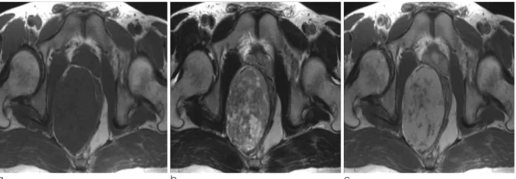
a well circumscribed mass of homogeneous iso-signal intensity relative to the adjacent muscle, without invasion of adjacent soft tissue (Fig. 2A). The mass was heterogeneous high and intermediate signal intensity including curvilinear hypointense areas on T2 weighted images (Fig. 2B). Gadolinium enhanced T1 weighted images revealed heterogeneous enhancement, and these variable enhancements were suggestive of mass containing various components such as cyst, myxoid, and necrosis (Fig. 2C). There was no evidence of enlarged lymph node and distant metastasis in the
peritoneal and retroperitoneal space. Tubular structures, which were considered to be collateral feeding vessels (Fig. 3), were revealed in inferior aspects of mass. These vessels were originated from the subcutaneous fatty tissue of the perineum. The preoperative radiologic differential diagnosis was a neurogenic tumor, leiomyoma, desmoid tumor as preoperative diagnosis.
On laparotomy, a well circumscribed, entirely encapsulated tumor was found in the pelvic cavity and protruding to right buttock area. Partial adhesion with adjacent muscle was completely dissected and there was no evidence of direct invasion. The cut surface showed a whitish yellow solid portion with multiple tiny cysts. Microscopic examination demonstrated the tumor was composed of primitive spindle cells with collageneous area and small vessels (Fig. 4A). Few mitosis less than 2/10 HPF (high power field) was found. Immunohistochemical analysis of the tumor cells had positive results for CD34, CD68, CD99, Bcl-2, but negative for cytokeratin, S-100 (Fig. 4B).
Discussion
SFT is an uncommon mesenchymal neoplasm. The extrapleural SFTs have been increasingly reported, and the incidence of extrapleural SFTs is higher than that of pleural lesions (1, 2). To our knowledge, a few reports have described the detailed MR findings of retroperitoneal SFT (3-6).
a b c
Fig. 2. Axial T1 weighted (a), T2 weighted (b), and contrast enhanced T1 weighted (c) images show soft tissue mass in right ischiorectal fossa. Mass exhibits homogeneous high signal intensity in (a) and heterogeneous high signal in (b), and heterogeneous enhancement in (c). The hyperintense area on T2-weighted images within mass revealed varying degree of enhancement.
Fig. 1. Contrast enhanced coronal reformatted CT shows large, well defined mass with heterogeneous attenuation in the right ischiorectal fossa.
SFTs usually occur in middle-aged adults with no sex predilection (1). Most thoracic SFTs are asymptomatic at presentation, but extrathoracic SFTs are usually symptomatic. The manifestations are a painless mass or local pressure effects (7). SFTs are usually benign, but 10% present cellular atypia, hypercellularity, although these findings do not predict clinical aggressiveness (8).
The behavior of extrapleural SFTs is unpredictable, and 10-15% of them show recurrent or metastatic diseases,
and surgical excision is curative in almost all cases (1, 7).
Microscopically, SFT has been described as a
‘patternless’ pattern, which means a wide range of morphological patterns, from predominantly fibrous lesions to more cellular lesions. The histological findings are consistent regardless of the involved organ (1, 2). These characteristic findings are reflected on MR imagings, especially on the T2 weighted images, rather than CT images. Based on previous published reports, SFTs usually exhibit low signal intensity relative to muscle on T1 weighted images and variable signal intensity on T2 weighted image. The linear or curvilinear hypointense areas on T2 weighted images are correlated with the hypocellular and collagenous sclerotic area (9, 10). These findings were seen in our case, and corresponded well to the abundant collagenous stroma for the pathologic correlation. The high signal intensity on T2 weighted images is correlated with more cellular tissues. Myxoid or cystic changes also show a high signal intensity on T2 weighted images (3, 11, 12). Our case showed multiple cystic areas on the histologic examination, and these seem to be related to the high signal intensity on T2 weighted images.
The well enhancement pattern of SFTs corresponds with prominent hemangiopericytic vascular spaces that are encountered histologically (5). A degree of enhancement of SFTs depends not only on a vascular structure but also on histologic factors such as Fig. 3. Sagittal contrasted enhanced T1 weighted images
demonstrate multiple tubular structures arising from the subcutaneous fat of perineum. The low signal intensity of these structures, caused by a flow void effect, is suggestive of collateral feeding vessels (arrow).
a b
Fig. 4. (a) The tumor is composed of short spindle cells with abundant stromal collagen bundles and ectactic vessels (hematoxylin and eosin staining, ×200). (b) The tumor cells are diffuse strong positive for CD34 staining (hematoxylin and eosin staining, ×200).
cellularity, myxoid tissue. Thus variable degree of enhancement may be revealed on the contrast enhanced CT and MR images. Our case also demonstrated prominent small vessels with branching patterns, which caused heterogeneous enhancement on the contrast enhanced images.
Large feeding vessels arising from the subcutaneous fatty tissue of the perineum were additionally demonstrated in our case (Fig. 3). According to a recent published report, large collateral feeding vessels are shown in 35% of SFTs arising from soft tissues (4).
These collateral vessels may be helpful for narrowing the differential diagnosis. However, the presence of the collateral vessels is not specific and not seem to be related to histologic subtype of the tumor, and other hypervascular tumors also show these findings (13, 14).
The ischiorectal fossa is a pyramid shaped space bounded medially by the levator ani muscle and external sphincter muscle. The anterior border and the apex are formed by perineal muscle and obturator fascia, respectively. The tumor involvement of the ischiorectal fossa is usually secondary to direct invasion of primary anorectal, prostate, and sacral tumors, but primary tumor is rare (15). The primary tumor arising from the ischiorectal fossa is vast, but our tumor in our case should be differentiated from the other solid mass, including neurogenic tumor, desmoid tumor, inflammatory pseudotumor, angioleiomyoma, leiomyoma, and lymphoma (5, 11). Desmoid tumors are often intramuscular and locally aggressive patterns, which are usually not seen in the SFTs, and a clinical history such as a previous operation and a postpartum period should be considered (16). Although retroperitoneal schwannomas can be larger tumors, schwannomas are usually less than 5 cm in diameter (17). However, the imaging finding of above mentioned tumors may overlap that of each other, and SFTs can be considered in the differential diagnosis.
In conclusion, we reports a rare case of an SFT of the ischiorectal fossa that appeared as a well circumscribed, large, solid mass with heterogenously enhancement at CT and MRI. Although the SFT of the ischiorectal fossa is rare, we suggest that SFTs be included as part of the differential diagnosis of the ischiorectal fossa, especially when the tumor shows a hypervasularity and a heterogeneous, high signal intensity on T2 weighted images.
References
1.Gengler C, Guillou L. Solitary fibrous tumour and haemangiopericytoma: evolution of a concept. Histopathology 2006;48:63-74
2.Goodlad JR, Fletcher CD. Solitary fibrous tumour arising at unusual sites: analysis of a series. Histopathology 1991;19:515-522
3.Chun HJ, Byun JY, Jung SE, Kim KH, Shinn KS. Benign solitary fibrous tumour of the pre-sacral space: MRI findings.
Br J Radiol 1998;71:677-679
4.Wignall OJ, Moskovic EC, Thway K, Thomas JM. Solitary fibrous tumors of the soft tissues: review of the imaging and clinical features with histopathologic correlation. AJR Am J Roentgenol 2010;195:W55-62
5.Rosenkrantz AB, Hindman N, Melamed J. Imaging appearance of solitary fibrous tumor of the abdominopelvic cavity. J Comput Assist Tomogr 2010;34:201-205
6.Vossough A, Torigian DA, Zhang PJ, Siegelman ES, Banner MP. Extrathoracic solitary fibrous tumor of the pelvic peritoneum with central malignant degeneration on CT and MRI. J Magn Reson Imaging 2005;22:684-686
7.Gold JS, Antonescu CR, Hajdu C, Ferrone CR, Hussain M, Lewis JJ, et al. Clinicopathologic correlates of solitary fibrous tumors. Cancer 2002;94:1057-1068
8.Vallat-Decouvelaere AV, Dry SM, Fletcher CD. Atypical and malignant solitary fibrous tumors in extrathoracic locations:
Evidence of their comparability to intra-thoracic tumors. Am J Surg Pathol 1998;22:1501-1511
9.Jeong AK, Lee HK, Kim SY, Cho KJ. Solitary fibrous tumor of the parapharyngeal space: MR imaging findings. AJNR Am J Neuroradiol 2002;23:473-475
10.Kim HJ, Lee HK, Seo JJ, Shin JH, Jeong AK, Lee JH, et al. MR imaging of solitary fibrous tumors in the head and neck.
Korean J Radiol 2005;6:136-142
11.Kakihara D, Yoshimitsu K, Eto M, Matsuura S, Honda H.
MRI of retroperitoneal solitary fibrous tumor in the suprarenal region. AJR Am J Roentgenol 2007;188:W512-514 12.Casas JD, Balliu E, Sa′nchez MC, Mariscal A. Benign solitary
fibrous tumor of the retroperitoneum: radiological features.
CMIG Extra: Cases 2004;28:50-53
13.Hide IG, Baudouin CJ, Murray SA, Malcolm AJ. Giant ancient schwannoma of the pelvis. Skeletal Radiol 2000;29:538-542
14.Totterman S, Reitamo JJ. Desmoid tumour: an angiographic study of five cases. Br J Radiol 1979;52:936-941
15.Llauger J, Palmer J, Perez C, Monill J, Ribe J, Moreno A. The normal and pathologic ischiorectal fossa at CT and MR imaging. Radiographics 1998;18:61-82; quiz 146
16.Lee JC, Thomas JM, Phillips S, Fisher C, Moskovic E.
Aggressive fibromatosis: MRI features with pathologic correlation. AJR Am J Roentgenol 2006;186:247-254
17.Rha SE, Byun JY, Jung SE, Chun HJ, Lee HG, Lee JM.
Neurogenic tumors in the abdomen: tumor types and imaging characteristics. Radiographics 2003;23:29-43
통신저자 : 차상훈, (425-707) 경기도 안산시 단원구 고잔1동 516, 고려대학교 의과대학 영상의학교실 Tel. 82-31-412-5227 Fax. 82-31-412-5224 E-mail: [email protected]
좌골직장와에서 기원하는 고립섬유종양: CT, MR 영상소견의 증례 보고
고려대학교 안산병원 영상의학과 김기환∙차상훈∙염석규∙이승화∙정환훈
고립섬유종양은 드문 종양으로써 주로는 흉막기원의 형태로 발생하지만, 드물게 다양한 위치에서 중간엽세포 기원 의 종양으로 발생할 수 있다. 하지만 현재까지 좌골직장와에서 발생한 고립섬유종양의 영상적 보고는 매우 드물게 보 고되었다. 저자들은 36세 남자환자의 좌골직장와에서 발생한 고립섬유종양의 증례를 자기공명 소견을 중심으로 기술 하고자 한다. 종양은 T1 강조영상에서 주변 근육조직과 비슷한 균일한 신호강도를 보이고, T2 강조영상에서는 혼합 된 고신호강도를 보이며, 조영증강 후 T1 강조영상에서는 불균일한 조영증강 소견을 보였다.
대한자기공명의과학회지 15:72-76(2011)