• 검색 결과가 없습니다.

Pulmonary Thromboembolectomy for Acute Pulmonary Thromboembolism

N/A
N/A
Protected

Academic year: 2021

Share "Pulmonary Thromboembolectomy for Acute Pulmonary Thromboembolism"

Copied!
5
0
0

로드 중.... (전체 텍스트 보기)

전체 글

(1)

http://dx.doi.org/10.5090/kjtcs.2011.44.5.343 ISSN: 2233-601X (Print) ISSN: 2093-6516 (Online)

*Department of Thoracic and Cardiovascular Surgery, Mokpo Hankook Hospital

**Department of Thoracic and Cardiovascular Surgery, Kyung Hee University Hospital

***Department of Thoracic and Cardiovascular Surgery, Kyung Hee University Hospital at Gangdong

†This journal was presented at the 35th fall conference.

Received: August 31, 2010, Revised: September 28, 2010, Accepted: June 21, 2011

Corresponding author: Joo Chul Park, Department of Thoracic and Cardiovascular Surgery, Kyung Hee University Hospital, 1, Hoegi-dong, Dongdaemun-gu, Seoul 130-872, Korea

(Tel) 82-2-958-8415 (Fax) 82-2-958-8410 (E-mail) [email protected]

C

The Korean Society for Thoracic and Cardiovascular Surgery. 2011. All right reserved.

CC
This is an open access article distributed under the terms of the Creative Commons Attribution Non-Commercial License (http://creative- commons.org/licenses/by-nc/3.0) which permits unrestricted non-commercial use, distribution, and reproduction in any medium, provided the original work is properly cited.

Pulmonary Thromboembolectomy for Acute Pulmonary Thromboembolism

Inho Yi, M.D.*, Joo Chul Park, M.D.**, Kyu Seok Cho, M.D.**, Bum Shik Kim, M.D.**, Soo-Cheol Kim, M.D.**, Dae Hyun Kim, M.D.***, Jung-Heon Kim, M.D.**, Hyo Chul Youn, M.D.**

Background: Acute pulmonary thromboembolism is fatal because of abruptly occurring hypoxemia and right ven- tricular failure. There are several treatment modalities, including anticoagulation, thrombolytics, ECMO (extracorporeal membrane oxygenator), and thromboembolectomy, for managing acute pulmonary thromboembolism. Materials and Methods: Medical records from January 1999 to December 2004 at our institution were retrospectively reviewed for pulmonary thromboembolectomy. There were 7 patients (4 men and 3 women), who underwent a total of 8 oper- ations because one patient had post-operative recurrent emboli and underwent reoperation. Surgery was indicatedfor mild hypoxemia and performed with CPB (cardiopulmonary bypass) in a beating heart state. Results: The patients had several symptoms, such as dyspnea, chest discomfort, and palpitation. Four patients had deep vein throm- boembolisms and 3 had psychotic problems, specifically schizophrenia. Post-operative complications included hemo- thorax, pleural effusion, and pericardial effusion. There were two hospital deaths, one each by brain death and right heart failure. Conclusion: Emergency operation should be performed when medical treatments are no longer effective.

Key words: 1. Pulmonary thromboembolism 2. Thromboembolectomy

INTRODUCTION

Acute pulmonary thromboembolism is a fatal disease that can cause patients to take a sudden turn for the worse from the moment of symptom manifestation due to right heart dys- function, hypotension, and hypoxia, if there is no precise and fast diagnosis and treatment for it [1]. For medical therapy of acute pulmonary thromboembolism, we can use anticoagulants and thrombolytics. However, when it comes to patients who have unstable vital signs or for whome medical therapy is not

indicated, ECMO (extracorporeal membrane oxygenator) ap- plication and emergent thromboembolectomy can be consi- dered. On the basis of the authors’ surgeries, we wanted to determine the indications and results of thromboembolectomy.

MATERIALS AND METHODS

Eight patients underwent surgery due to acute pulmonary

embolisms from January 1999 to December 2004. Among

them, we reviewed 7 patients who could be investigated by

(2)

Table 1. Characteristics of patients

Sex Age Symptoms Combined disease AaDO2*

(mmHg) DVT

Preoperative heparinization

Preoperative inotropics

Preoperative CPR

M

F M

M F M F

23

52 25

32 39 54 68

Dyspnea

Dyspnea

Chest discomfort, Syncope Chest pain Palpitation Dyspnea Chest pain

Nephrotic syndrome, mental retardation Schizophrenia

CVA

§

, PFO

Schizophrenia TKR

, Lt TKR, both

42.9  

60.9

229.3 52.6 67.6 63.8

O

O O O

O O

O

O

O

O

O

*AaDO2=Alveolar arterial oxygen difference;

DVT=Deep vein thrombosis;

CPR=Cardiopulmonary resuscitation;

§

CVA=

Cerebrovascular attack;

PFO=Patent foramen ovale;

TKR=Total knee replacement.

medical records. One of these underwent reoperation due to recurrence of pulmonary embolism on the 13th postoperative day.

Patients who had needed vasopressors for hypotension or had shown right heart dysfunction by echocardiography un- derwent surgery. We performed median sternotomy under general anesthesia and then performed cardiopulmonary by- pass without cardiac arrest. After making incisions to the pul- monary arteries longitudinally, we removed the thromboembo- lisms and cautiously compressed both lungs after opening both pleurae using an uncapped Yankauer sucker. For artery cannulation, we used the ascending aorta in 7 cases and the femoral artery in one case. On the other hand, for venous cannulation, we used both the Superior Vena Cava (SVC) and Inferior Vena Cava (IVC) in 7 cases and the femoral vein in one case. The one case of using the femoral artery and vein was the patient who had suffered from hypotension in the operating room, so we cannulated the femoral artery and vein and ran the CPB first and then continued the surgery. The mean CPB time was 80.5±48.9 minutes. The mean pharyngeal temperature was 34.4

o

C during the CPB time.

RESULTS

The mean duration from admission to operation was 3 days, which included the cases referred from other departments. The mean age was 41.9 years old (23 to 68).

The total patients were 7 cases (4 males, 3 females). Because one female had a reoperation on the 13

th

postoperative day, there were a total of 8 operations.

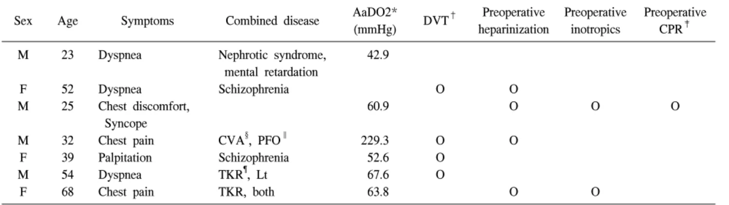
The main symptoms were dyspnea in 4 cases, chest pain and discomfort in 2 cases, palpitation in one case, and syn- cope in one case. Four of six cases showed deep vein throm- bosis in the lower extremity sonography. The preoperative blood pressure in 5 cases were in the normal range.

However, a vasopressor was infused into the two patients

who had suffered from hypotension. The mean respiratory

rate was 27 breaths per minute (22 to 36) and the mean

pulse rate was 113 beats per minute (98 to 120). The body

temperature was 38

o

C in one patient and normal in the

others. We diagnosed the disease using chest computed to-

mography and lung perfusion scan, echocardiography and pul-

monary angiography. In the chest computed tomography

scans, more than two lobar pulmonary arteries were plugged

with thromboembolisms. Echocardiography showed right heart

dilatation in 6 cases, and moderate Tricuspid regurgitation

was noted in 3 of them. A preoperative check of D-dimer

was performed in 4 cases, which were found to be elevated

to >1.0, 3.28, 1.84, and 16.8 mcg/mL. Troponin I was ele-

vated to 2, 2, and 3.43 ng/mL in 3 cases. We calculated the

difference in oxygen pressure (AaDO2) and the values were

elevated in 6 cases (Table 1). The causes of pulmonary artery

thromboembolism were deep vein thrombosis in 4 cases, pre-

vious knee transplantation in 2, cases and schizophrenia in 2

cases. Of the remaining cases, one each was caused by right

(3)

Table 2. Managements and outcomes of patients

Sex Age IVC*

filter Recurrence Death CPB time (min)

CPB BT (

o

C)

M F M M F M F

23 52 25 32 39 54 68

O

O O O

O

O

O

68 60 180 70 38 43 105

36 31 32.5 35 35 36 35.9

*IVC=Inferior vena cava (IVC filters were inserted when pa- tients were transferred from the intensive care unit to general ward after thromboembolectomy).

atrial thrombosis, patent foramen ovale (PFO) with cerebral stroke (for which we performed thromboembolectomy with PFO primary closure) and steroid taking due to renal failure.

There were two patients who had used vasopressors before surgery and one of them underwent cardiopulmonary resusci- tation. We infused heparin to 4 patients before surgery, and we inserted an IVC filter below the renal vein in 4 cases.

After that, we continued anticoagulation with warfarin.

The mean hospital day was 33.2 days (16 to 67). Hemo- throax occurred in one case and was cured by reoperation.

One patient suffered from pleural and pericardial effusion, but went home without any special treatment. The patients who had a long hospital stay needed treatment for psychotic prob- lems, and reoperation made for a long hospitalization. Two patients died, one on the operative day and the other on the first postoperative day. One was in brain death after CPR, and the other died of right heart failure with myocardial infarction. The patient in brain death underwent the operation due to the family’s insistence (Table 2). Four patients who came back for follow-up among the five surviving cases lived at least 5 years after their operations.

DISCUSSION

Acute pulmonary thromboembolism can be fatal. In our cases, the main clinical symptoms were acute dyspnea and chest comfort, and other symptoms are hypoxemia and hypo- tension without specific previous disease. Twenty percent of

patients show the normal range of arterial oxygen pressure and the normal difference between alveolar and arterial oxy- gen pressure (AaDO2). Hemodynamic changes can be shown in the time when 30∼50% of pulmonary arteries are plugged with thrombi [4].

Acute pulmonary thromboembolism can be categorized into massive and sub-massive groups. In massive acute pulmonary thromboembolism, there can be cases of systolic blood pres- sure below 90 mmHg, systolic blood pressure decreases of more than 40 mmHg over the course of 15 minutes or more, and cardiogenic shock. On the other hand, sub-massive acute pulmonary thromboembolism evidences right heart dysfunc- tion even when it shows a hemodynamically stable state [2].

The risk factors of acute pulmonary thromboembolism in- clude old age, history of deep vein thrombosis, four-limb lim- itation due to neurogenic problems, long bedridden periods due to cardiac or acute pulmonary injury, neoplasm, hormonal therapy, congenital or acquired thrombophilia, oral contra- ception, pericarditis, and history of pulmonary thromboem- bolism. In patients with deep vein thrombosis in the proximal lower extremity without chest symptoms, pulmonary embo- lism can be detected in 50% of cases by a lung perfusion scan. Seventy percent of the detected pulmonary throm- boembolisms are combined with deep vein thrombosis in the lower extremities. Among these combined cases, limitation of movement due to neurogenic disease, being over 75 years old, and cardiopulmonary disease or neoplasm are risk factors for death by acute pulmonary thromboembolism [3,4]. Danilo et al. reported that moderately depressive patients with move- ment limitations were in danger of pulmonary thromboembo- lism [10].

The revised Geneva score can be used clinically for pul-

monary embolism classification. The score takes into account

age over 65 years, history of arrhythmia, thrombus and pul-

monary embolism, history of operation within the most recent

one month, history of fracture, progressive cancer, pain of a

unilateral lower extremity, hemoptysis, heart rate over 75

beats per minute, pain on palpation of the lower extremity

vein, and unilateral edema in the lower extremity. Clinical

possibilities can be classified into three grades (low, inter-

mediate, high) according to the grade of individual risk

factors. Other diagnostic tools are chemical tests such as

(4)

pro-BNP, BNP, troponin, echocardiography, chest computed tomography, and lung perfusion scan [4].

Echocardiography can reveal the level of pressure overload to the right ventricle, through the signs of a diameter ratio between the right and left ventricle of more than 1, right ven- tricular dilatation, right ventricular hypokinesis, and the de- gree of ventricular septal bowing. In particular, it is said that the degree of ventricular septal bowing is useful in predicting death [3,5].

Among the patients diagnosed with pulmonary throm- boembolism, hemodynamically unstable patients should be treated immediately with thrombolytics, operations, and cathe- terized embolectomy. However, hemodynamically stable pa- tients without right heart dysfunction can be treated with anticoagulation. If there is an abnormal finding of the right heart, the patient can be treated with thrombolytics under close observation.

Initial therapies include anticoagulation (unfractionated hep- arin, low molecular weight heparin, fondaparinux, warfarin), thrombolytics, percutaneous thromboembolectomy, and oper- ative treatment. Warfarin therapy must be continued for three months (Target INR: 2.0∼3.0). In the cases with risk factors such as male gender, old age, malignancy, and pulmonary embolism without special preceding causes, however, warfarin therapy must be continued at least three months due to the increased possibility of recurrence. Thrombolytics cannot be used in cases of cerebral diseases, intractable hypertension, or within three weeks post-operatively [3].

An IVC filter is indicated for cases of contraindication of anticoagulation therapy, the failure of anticoagulation therapy, cardiopulmonary dysfunction with arrhythmogenic thrombosis, high risk patients for operation and the patient taking throm- bolytics due to proximal arrhythmogenic thrombosis [6].

Pulmonary thromboembolectomy has to be performed whenever several medical treatments do not show the effect in the cases of unstable blood pressure, hypoxemia, imbalance of electrolytes, or acidosis. If an emergency operation is diffi- cult to perform, the application of ECMO can be helpful to maintain survival [8].

The death rate from pulmonary thromboembolectomy has been reported to be 20%. We can perform it as follows. The first indication is the case of confirmed massive pulmonary

thromboembolism with hemodynamic changes. In this case, right heart dysfunction or the need for vasopressors becomes evident. The second one is hemodynamic instability such as shock, in spite of heparin infusion or supportive care for sta- ble vital signs. The last one is the case of a contraindication or intractable state of anticoagulation [7]. If there is a mas- sive thrombus in the proximal lower limb and a percutaneous thromboembolectomy using a catheter cannot be performed, an operation has to be considered even though the vital signs are stable [9].

CONCLUSION

Although noninvasive therapies such as anticoagulation and thrombolytics tend to gradually replace surgical interventions, in light of this study, we suggest that emergency pulmonary thromboembolectomies are needed for patients who are in a hemodynamically unstable state or have a right heart dysfunc- tion. Furthermore, patients whose movements are limited due to psychotic problems are in danger of pulmonary throm- boembolism. Therefore, careful observation of such patients is necessary.

REFERENCES

1. Lee S, Song SW, Yi G, Youn YN, Yoo KJ, Chang BC.

Open pulmonary thromboembolectomy in patients with major pulmonary thromboembolism. Yonsei Med J 2008;31;49:973- 7.

2. Digonnet A, Moya-Plana A, Aubert S, et al. Pulmonary em- bolism: a current surgical approach. Interact Cardiovasc Thorac Surg 2007;6:27-9.

3. Agnelli G, Becattini C. Acute pulmonary embolism. N Engl J Med 2010;363:266-74.

4. Torbicki A, Perrier A, Konstantinides S, et al. Guidelines on the diagnosis and management of acute pulmonary embo- lism: the task force for the diagnosis and management of acute pulmonary embolism of the European Society of Cardiology. Eur Heart J 2008;29:2276-315.

5. Araoz PA, Gotway MB, Harrington JR, Harmsen WS, Mandrekar JN. Pulmonaryembolism: prognostic CT findings.

Radiology 2007;242:889-97.

6. Young T, Tang H, Hughes R. Vena caval filters for the pre- vention of pulmonary embolism. Cochrane Database Syst Rev 2010;17;2:CD006212.

7. Dauphine C, Omari B. Pulmonary embolectomy for acute

(5)

massive pulmonary embolism. Ann Thorac Surg 2005;79:

1240-4.

8. Hori D, Tanaka M, Kohinata T, et al. Successful usage of extracorporeal membrane oxygenation as a bridge therapy for acute pulmonary embolism between hospitals. Gen Tho-

rac Cardiovasc Surg 2010;58:283-6.

9. Stein PD, Alnas M, Beemath A, Patel NR. Outcome of pul- monary embolectomy. Am J Cardiol 2007;99:421-3.

10. Arnone D, Hansen L, Davies G. Pulmonary embolism and

severe depression. Am J Psychiatry 2002;159:873-4.

수치

Table 1. Characteristics of patients
Table 2. Managements and outcomes of patients

참조

관련 문서

Percutaneous polymethylmethacrylate vertebroplasty in the treatment of osteoporotic vertebral body compression fractures: technical aspects. Pulmonary embolism caused

In order to defeat these problems and enhance patient’s convenience, we designed fluidal lipid matrix (FLM) for long-acting injection that can maintain the long efficacy

By simple analysis to fine the relation between the characteristics of subjects and use of PERS for the recent 6 months, marital status, religion,

Undiagnosed malignancy in patients with deep vein thrombosis: incidence, risk indicators, and diagnosis.. Monreal M, Fernandez-Llamazares J, Perandreu

The positive change pattern of CK-MB according to time on acute myocardiac infarction patient.. The positive change pattern of troponin-T according to time on

(Background) Gallbladder wall thickening(GWT) and gallbladder contraction are often observed in patients with acute hepatitis.. The incidence of acute hepatitis A

In detail, economic activity and income level in socio-demographic factors showed statistic significance, preparation for the old age in personal factors showed

Suggesting their wishing program for the future is a old man's role in church, financial life for old age, special activity, take care of old age's stress and the way how to