ISSN 0378-6471 (Print)⋅ISSN 2092-9374 (Online)
http://dx.doi.org/10.3341/jkos.2015.56.11.1789
Case Report
5% Imiquimod Cream으로 치료한 눈썹부위 바닥세포암 증례 1예
A Case Report of Eyebrow Basal Cell Carcinoma Successfully Treated with 5% Imiquimod Cream
강병수1⋅김남주1,2⋅정호경1,3⋅곽상인1
Byeong Soo Kang, MD1, Nam Ju Kim, MD1,2, Ho Kyung Choung, MD1,3, Sang In Khwarg, MD1 서울대학교 의과대학 안과학교실1, 서울대학교 의과대학 분당서울대학교병원 안과학교실2,
서울대학교병원운영 서울특별시보라매병원 안과학교실3
Department of Ophthalmology, Seoul National University College of Medicine1, Seoul, Korea
Department of Ophthalmology, Seoul National University Bundang Hospital, Seoul National University College of Medicine2, Seongnam, Korea Department of Ophthalmology, Seoul Metropolitan Government Seoul National University Boramae Medical Center3, Seoul, Korea
Purpose: Basal cell carcinoma is the most common malignant neoplasm of the skin. Periorbital basal cell carcinoma needs to be treated for cosmetic improvement and prevention of intraorbital invasion. Although surgical excision is a standard treatment, nonsurgical treatment using imiquimod cream, a topical immunomodulator, has been attempted. The authors report the first case of periorbital basal cell carcinoma successfully treated with imiquimod cream; to the best of our knowledge, similar cases have not been published to date in the Korean ophthalmology journals.
Case summary: A 73-year-old female visited our hospital for treatment of a biopsy-proven basal cell carcinoma in the right eyebrow. A flat, elevated, 1 x 1 cm-sized mass having central indentation was observed in the centromedial area of the right eyebrow. She was treated with 5% imiquimod cream once daily at night, 5 days per week for 6 weeks. Eight weeks after the treat- ment with imiquimod cream was discontinued, the mass was flattened and the cilia began to grow on the mass site. Under the impression of a partial regression, an additional 4 weeks of application was recommended. The mass disappeared more and her eyebrow became more normal. At the final follow-up 31 months after treatment, her right eyebrow appeared normal.
Conclusions: Imiquimod cream may be a useful medication which can be used for complex periorbital basal cell carcinomas that are difficult to reconstruct after resection or surgical candidates with poor general condition.
J Korean Ophthalmol Soc 2015;56(11):1789-1793
Key Words: Imiquimod cream, Periorbital basal cell carcinoma
■Received: 2015. 5. 29. ■ Revised: 2015. 6. 22.
■Accepted: 2015. 9. 4.
■Address reprint requests to Sang In Khwarg, MD Department of Ophthalmology, Seoul National University Hospital, #101 Daehak-ro, Jongno-gu, Seoul 03080, Korea Tel: 82-2-2072-2879, Fax: 82-2-741-3187
E-mail: [email protected]
ⓒ2015 The Korean Ophthalmological Society
This is an Open Access article distributed under the terms of the Creative Commons Attribution Non-Commercial License (http://creativecommons.org/licenses/by-nc/3.0/) which permits unrestricted non-commercial use, distribution, and reproduction in any medium, provided the original work is properly cited.
바닥세포암은 피부악성종양 중 가장 흔한 것으로 느린 성장 속도와 전이가 잘 안 되는 성질로 사망률은 낮은 질환 이지만 눈 주변부에 발생하는 경우 미용적인 관점과 안와
내로의 침범 예방을 위하여 치료가 필요하다.1-3 가장 보편 적으로 시행되는 치료는 외과적 절제술이지만, 냉동치료술 이나 국소적 약제 도포 등 다양한 비수술적인 방법들이 시 도되고 있다.4 최근 여러 다양한 피부 질환에 광범위하게 사용되고 있는 imiquimod는 면역반응조절제로 종양세포의 apoptosis를 유발함으로써 바닥세포암의 치료에도 효과가 있다는 것이 보고되어 왔다.5,6
국내에서는 imiquimod의 사용허가 범위에 바닥세포암이 들어가 있지 않고, 눈 주변의 바닥세포암에 대한 imiquimod 국소요법의 증례가 국내 안과 관련 학회지에서는 보고된
Figure 1. External photography and the histopathological slides at her first visit. (A) An 10 × 10-mm-sized, flat, elevated lesion with minor central excavation is observed in the right centromedial eyebrow at her first visit. (B) The histopathological findings, HE stain, ×1.25 magnification: the invasive mass lesion is observed from the basal layer of the epidermis to the dermis. (C) The histo- pathological findings, HE stain, ×100 magnification: the clefts between stroma and tumor cell rest and necrotic lesion inside tumor cell are observed. (D) The histopathological findings, HE stain, ×400 magnification: the tumor cell apoptosis and mitosis are fre- quently observed. The tumor cell polymorphism is mildly observed. The basaloid cells that present thick granulated chromatins of nucleus surrounding thin pale cytoplasm with palisade arrangement of peripheral cells were observed. HE stain= Hematoxylin and eosin stain.
바가 없어 이에 대한 증례 1예를 보고하고자 한다.
증례보고
73세 여자 환자가 10년 전에 발생한 우측 눈썹의 종양이 1년 전부터 서서히 커지는 듯하여 2주 전 타 대학병원 피부 과에서 조직검사를 받은 후 바닥세포암이므로 수술이 필요 하다는 이야기를 듣고 서울대학교병원에 왔다. 우측 눈썹 의 중앙부 안쪽에 편평하게 융기된 양상의 10×10 mm 크기 의 종양이 관찰되었고 종양의 중심부는 경도로 함몰되어 있었으며 일부 눈썹이 빠진 모습이었다(Fig. 1A). 타 병원 에서 가져온 병리슬라이드를 재판독하였을 때, 표피의 바 닥세포부터 진피까지 침윤된 병변이 관찰되고, 굵게 과립 화된 염색질 양상의 핵을 둘러싸고 있는 얇고 옅은 세포질 을 가진 기저양 세포와 함께 울타리배열을 하고 있는 주변
부 세포 소견들이 관찰되며, 세포의 유사분열 및 경도의 다 형성증이 보여 기저세포암으로 진단하였다(Fig. 1B, C, D).
수술적 절제를 하였을 때 우측 눈썹이 흉한 모습으로 변할 가능성을 설명하고 일단 5% imiquimod 연고를 바르는 비 수술적인 방법으로 치료를 시도해 보고 효과가 없을 경우 수술적으로 절제하는 방안을 설명하였으며 환자의 동의를 구하였다. 바닥세포암에 대한 imiquimod 연고가 바닥세포 암 치료 효과가 있다는 연구보고가 있지만 바닥세포암에 대한 imiquimod 연고의 국민건강보험 요양급여 사용이 아 직 허가되어 있지 않다는 것을 설명하고, 허가 범위 초과약 제 비급여 사용 승인 신청을 하여 승인을 받은 후 치료를 시작하기로 하였다.
내원 2개월째 비급여 사용승인을 받은 후 5% Imiquimod cream (Aldara 250 mg/pkg cream, MEDICIS®; Valeant Pharmaceuticals, North America LLC Bridgewater, NJ,
A B
C D
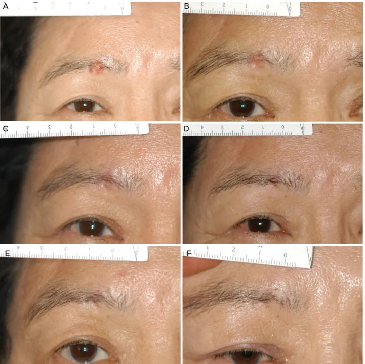
Figure 2. External photography after medication. (A) Post-medication photo: at 1 week after starting 5% Imiquimod cream. A focal redness is observed in the right eyebrow lesion, but there is no other complication or symptom. (B) Post-medication photo: at 8 weeks after 6 weeks’ 5% Imiquimod cream application and just before additional treatment. Her eyelid lesion is flattened and the cil- ia of the eyebrow begun to grow on the lesion site. However, the lesion still remains giving an impression of a partial regression.
(C) Post-medication photo: at 4 week after additional 4 weeks’ 5% imiquimod cream. Her eyebrow lesion disappeared more. (D) At 6 months after the last application of 5% imiquimod cream. The eyebrow is like normal appearance. (E) At 9 months after last application. (F) At 31 months after last application (final follow up), the eyebrow looks normal.
USA)을 일주일에 5일 동안 잠자기 전에 우측 눈썹 종양 부 위에 바르고 아침에 일어나서 물로 씻어내도록 하였으며, 혹시 imiquimod가 눈 안으로 들어가는 것을 방지하기 위하 여 Dura tears 연고를 피부연고를 바르기 전에 눈 안에 넣도 록 하였다. 일주일 중 나머지 2일 동안은 연고를 바르지 않
도록 하여 6주간 같은 방법으로 시행하도록 하였다. 약제를 투여하고 첫 일주일 후 관찰하였을 때 종양 부위에 국한된 발적이 관찰되었으나 다른 이상소견이나 불편 증상은 관찰 되지 않았다(Fig. 2A). 6주간 약제 사용이 끝나고 8주가 지 났을 때(Fig. 2B) 종괴의 융기는 사라져서 주변 피부와 편
A B
C D
E F
평하게 되었고 소실된 속눈썹도 자라난 모습이었으나 종괴 가 완전히 사라졌다고 판단되지 않아 추가로 4주간 5%
imiquimod 연고를 투여하였다. 추가 4주간 약제 사용이 끝 나고 4주째 관찰소견(Fig. 2C)에서 병변이 좀 더 감소한 것 으로 보여 경과만 관찰하기로 하였다. 6개월과 9개월 후 (Fig. 2D, E) 병변 부위는 점점 정상 피부 모습으로 변하였 고 최종 추적관찰한 31개월 후(Fig. 2F)에는 정상 눈썹 피 부 모습이 관찰되었다.
고 찰
Imiquimod는 화학적으로 합성한 imidazoquinolinamine 계통의 물질로 cytokine을 유도하여 선천성, 후천성 면역반 응을 활성화하고 면역 조절 및 항바이러스, 항종양 효과를 나타내는 물질이다. 항원 전달 세포의 표면에 있는 Toll-like receptors-7 (TLR-7)과 결합하여 작용함으로써 랑게르한스 세포, 단핵구, 대식세포, 수지상세포로부터 많은 수의 내인 성 cytokine을 합성 및 분비하게 한다. Interferon-alpha (IFN-α), Tumor necrosis factor-α (TNF-α), Interleukines (IL) 1, 6, 8, 10, 12 등이 주요 cytokine이고 이들은 CD4와 CD8의 세포성 면역 반응을 활성화시킨다. Imiquimod는 또한 NK 세포도 자극하여 1형 T 보조 세포 및 세포독성 T 림프 구를 활성화시키고 결국 바이러스에 감염된 세포 또는 종 양 세포를 사멸시키는 역할을 한다.5 이러한 기전으로 활성 화된 선천성 및 후천성 면역 반응으로 바닥세포암을 세포 자멸사시키는 imiquimod는 바닥세포암, 광선각화증, 편평세 포암, 전이성 악성흑점자흑색종, 균상식육종, 각질극세포종, 유방외 파제트병에 대해 임상시험이 진행되었다. 상체, 몸 통, 목 등 여러 부위의 얕은 바닥세포암과 결절 바닥세포암 에 대한 imiquimod 사용의 치료 효과는 Beutner et al7에 의 해 처음 보고되었고, 그 이후로 Marks et al8, Shumack et al9 에 의해 보고되었다.
5% imiquimod cream은 하루에 한 번씩 일주일에 다섯 번 병변에 도포하며 6-16주간 치료하게 된다. 보통 병변에 밤에 도포하고 아침에 씻어내는 방식으로 한 번 도포할 때 8-10시간 정도 유지되도록 한다. 눈 주위에 점안할 경우 결 막에 약제가 반복적으로 닿게 되면 불편감 및 통증을 느끼 거나 발적을 나타낼 경우가 있고, 이를 예방하기 위하여 인 공누액 등의 안연고를 결막낭에 넣고 눈 안에 들어가지 않 도록 교육하면 이러한 부작용을 완화시킬 수 있다.10 병변 부위 피부에 나타나는 부작용으로는 염증 반응을 일으키지 만, 이는 치료 기간 동안 가장 강하게 나타나며, 약제 사용 을 중단할 경우 완화된다.10
정기적인 병변의 활동성 및 크기 등을 경과 관찰하여 임 상적으로 완치됨을 판정할 수 있고, 조직검사를 통해 병리 학적으로 완치됨을 판정할 수도 있다. 이 증례에서는 크림 사용 초기에 종괴부의 발적은 관찰되었지만 주변 피부의 발적은 관찰되지 않았고 눈의 불편감 등의 부작용은 일어 나지 않았다. 종괴의 완치 확인을 위하여 조직검사를 시행 하지는 않았지만 31개월의 관찰 후에도 병변 재발의 소견 이 보이지 않아 완치되었다고 판단할 수 있을 것이다.
최근 연구에서 imiquimod cream은 비용 면에서 효과적 이므로 수술보다 좋은 평가를 받고 있고, 피부과 영역에서 도 작고 피상적인 병변에서 수술적 치료에 앞서 시도되며 추천되고 있다.11 이 증례와 같이 수술적 제거 후 재건이 쉽 지 않거나 전신상태 등의 문제로 수술이 어려운 경우 유용 하게 시도해 볼 만한 치료 방법이라고 생각한다.
참고문헌
1) Lomas A, Leonardi-Bee J, Bath-Hextall F. A systematic review of worldwide incidence of nonmelanoma skin cancer. Br J Dermatol 2012;166:1069-80.
2) Chinem VP, Miot HA. Epidemiology of basal cell carcinoma. An Bras Dermatol 2011;86:292-305.
3) Kim HS, Cho EA, Bae JM, et al. Recent trend in the incidence of premalignant and malignant skin lesions in Korea between 1991 and 2006. J Korean Med Sci 2010;25:924-9.
4) Rubin AI, Chen EH, Ratner D. Basal-cell carcinoma. N Engl J Med 2005;353:2262-9.
5) Carneiro RC, de Macedo EM, Matayoshi S. Imiquimod 5% cream for the treatment of periocular basal cell carcinoma. Ophthal Plast Reconstr Surg 2010;26:100-2.
6) David CV, Nguyen H, Goldenberg G. Imiquimod: a review of off-label clinical applications. J Drugs Dermatol 2011;10:1300-6.
7) Beutner KR, Geisse JK, Helman D, et al. Therapeutic response of basal cell carcinoma to the immune response modifier imiquimod 5% cream. J Am Acad Dermatol 1999;41:1002-7.
8) Marks R, Gebauer K, Shumack S, et al. Imiquimod 5% cream in the treatment of superficial basal cell carcinoma: results of a multi- center 6-week dose-response trial. J Am Acad Dermatol 2001;44:
807-13.
9) Shumack S, Gebauer K, Quirk C, et al. 5% imiquimod cream for the treatment of large superficial basal cell carcinoma. Arch Dermatol 2004;140:1286-7.
10) Geisse J, Caro I, Lindholm J, et al. Imiquimod 5% cream for the treatment of superficial basal cell carcinoma: results from two phase III, randomized, vehicle-controlled studies. J Am Acad Dermatol 2004;50:722-33.
11) Vanaclocha F, Daudén E, Badía X, et al. Cost-effectiveness of treat- ment of superficial basal cell carcinoma: surgical excision vs. imi- quimod 5% cream. Br J Dermatol 2007;156:769-71.
= 국문초록 =
5% Imiquimod Cream으로 치료한 눈썹부위 바닥세포암 증례 1예
목적: 바닥세포암은 가장 흔한 피부악성종양으로 눈 주변부에 발생하는 경우 미용적인 관점과 안와 내로의 침범 예방을 위하여 치료 가 필요하다. 이에 대한 치료 중 면역반응조절제인 imiquimod 연고를 통한 비수술적 치료의 효과가 보고되어 왔다. 눈 주변의 바닥세 포암에 대한 imiquimod 국소요법의 증례가 국내 안과 관련 학회지에서는 보고된 바가 없어 이에 대한 증례 1예를 보고하고자 한다.
증례요약: 73세 여자 환자가 서서히 커지는 우측 눈썹의 종양으로 시행한 조직검사상, 바닥세포암으로 진단 받고 서울대학교병원에 방문했다. 내원 시 우측 눈썹의 중앙부 안쪽에 편평하게 융기되고 중심부는 경도로 함몰된 양상의 1x1 cm 크기의 종양이 관찰되었다.
5% Imiquimod 연고를 1주에 5일 동안 수면 시간에 우측 눈썹 종양 부위에 6주간 바르도록 하였다. 이후 8주가 지났을 때 종괴는 호전된 양상이었으나 완전 관해로 판단되지 않아 추가로 4주간 투여하였고, 점점 정상 피부 모습으로 변하였으며, 최종 추적관찰한 31개월 후에는 정상 눈썹 피부 모습이 관찰되었다.
결론: Imiquimod 연고는 종양의 수술적 제거 후 재건이 어렵거나 수술이 어려운 경우 유용하게 시도될 수 있는 치료제로 생각한다.
<대한안과학회지 2015;56(11):1789-1793>