Korean J Gastroenterol Vol. 77 No. 3, 141-144 https://doi.org/10.4166/kjg.2021.026 pISSN 1598-9992 eISSN 2233-6869
IMAGE OF THE MONTH
Korean J Gastroenterol, Vol. 77 No. 3, March 2021 www.kjg.or.kr
전이성 담낭암으로 오인된 황색육아종성 담낭염
한재호, 안상정
1, 조인래
2,3가톨릭관동대학교 의과대학 국제성모병원 소화기내과, 가톨릭관동대학교 의과대학 국제성모병원 병리과1, 서울대학교 의과대학 내과학교실 및 간연구소2, 서울대학교병원 소화기내과3
Xanthogranulomatous Cholecystitis Suspected as Metastatic Gallbladder Cancer
Jaeho Han, Sangjeong Ahn1 and In Rae Cho2,3
Division of Gastroenterology, Department of Internal Medicine, International St. Mary’s Hospital, Catholic Kwandong University College of Medicine, Incheon; Department of Pathology, International St. Mary’s Hospital, Catholic Kwandong University College of Medicine1, Incheon;
Department of Internal Medicine and Liver Research Institute, Seoul National University College of Medicine2, Seoul; Division of Gastroenterology, Department of Internal Medicine, Seoul National University Hospital3, Seoul, Korea
CC This is an open access article distributed under the terms of the Creative Commons Attribution Non-Commercial License (http://creativecommons.org/licenses/
by-nc/4.0) which permits unrestricted non-commercial use, distribution, and reproduction in any medium, provided the original work is properly cited.
Copyright © 2021. Korean Society of Gastroenterology.
교신저자: 조인래, 03080, 서울시 종로구 대학로 101, 서울대학교병원 소화기내과
Correspondence to: In Rae Cho, Division of Gastroenterology, Department of Internal Medicine, Seoul National University Hospital, 101 Daehak-ro, Jongno-gu, Seoul 03080, Korea. Tel: +82-2-2072-1489, Fax: +82-2-762-9662, E-mail: [email protected], ORCID: https://orcid.org/0000-0001-9874-5526
Financial support: None. Conflict of interest: None.
A
A B
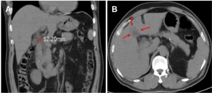
Fig. 1. Abdomen computed tomography findings. (A) Impacted distal common bile duct stone with proportional biliary dilatation. (B) Collapsed gallbladder, suspicious of chronic cholecystitis (arrows).
증례: 63세 남자 환자가 상복부 통증으로 외부 병원에 내원 하여 시행한 혈액 검사와 복부 컴퓨터단층촬영(CT) 검사에서 폐쇄성 황달과 급성 담관염을 동반한 총담관결석이 의심되어 내원하였다. 내원 당시 활력징후는 혈압 130/85 mmHg, 맥박 72회/분, 체온 37.5℃였으며, 말초혈액 검사에서 백혈구(white blood cell count) 13,330/μL (참고치 4,000-10,000), 혈색소 (hemoglobin) 14.2 g/dL (참고치 13-17), 혈소판(platelet count) 223,000/μL (참고치 140,000-400,000), 혈청 생화학 검사에서 AST 87 IU/L (참고치 0-40), ALT 208 IU/L (참고치 0-40), ALP 159 IU/L (참고치 38-110), γ-GTP 426 IU/L (참고 치 0-55), total bilirubin 5.9 mg/dL (참고치 0.2-1.4), CRP 113.66 mg/L (참고치 0-5), CA 19-9 65.97 U/mL (참고치 0-35)로 확인되었다. 과거력상 고혈압과 일과성 허혈발작 (transient ischemic attack)의 병력이 있었으며, 문진 시 약물 복용력은 확인되지 않았다.
외부 병원에서 시행한 CT 상 총담관 원위부에 0.5 cm 크기의 총담관결석과 담관확장이 확인되었고(Fig. 1A), 주변 간 실질과 경계가 뚜렷하지 않은 만성 담낭염 소견(collapsed gall-
bladder)이 보이는 것을 확인하였다(Fig. 1B). 내시경적 역행성 담췌관 조영술(ERCP)을 통해 담관결석 제거 및 스텐트 삽입술 을 시행하였으며 (Fig. 2), 담낭 병변의 감별 진단을 위해 자기공 명영상(MRI)을 시행하였다. MRI 상 수축된 담낭 내부에 담석이 확인되었고, 불규칙한 조영증강을 보이는 담낭벽의 비후가 확 인되었다. 담낭 병변은 확산 제한(diffusion restriction)을 보였 으며, 담낭과 인접한 간우엽 앞쪽아래구역(S5)에 약 1.5 cm
142
한재호 등. 전이성 담낭암으로 오인된 황색육아종성 담낭염The Korean Journal of Gastroenterology A
A B
Fig. 3. Magnetic resonance imaging findings. (A) Irregular enhanced wall thickening of the gallbladder with diffusion restriction and direct invasion of the adjacent liver parenchyma (arrows), suspicious of gallbladder cancer with direct invasion (T3 vs. T4Nx). (B) Multiple subcentimeter peripherally enhancing lesions in the liver (arrows).
A
A B C
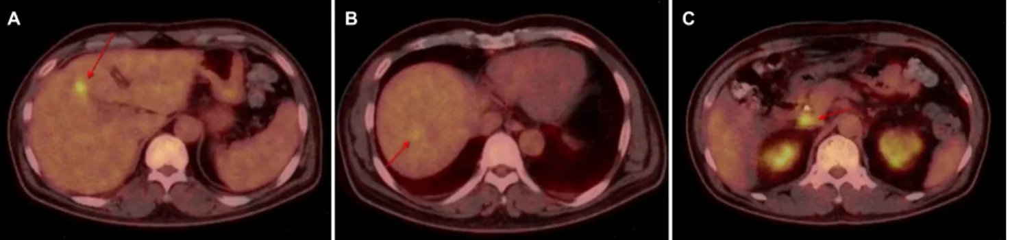
Fig. 4. Positron emission tomography-computed tomography shows (A) increased FDG uptake in the gallbladder (arrow), (B) multifocal mildly increased FDG uptake in the liver (arrow), and (C) focally increased FDG uptake in the portocaval space (arrow).
A
A B
Fig. 2. (A) Endoscopic retrograde cholangiopancreatography showing mild common bile duct dilatation without a definite filling defect. (B) Stone extraction using balloon catheter and biliary stent insertion were performed.
크기의 간담도기 결손(hepatobiliary phase defect) 소견이 확인되어 담낭암의 간 침범(gallbladder cancer with direct liver invasion)으로 의심되었다(Fig. 3A). 또한 hepatobiliary phase에서 간실질에 1 cm 미만의 병변들이 다수 확인되었는 데, 확산제한 소견을 보이고, T2 강조영상에서 중등도 신호강도 로 관찰되며 병변 주위의 조영증강(perilesional enhance-
ment)을 보여 다발성 간전이(multiple liver metastases)와 호산구성 간농양(eosinophilic abscess)의 감별이 필요하였다 (Fig. 3B). 악성 질환의 감별을 위해 시행한 양성자방출단층촬영 (18F-fluoro-deoxyglucose [FDG] PET-CT)에서 담낭병변의 FDG 흡수 증가(SUVmax 6.7) 소견이 확인되었고, 간비면 부위 (liver dome)를 비롯하여 간우엽의 여러 곳에서 경미한 FDG 흡수 증가(SUVmax 3.6)가 관찰되었다(Fig. 4A, B). FDG 흡수 증가가 관찰되는 부위는 MRI에서 관찰되었던 간병변에 부합하 여 간전이의 가능성을 우선적으로 생각하였으며, 문맥-하대정 맥 공간(portocaval space)에서도 임파선 비대소견 및 in- creased FDG uptake (SUVmax 5.9)가 확인되어 전이성 림프절 병증(metastatic lymphadenopathy)의 가능성을 고려하였다 (Fig. 4C).
악성 질환이 의심되는 담낭 및 림프절의 조직학적 진단을 위해 외과 협진 하에 진단적 복강경술(exploratory laparo- scopy)을 시행하였다. 담낭 장막층, 담낭과 맞닿는 간 부위와 문맥-하대정맥 림프절(portocaval lymph node)에서 시행한 동결절편생검(frozen section biopsy)에서 악성종양 소견은 확인되지 않았으며, 이에 담낭절제술을 시행하였다. 담낭절제
Han J, et al. Xanthogranulomatous Cholecystitis Suspected as Metastatic Gallbladder Cancer
143
Vol. 77 No. 3, March 2021 A
A BB
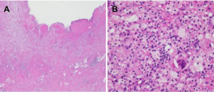
Fig. 5. Microscopically, (A) thickened gallbladder wall revealed a relatively preserved mucosal epithelium and partly disrupted muscular layers (left side), in which collections of lipid-laden macrophages were found admixed with mixed inflammatory cells and fibrosis (H&E, ×40). (B) Xanthogranuloma, a characteristic feature of xanthogranulomatous cholecystitis, was noted, which contained foamy histiocytes, multinucleated giant cells, and lymphoplasmacytic cells (H&E, ×200).
술 후 시행한 병리학적 검사 상, 섬유증과 다양한 염증 세포의 침윤으로 비후된 담낭벽이 관찰되었고(Fig. 5A), 두꺼워진 담 낭벽 사이에 포말성 대식세포(foamy macrophages)로 구성 된 황색육아종성 병변 (xanthogranulomatous lesion)이 특 징적으로 확인되었다(Fig. 5B). 이에 본 증례는 병리학적으로 황색육아종성 담낭염(xanthogranulomatous cholecystitis) 으로 진단되었다.
이후 환자는 담관염 및 담낭염에 대한 항생제 치료를 시행 받았으며, 약 2주 후 시행한 CT 검사에서 이전 MRI와 PET-CT에서 간우엽에 다발성으로 발견되었던 병변은 확인 되지 않았다. 이에 담관결석으로 인해 발생한 폐쇄성 담관염 으로 인해 발생한 간 미세농양이 항생제 치료 후 회복된 것으 로 판단하고 치료를 종료하였다.
진단: 황색육아종성 담낭염
황색육아종성 담낭염은 비교적 드문 담낭의 염증성 질환으 로, 발생빈도는 1% 미만에서 10%에 이르기까지 보고자에 따 라 다양하게 나타나는데, 담낭염으로 담낭절제술을 시행한 환 자의 약 1% 내외일 것으로 추정하고 있다.1-3
황색육아종성 담낭염은 담석 등으로 인한 담낭관의 폐색으 로 급성 염증반응이 발생하면서 Rokitansky-Aschoff sinus 의 파열이나 점막의 궤양성 변화가 초래되고, 이를 통해 담낭 주변 조직으로 담즙이 유출되면서 발생한다. 유출된 담즙에 의해 담낭 주변조직에서 염증반응이 일어나는 과정에서 섬유 아세포(fibroblast)와 대식세포(macrophage)를 포함한 다양 한 염증세포가 증식하고, 대식세포는 콜레스테롤 및 인지질과 같은 담즙 지질을 탐식하여 지질 함유 혹은 포말성 대식 세포 (lipid-laden macrophages, foamy macrophages)로 변형되 고, 해당 세포들이 증식하여 병리조직학적으로 특징적인 황색 육아종(xanthogranuloma)을 형성한다(Fig. 5B).4육안적으로
담낭벽의 황색결절을 동반하는 불규칙한 담낭벽의 비후소견 이 특징이며, 병리조직학적으로는 염증세포와 황색육아종의 증식을 특징으로 한다.5 주위 장기로의 침윤, 유착을 일으키는 경우가 많고, 담낭벽의 비후나 종괴를 흔히 동반하므로 악성 종양과의 감별이 쉽지 않다.
황색육아종성 담낭염에 특징적인 임상증상이나 징후는 없 으며, 대개 급성이나 만성 담낭염의 임상 양상과 유사하고, 복통, 황달, 발열, 식욕부진 등의 임상적 증상이 담낭암보다 흔하다고 알려져 있다.6,7 CA 19-9는 췌담도의 악성 질환이 의심될 때 측정해 볼 수 있는데 황색육아종성 담낭염과 같은 양성 질환의 환자에서도 상승할 수 있고, 일관적이지 않아 두 질환의 감별에 큰 도움이 되지 않는다.8
복부 초음파 검사에서 황색육아종성 담낭염은 담낭오니 (gallbladder sludge) 및 담석을 흔히 동반하고, 담낭의 종괴나 주변의 액체저류, 간과의 경계 소실을 보일 수 있다. CT에서도 주변 조직을 침윤하는 불규칙한 담낭벽의 비후나 결절, 종괴 소견이 확인되므로 영상 검사만으로 담낭암과 감별 진단을 시 행하는 것은 쉽지 않으나, 담낭벽 내의 저음영 결절(hypoechoic nodules or bands)이 담낭암에 비해 높은 빈도로 관찰되며, 담낭암과 달리 미만성 담낭벽 비후(diffuse gallbladder wall thickening)를 보이고, 연속적인 내강 점막의 조영증강을 보이 거나 국소적인 소실만 보이는(luminal surface enhancement with continuous mucosal lines or mucosal lines with focal breach) 경우가 많은 것으로 알려져 있다.9-12
황색육아종성 담낭염은 심한 염증반응으로 인해서 주변 장 기로의 침범이나 인근조직과의 유착을 일으키는 경우가 많고, 장막이 파열되어 간, 횡행결장, 십이지장 등에 유착을 초래할 수 있다. 이로 인해 다른 원인에 의한 담낭염으로 담낭절제술 을 시행한 환자들에 비해서 담즙유출, 담즙복막염, 담낭출혈, 간농양, 상처감염 등의 수술 후 합병증이 많이 발생하며, 심한 만성 염증반응이 간문(hepatic hilum)으로 파급되었을 때, 주 변 장기(adjacent organs)와 간외담관(extrahepatic bile duct)의 절제술이 필요할 수 있다.13,14
황색육아종성 담낭염의 약 30%에서 담낭암이 동반되었다는 보고가 있어 담낭암의 동반 가능성에 대한 고려가 필요하며, 담낭암과의 정확한 감별을 위해서 수술 중 동결절편 조직생검 (frozen section)을 적극적으로 시행하고 그 결과에 맞추어 수술 범위 확장이나 개복 담낭절제술(open cholecystectomy)로의 전환을 고려해야 한다.15 또한 황색육아종성 담낭염에 담낭암이 동반되는 경우에는 암조직이 비후된 담낭벽 전체에 있는 것이 아니고 특정 부위에 암병소를 내포할 수 있기 때문에 가능한 육아종성 염증조직을 완전하게 제거해서 동결절편 조직생검을 시행하는 것이 적절하다.
저자들은 담낭암과 감별이 어려운 황색육아종성 담낭염이
144
한재호 등. 전이성 담낭암으로 오인된 황색육아종성 담낭염The Korean Journal of Gastroenterology
폐쇄성 담관염으로 인해 발생한 다발성 미세 간농양과 병발하 여 전이성 담낭암으로 오인되었던 증례를 경험하며 진단 및 치료과정에서 다양한 가능성에 대한 고찰이 필요함과 적극적 인 조직 검사가 중요함을 확인하였기에 영상 및 문헌과 함께 보고한다.
REFERENCES
1. Ros PR, Goodman ZD. Xanthogranulomatous cholecystitis ver- sus gallbladder carcinoma. Radiology 1997;203:10-12.
2. Kang J, Lee SH, Lee JW, et al. A case of xanthogranulomatous cholecystitis decreased in size after steroid treatment and avoid- ed extended resection. Korean J Pancreas Biliary Tract 2015;
20:37-41.
3. 전훈배, 이승규, 이영주, et al. Xanthogranulomatous cholecystitis.
Korean Journal of HBP Surgery 1999;3:203-209.
4. Goodman ZD, Ishak KG. Xanthogranulomatous cholecystitis. Am J Surg Pathol 1981;5:653-659.
5. Joo YE, Lee JJ, Chung IJ, et al. A case of xanthogranulomatous cholecystitis. Korean J Intern Med 1999;14:90-93.
6. Yang T, Zhang BH, Zhang J, Zhang YJ, Jiang XQ, Wu MC. Surgical treatment of xanthogranulomatous cholecystitis: experience in 33 cases. Hepatobiliary Pancreat Dis Int 2007;6:504-508.
7. Chang BJ, Kim SH, Park HY, et al. Distinguishing xanthogranulom- atous cholecystitis from the wall-thickening type of early-stage gallbladder cancer. Gut Liver 2010;4:518-523.
8. Spinelli A, Schumacher G, Pascher A, et al. Extended surgical re- section for xanthogranulomatous cholecystitis mimicking ad- vanced gallbladder carcinoma: a case report and review of literature. World J Gastroenterol 2006;12:2293-2296.
9. Singh VP, Rajesh S, Bihari C, Desai SN, Pargewar SS, Arora A.
Xanthogranulomatous cholecystitis: what every radiologist should know. World J Radiol 2016;8:183-191.
10. Chun KA, Ha HK, Yu ES, et al. Xanthogranulomatous chol- ecystitis: CT features with emphasis on differentiation from gall- bladder carcinoma. Radiology 1997;203:93-97.
11. Kim PN, Ha HK, Kim YH, Lee MG, Kim MH, Auh YH. US findings of xanthogranulomatous cholecystitis. Clin Radiol 1998;53:
290-292.
12. Kim PN, Lee SH, Gong GY, et al. Xanthogranulomatous chol- ecystitis: radiologic findings with histologic correlation that fo- cuses on intramural nodules. AJR Am J Roentgenol 1999;172:
949-953.
13. Nacif LS, Hessheimer AJ, Rodríguez Gómez S, Montironi C, Fondevila C. Infiltrative xanthogranulomatous cholecystitis mim- icking aggressive gallbladder carcinoma: a diagnostic and ther- apeutic dilemma. World J Gastroenterol 2017;23:8671-8678.
14. Seo SH, Park JI, Kim JS, Kim KH, Choi CS, Choi YK.
Xanthogranulomatous cholecystitis: a retrospective analysis of 36 cases. J Korean Surg Soc 2009;76:317-377.
15. Kwon AH, Sakaida N. Simultaneous presence of xanthogranu- lomatous cholecystitis and gallbladder cancer. J Gastroenterol 2007;42:703-704.