pISSN: 0378-6471 eISSN: 2092-9374 DOI : 10.3341/jkos.2010.51.458
= 증례보고 =
변형된 광역학 요법으로 치료한 유방의 침윤성 관상암종의 맥락막 전이 1예
조윤정⋅박성표
한림대학교 의과대학 강동성심병원 안과학교실
목적: 유방의 침윤성 관상암종 환자에서 발생한 맥락막 전이 종양을, 광역학 요법을 사용하여 효과적인 종양 크기의 감소를 경험하였 기에 이를 보고하고자 한다.
증례요약: 45세 여자 환자가 우안 아래쪽의 시야 흐림을 주소로 내원하였다. 기저질환은 없었고 1년 전부터 지속되는 요통을 호소하였다.
최대교정시력은 양안 모두 20/20이었다. 안저검사에서 우안의 상비측 부위에 노란색의 융기된 병변이 관찰되어 초음파 및 형광안저혈 관조영술을 시행하였다. 초음파 검사에서 중등도 이상의 내부 반향과 증가된 에코음영을 보이는 융기된 종양이 관찰되었으며, 형광안 저혈관조영술에서는 병변 부위에 초기에는 저형광, 후기에는 고형광의 특징을 보였다. 맥락막 전이의 감별을 위해 시행한 방사선 검사 에서 유방암 및폐, 척추, 난소로의 전이가 확인되었고, 전신적 항암치료를 시작하였다. 이에 유방암의 맥락막 전이로 진단하고 맥락막 종양의 치료를 위해 변형된 광역학 치료를 시행하였다. 치료 6일째 종양 크기의 변화는 없었으며 아래쪽 안저와 황반부위에 장액망막 박리가 발생하였다. 그러나 치료 22일째 시행한 검사상 현저한 종양크기의 감소와 함께 아래쪽에 발생하였던 장액망막박리도 호전된 양상을 보였다.
<대한안과학회지 2010:51(3):458-462>
■ 접 수 일: 2008년 11월 24일 ■ 심사통과일: 2009년 2월 4일
■ 통 신 저 자: 박 성 표
서울특별시 강동구 길동 445 강동성심병원 안과
Tel: 02-2224-2274, Fax: 02-470-2088 E-mail: sungpyo@hananetnet
Figure 1. The fundus photography of the right eye shows elevated mass-like lesion superonasal to the optic disc.
안구의 전이성 종양은 안구 내에 발생되는 종양 중 가장 흔하며, 암 환자 중 2~7%에서 안구 내 전이가 동반된다고 알려져 있다.1,2,3 안구로의 전이는 주로 혈행성으로, 안구 내의 어떤 부위에서도 전이성 병변이 발생될 수 있으나 맥락 막이 많은 혈관분포와 비교적 늦은 혈류 속도를 갖는 특징에 의해 전이의 발생 빈도가 가장 높다.3여성에서는 유방암, 남 성에서는 폐암이 맥락막 전이성 종양의 가장 흔한 원발병 소인 것으로 알려져 있으며 안구 내 종양이 있는 여성 환자의 80~90%가 유방암에 의한 전이성 병변이라는 보고가 있다.4,5
맥락막으로 전이된 종양은 안내 종양 위치나 시각 증상 유무, 원발 종양의 부위 등 여러 가지 요인을 고려하여 단순 히 경과 관찰만을 시행할 수도 있으며 혹은 전신 항암치료, 호르몬 치료, 방사선 치료, 냉동 치료, 레이저 광응고술, 경 동공 온열요법, 수술적 절제술 등의 방법을 이용하여 치료할 수 있다.6 또한 Isola et al7은 유방암의 맥락막 전이 종양의 치료로서 전신적인 항암치료와 함께 보조적 광역학치료를 사 용하여 효과적인 시력 향상 및 종양의 퇴행을 보고한바 있다.
저자들은 맥락막 종양이 발견된 환자에서 전신적 검사를
통해 척추와 폐, 난소에 다발성 전이를 일으킨 유방의 침윤성 관상암종을 진단하였으며 변형된 광역학치료를 시행하여 현저한 맥락막 종양의 크기 감소를 경험하였다. 맥락막 전이 종양에 대한 변형된 광역학치료는 아직 국내에서 보고된 바 없는 증례로 이에 보고하고자 한다.
증례보고
45세 여자환자가 한 달 전부터 발생한 우안의 시야장애를
Figure 2. Ultrasonography of the right eye revealed a highly echogenic choroidal mass with moderate to high internal reflectivity.
Figure 3. Fluorescein angiography shows generalized hypofluorescence with pinpoint hyperfluorescence during the prearterial and arteriovenous phase (Top) and hyper- fluorescence during the venous phase (Bottom) due to leakage.
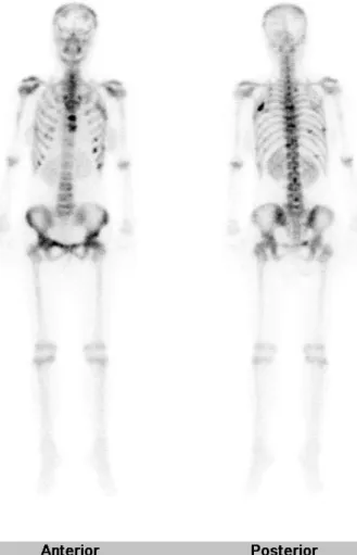
Figure 4.The 99mTc bone scan revealed multiple active bone lesions in the thoracic spine and the ribs on both sides.
주소로 내원하였다. 기저질환은 없었으나 1년 전부터 지속 되는 요통을 호소하였다. 최대교정시력은 양안 20/20이었 으며 안저검사상 우안 상비측부위에 노란색의 융기된 병변이 약 8 유두직경 크기로 관찰되었다(Fig. 1). 병변은 초음파 에서 중등도 이상의 내반향파와 증가된 에코음영을 보이는 고형 종괴로 관찰되었다(Fig. 2). 형광안저혈관조영술에서 초기에 부분적 과형광부위가 동반된 전반적인 저형광을 보 였고 후기로 갈수록 병변 부위와 일치하는 과형광이 나타 났다(Fig. 3). 결과를 토대로 맥락막의 전이성 종양이 의심 되어 전신 검사를 시행하였다. Bone scan에서 요추의 다발성 전이 병변이 발견되었고(Fig. 4), 흉부전산화단층촬영에서 조영 증강되는 유방의 결절이 보여 조직검사 후 침윤성 관상 암종으로 진단되었다(Fig. 5). 또한 복부전산화단층촬영 및 척추자기공명영상에서 폐, 척추, 난소로의 다발성 전이가 확인되었다. 이에 유방암에서 전이된 맥락막 종양으로진단 하였고, 전신적 항암치료를 시작하였다. 항암치료 6주 뒤 맥락막 종양 크기의 변화는 없었고 환자의 불편이 지속되어
Figure 5.Pathology of primary breast cancer. The in- vasive ductal carcinoma infiltrating into the adjacent stromal tissue.
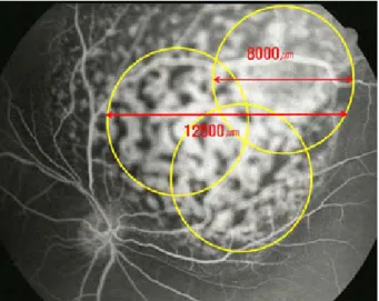
Figure 6.The diameter of the treatment spot is calculated based on the lesion size measured on the pretreatment fluorescein angiogram. Fundus drawing demonstrating photodynamic therapy of the choroidal tumor using three overlapping spot (yellow circles). The greatest linear dimension of the retinal lesion is 12,500 μm. And each treatment spot size is 8,000 μm.
Figure 7.At 6 days after photodynamic therapy, atrophic change started on the tumor surface. And serous elevated lesion appeared surrounding the macula and inferior retina.
Figure 8.At 22 days after photodynamic therapy, the fundoscopy shows resolution of serous retinal detatchment and marked tumor shrinkage.
변형된 광역학치료를 시행하였다. Verteporfin 6 mg/m2를 2분간 정맥주사 후, 다이오드레이저(690 nm)를 종양 표면에 166초씩 총 3회 부분 중복시켜(600 mW, 100 J/cm2) 조사 하였다(Fig. 6). 광역학치료 6일째 최대교정시력우안 20/40 이었고 종양의 위축성 변화가 보였으나 크기에는 변화가 없었다. 또한 아래쪽 안저와 황반부위에 장액망막박리가 발 생하였다(Fig. 7). 이에 매일 경구 prednisolone을30 mg씩 처방하였다. 광역학치료 22일째 우안의 최대교정시력은 20/25로 향상되었으며 종양의 크기는 약 6 유두직경으로 감소하였고 장액망막박리 역시 호전된 양상을 보였다(Fig. 8).
환자는 이후 더 이상 안과적 검사를 시행하지 못하였으며 맥 락막 전이 종양이 발견된 이후 약 3개월 뒤에 사망하였다.
고 찰
안구의 조직 중 맥락막은 전이성 종양의 가장 흔한 침범 부위로 주로 맥락막의 후극부가 침범된다.2 Shields et al4 의 보고에 따르면 맥락막 전이가 있는 420명의 환자에서 가장 흔하게 발견된 원발성암의 병소는 유방암(47%), 폐 암(21%), 그리고 위장관 암(4%)이었다. 맥락막 전이 암은 무증상인 경우도 있으나 무통성의 시력장애가 가장 흔하며 이차성 녹내장으로 인한 통증, 시야결손 및 비문증 등을 호 소할 수 있다.2,8병변은 주로 양안에 황색 혹은 황백색으로 나타나고 다발성이며 상대적으로 편평하고 약간의 융기된 판 모양을 보이며 장액성 망막박리를 동반하기도 한다.5,9 원발암의 기왕력이 있는 경우 전이종양의 진단상 도움 요 소가 될 수 있으나 맥락막모반, 무멜라닌성흑색종, 후부공 막염, 맥락막골종, 맥락망막염, 열공망막박리, 하라다병, 중 심성장액망막병증 등을 감별하여야 하며10형광안저혈관조 영술, indocyanine green (ICG) 형광안저혈관조영술, 초음 파검사 등이 보조적인 검사로 도움이 될 수 있다. 형광안저 혈관조영술에서는 대체로 초기에 종양에 의해 맥락막순환이 차단되어 저형광이 나타나고 후기로 갈수록 망막색소상피로 부터 다양한 형태의 누출이 나타나 과형광을 보이게 된다.
대조적으로 ICG 형광안저혈관조영술에서는 초기부터후기 까지 지속적인 저형광을 나타낸다. 초음파 검사상 A-scan 에서는 중등도에서 고도의 내부 반향을 보이며 B-scan에 서는 고도의 방사성을 가진 망막하 종양을 관찰할 수 있 다.11-13
치료는 환자의 전신상태 및 시력, 안구의 상태 등을 종합 해서 고려하며 증상이 없는 경우에는 전신적 원발암 치료 만을 시행하고 안구 병변은 단순히 경과 관찰을 시행하기도 한다. 그러나 시력 저하나 그 위험이 있는 경우에, 또한 전신 치료에도 불구하고 병변이 계속 성장할 때에는 국소적 치 료가 권유된다.12,14본 증례에서는 전신적 항암요법에도 맥 락막 종양의 크기 변화가 보이지 않았고, 환자의 증상도 지속 되어 국소치료를 결정하였다. 국소치료 방법으로는 방사선 치료, 냉동치료, 레이저 광응고술, 경동공 온열요법, 광역학 치료, 수술적 절제술 등의 방법을 이용할 수 있다.
Verteporfin을 이용한 광역학치료는 망막 신경섬유층을 보존하면서 혈관 내피세포만을 선택적으로 광화학적 파괴를 하는 치료법으로 파괴된 내피세포막이 혈소판 응집 및 분해 를 일으켜 목표로 한 혈관구조에 혈전이 생기게 만들어 비정 상적인 혈관의 폐쇄를 유도하여 혈관조직의 섬유화를만들고 이차적으로 종양의 크기를 감소시킨다.15,16TAP (Treatment of Age-Related Macular Degeneration with Photodynamic therapy) 및 VIP (Verteporfin in Photodynamic therapy)
연구에서는 광역학치료의 표준 방법으로 10분간 verteporfin 6 mg/m2를 정맥 주사한 뒤 5분 후 다이오드 레이저(689 nm) 를 종양의 표면에 83초간 600 mW, 50 J/cm2의 강도로 조 사하는 것을 권유하고 있으나 광역학치료를 시행하는 기저 질환의 병리학적 특징 및 각각의 증례에 적용하여 여러 사 례에서 변형된 형태로 이용되었다.17 본 증례에서는 거대 맥락막 종양을 보이고 있었기에 광역학 치료 효과의 증대를 위하여 verteporfin 정맥주사 주입 시간을 2분으로 줄여 단 시간 내에 종양 내부로의 약물 농도를 상승시키면서 레이저 강도를 100 J/cm2로 증가시켰으며 종양을 3부위로 나누어 각각 166초씩 레이저를 3차례 조사하는 방법을 사용하였다.
저자들은 여러 연구 등을 종합하여 변형된 광역학 치료를 시행하였고 광역학 치료 후에 장액성 망막 박리 외에는 큰 합병증 없이 종양의 크기가 감소되는 효과를 얻었다. 비록 환자의 사망으로 광역학 치료 후의 장기적 경과 관찰은 시 행되지 못하였지만 유방암에 의해 발생된 맥락막의 전이성 거대 종양에 대하여 변형된 광역학 치료가 효과를 나타낼 수 있다는 결과를 얻었기에 문헌 고찰과 함께 보고하는 바 이다.
참고문헌
1) Bloch RS, Gartner S. The incidence of ocular metastatic carci- noma. Arch Ophthalmol 1971;85:673-75.
2) Ferry AP, Font RL. Carcinoma metastatic to the eye and orbit.
I.A clinocopathologic study of 227 cases. Arch Ophthalmol 1974;
92:276-86.
3) Mejia-Novelo A, Alvarado-Miranda A, Morales-Vázquez F, et al.
Ocular metastases from breast carcinoma. Med Oncol 2004; 21:
217-21.
4) Shields CL, Shields JA, Gross NE, et al. Survey of 520 eyes with uveal metastases. Ophthalmology 1997;104:1265-76.
5) Freedman MI, Folk JC. Metastatic tumors to the eye and orbit:
patient survival and clinical characteristics. Arch Ophthalmol 1987;105:1215-9.
6) Lee SJ, Kim SY, Kim SD. A case of diode laser photocoagulation in the treatment of choroidal metastasis of breat carcinoma.
Korean J Ophthalmol 2008;22:187-9.
7) Isola V, Pece A, Pierro L. Photodynamic therapy with verteporfin of choroidal malignancy from breast cancer. Am J Ophthalmol 2006;142:885-7.
8) Ferry AP, Font RL. Carcinoma metastatic to the eye and orbit. A clinicopathologic study of 26 patients with carcinoma metastatic to the anterior segment of the eye. Arch Ophthalmol 1975;93:
472-82.
9) Stephens RF, Shields JA. Diagnosis and management of cancer metastatic to the uvea: a study of 70 cases. Ophthalmology 1979;
86:1336-49.
10) Lee JS, Kim JH, Oum BS. A case of bilateral choroidal metastasis of breast invasive ductal carcinoma. J Korean Ophthalmol Soc 1996;37:1211-7.
=ABSTRACT=
A Case of Modified Photodynamic Therapy in the Management of Choroidal Metastasis of Breast Cancer
Yoon Jung Cho, MD, Sung Pyo Park, MD
Department of Ophthalmology, Kangdong Sacred Heart Hospital, Hallym University College of Medicine, Seoul, Korea
Purpose: To report a case of choroidal metastasis of breast cancer that was treated with modified photodynamic therapy.
Case summary: A 45-year-old woman visited our clinic with blurred vision of the right eye, which began 1 month before. The patient previously suffered from a low back pain for 1 year. The best corrected visual acuity was 20/20 in both eyes. Fundus examination revealed an elevated yellowish mass-like lesion at the superonasal area in the right eye. Ultrasonography of the right eye showed a highly echogenic choroidal mass with moderate to high internal reflectivity. Fluorescein angiography showed hypofluorescence during the prearterial and arteriovenous phase, and well circumscribed hyperfluorescence during the venous phase. Radiologic examination was performed upon suspicion of metastasis. The examination revealed breast cancer with lung, spine and ovary metastasis. Subsequently, biopsy of the breast mass revealed an invasive ductal carcinoma. Based on these results, the patient was diagnosed with choroidal metastasis from breast carcinoma. The patient received systemic chemotherapy, and modified photodynamic therapy (PDT) was performed on the metastatic choroidal mass. Six days after modified PDT, the mass size was unchanged, and serous retinal detachment developed at the macula and inferior retina. However, 22days after treatment, the mass size markedly decreased and the serous retinal detachment was improved.
J Korean Ophthalmol Soc 2010;51(3):458-462
Key Words: Breast cancer, Choroidal metastatic tumor, Photodynamic therapy
Address reprint requests to Sung Pyo Park, MD
Department of Ophthalmology, Kangdong Sacred heart Hospital, Hallym University College of Medicine
#445 Gil-dong, Gangdong-gu, Seoul 134-701, Korea
Tel: 82-2-2224-2274, Fax: 82-2-470-2088, E-mail: sungpyo@hananetnet 11) Shin JA, Yoo JS, Huh W. A case of radiotherapy of choroidal metastasis of breat carcinoma. J Korean Ophthalmol Soc 1993;
34:480-3.
12) Paul Chan RV, Young LH. Treatment options for metastatic tumors to the choroid. Semin Ophthalmol 2005;20:207-16.
13) Kim IT, Song HC. Fluorescein and Indocyanine green angio- graphy of choroidal tumors. J Korean Ophthalmol Soc 1999;
40:1866-76.
14) Amer R, Pe’er J, Chowers I, Anteby I. Treatment options in the management of choroidal metastases. Ophthalmologica 2004;218:
372-7.
15) Schmidt-Erfurth U, Baumann W, Gragoudas E, et al. Photo- dynamic therapy of experimental choroidal melanoma using lipoprotein delivered benzoporphyrin. Ophthalmology 1994;101:
89-99.
16) Schmidt-Erfurth U, Hasan T, Gragoudas E, et al. Vascular targeting in photodynamic occlusion of subretinal vessels. Ophthalmology 1994;101:1953-61.
17) Mennel S, Barbazetto I, Meyer CH, et al. Ocular photodynamic therapy-Standard applications and new indication (PartⅠ). Oph- thalmologica 2007;221:216-26.