http://dx.doi.org/10.3988/jcn.2014.10.2.133 J Clin Neurol 2014;10(2):133-139
Introduction
Vascular shear stress contributes to the site specificity of ath- erosclerotic lesions; however, the utility of arterial shear stress has been limited due to the technical difficulty of measuring this stress and uncertainty about the implications of the mech- anism for therapy. It is still unclear whether the ability to measure the arterial shear stress would improve diagnoses of vascular events. Improving the understanding of the patho-
physiological role of vascular shear stress requires more in- vestigations involving various methods and study designs.
The vascular-wall shear stress is the frictional force that acts tangentially. Vascular shear stresses within normal phys- iologic ranges are essential for maintaining endothelial-cell morphology (i.e., an elliptical shape with normal tight junc- tions) and function associated with the physiological release of nitric oxide.1 A low wall shear stress (<4 dyne/cm2) allows for active interactions between the cellular and plasmatic components of blood and the vessel walls,2 and potentially result in atherothrombosis through complex molecular and biomechanical mechanisms.3,4
The extracranial carotid artery was previously evaluated in calculations of arterial geometry-specific shear stress in pa-
Association between Ischemic Stroke and Vascular Shear Stress in the Carotid Artery
Seul-Ki Jeong,a Jun-Young Lee,a Robert S. Rosensonb
aDepartment of Neurology and Research Institute of Clinical Medicine, Chonbuk National University-Biomedical Institute of Chonbuk National University Hospital, Jeonju, Korea
bMount Sinai Heart, Mount Sinai School of Medicine, New York, NY, USA
Received March 25, 2013 Revised November 1, 2013 Accepted November 5, 2013 Correspondence Seul-Ki Jeong, MD, PhD
Department of Neurology & Research Institute of Clinical Medicine, Chonbuk National University- Biomedical Research Institute of Chonbuk National University Hospital, 20 Geonji-ro, Deokjin-gu, Jeonju 561-712, Korea
Tel +82-63-250-1590 Fax +82-63-251-9363 E-mail [email protected]
Background and PurposezzVascular shear stress is essential for maintaining the morphology and function of endothelial cells. We hypothesized that shear stress in the internal carotid artery (ICA) may differ between patients with ischemic stroke and healthy control subjects.
MethodszzICA shear stress was calculated in 143 controls and 122 patients with ischemic stroke who had a normal ICA or an ICA with <50% stenosis. The stroke group included patients who presented with a first-ever or recurrent ischemic stroke but excluded cardioembolic stroke and uncertain etiologies. Of the 122 patients, 107 (87.7%) and 15 (12.3%) patients were cate- gorized as first-ever and recurrent stroke, respectively.
ResultszzCarotid diameters were significantly larger, and both peak-systolic and end-diastolic velocities were significantly lower in patients with ischemic stroke than in controls (all p values
<0.05). Mean values of peak-systolic and end-diastolic shear stress in both ICAs were signifi- cantly lower in patients with ischemic stroke in models that adjusted for age, sex, and vascular risk factors (p for trend <0.05). The ICA shear stress was lowest in patients with recurrent stroke or the subtype of small-vessel occlusion. Higher peak-systolic and end-diastolic shear stresses in both ICAs were independently and negatively associated with ischemic stroke after adjusting for potential confounders (all p values <0.05).
ConclusionszzICA shear stresses were significantly lower in patients with ischemic stroke than in control subjects. Future studies should attempt to define the causal relationship between carotid arterial shear stress and ischemic stroke. J Clin Neurol 2014;10(2):133-139 Key Wordszz carotid artery, hemodynamics, ischemic stroke, shear stress.
Open Access
cc This is an Open Access article distributed under the terms of the Cre- ative Commons Attribution Non-Commercial License (http://creative- commons.org/licenses/by-nc/3.0) which permits unrestricted non-com- mercial use, distribution, and reproduction in any medium, provided the ori- ginal work is properly cited.
tients with vascular events.5 Other studies have investigated shear stress in the carotid artery in association with athero- thrombosis that caused ischemic stroke,6 and cardiovascular risk factors.7 A previous study found an association between shear stress in the common carotid artery (CCA) and isch- emic stroke, after measuring the patient’s blood viscosity.8
The carotid artery is divided into three segments: the CCA including the bulb, the internal carotid artery (ICA), and the external carotid artery. The ICA is a direct conduit for blood supply to cerebral tissues, and it exhibits a low resistance for cerebral autoregulation. The ICA shear stress is influenced by the cerebral blood flow, and may be more closely associated with stroke than are the other carotid segments.9 Stroke has a heterogeneous etiology, such as large-artery atherosclerosis (LAA) and small-vessel occlusion (SVO).10 A more compre- hensive vascular imaging approach may be needed to under- stand the etiologic heterogeneity of stroke.
We hypothesized that ICA shear stress differs between pa- tients with ischemic stroke and control subjects, and that it can provide discriminative information on the cerebral he- modynamics of ischemic stroke. To test this, we calculated the carotid artery shear stress along the carotid segments using duplex ultrasonography, and examined the association with ischemic stroke while considering potential confounders.
Methods
Study population
Consecutive patients referred to the Neurovascular Ultra- sound Laboratory of the Stroke Centre of Chonbuk National University Hospital from January 2007 to March 2008 were included in this study. Neurosonologic examinations were performed that included B-mode and Doppler ultrasonogra- phy for measurements of arterial diameter, peak-systolic (PS) flow velocities, and end-diastolic (ED) flow velocities along both carotid arteries. All participating subjects provided writ- ten informed consents, and the study was performed with the approval of the institutional ethics committee.
Assessment of risk factors and laboratory measurement
We collected demographic and clinical information on all of the study participants, including age, sex, history of ischemic stroke and stroke subtype, and major cardiovascular risk fac- tors. Hypertension was defined as a blood pressure of
≥140/90 mm Hg or treatment with antihypertensive medica- tion, diabetes mellitus was diagnosed by previous use of glu- cose-lowering medications or a fasting blood glucose of >7.0 mmol/L (126 mg/dL), and hyperlipidemia was defined as previous or current use of lipid-lowering medications or a to-
tal cholesterol of ≥6.2 mmol/L (240 mg/dL) or low-density lipoprotein (LDL) cholesterol at ≥4.1 mmol/L (160 mg/dL).
Blood samples were drawn in the morning after a 12-hour overnight fast.
Assessment of ischemic stroke and categorization Ischemic stroke was defined as a history of cerebral infarc- tion with evidence of an acute focal neurological dysfunction lasting more than 24 h, and cerebral ischemia diagnosed on brain imaging such as computed tomography and/or magnet- ic resonance imaging. Ischemic strokes are normally catego- rized into five groups: LAA, cardioembolism, SVO or lacu- nar infarction (LI), stroke of determined etiology, and stroke of undetermined etiology based upon the diagnostic criteria of the Trial of Org 10172 in Acute Stroke Treatment study.10 The present study only included patients with ischemic stroke of LAA or SVO, thereby excluding other subtypes such as cardioembolism. Recurrent stroke was defined at the time of enrollment by the occurrence of more than one ischemic stroke
>21 days after the previous stroke.11 The control group com- prised subjects with vascular risk factors but who had no his- tory of vascular events such as myocardial infarction, stroke, or peripheral arterial disease. The subjects in the control group were consulted in an interview with neurologists about head- ache, small-vessel disease, or silent lacunes while perform- ing regular health screening at Chonbuk National University Hospital. Those who agreed to participate in the study and underwent carotid ultrasonography were enrolled.
Ultrasonographic measurements of ICA stenosis Ultrasonography was performed with ECG triggering and using a high-frequency (5-to-12 MHz; 12L5) linear trans- ducer (Terason t3000, Teratech Corporation, Burlington, MA, USA) by a certified neurosonologist (S.K.J.). The ischemic stroke group only included patients who had received carotid duplex ultrasonographic examinations >2 weeks after the acute stroke (interval, 23.1±11.6 days, mean±SD) in order to evaluate patients with stable carotid hemodynamics. Carotid duplex ultrasonography was performed along the CCA and ICA on both the right and left sides. The carotid segments were distinguished in a longitudinal view that was strictly perpendicular to the ultrasound beam, with both vessel walls clearly visualized. The carotid bulb was defined as a segment from the diverging portion of the distal CCA to the begin- ning portion of the ICA, as recommended previously.12
An atherosclerotic plaque was defined as a focal structure encroaching the arterial lumen by at least 0.5 mm or 50% of the surrounding intima-media thickness (IMT), or as a struc- ture where the thickness measured from the media-adventitia interface to the intima-lumen interface was greater than 1.5
mm.12 Plaque echogenicity was classified into echolucent, echogenic, and calcified, while the plaque surface morphology was classified into smooth or irregular, as reported previous- ly.13 The degree of ICA stenosis in both grayscale and Doppler ultrasonography was stratified into the following six catego- ries according to the PS velocity and the presence of plaque:
1) normal (no stenosis), 2) <50% stenosis, 3) 50–69% steno- sis, 4) ≥70% stenosis to near occlusion, 5) near occlusion, and 6) total occlusion, as recommended previously.14
Both the internal diameter and the maximum centerline PS and ED velocities were measured, from which the shear rate was calculated along the carotid arterial segments as described previously.15 Video images were acquired in all carotid seg- ments over six consecutive cardiac cycles. The lumen diam- eter and IMT were measured at the peak of the R wave. For velocity measurements, a sample volume in the center of the flow was reduced to the smallest possible measurement size (approximately 1 mm), while the Doppler angle was generally maintained at 45±4 degrees, and never exceeded 60 degrees.
Calculation of shear stress in the carotid arteries Using the PS and ED velocities and diameters along the carot- id arterial segments, shear rates (s-1) were determined using the following definition of Newtonian flow:
Shear rate, γ˙=4
where V is the maximum centerline (PS or ED) flow velocity, assuming a parabolic velocity distribution across the arterial lumen, and D is the local lumen diameter in the ED phase.15 The blood viscosity (μ) was estimated from a validated mod- el using the hematocrit as follows:16
μ (centipoise)=1.4175+5.878H-12.98H2+31.964H3 where H represents the hematocrit [(%)/100]. Finally, the shear stress (dyne/cm2) was determined by multiplying the blood viscosity and shear rate as follows:17
Shear rate, τ=4μ
Exclusion criteria
Patients with the following characteristics were excluded: 1) ICA stenosis of >50% on at least one side, 2) carotid artery revascularization performed including carotid endarterecto- my and stenting, and 3) ischemic stroke other than LAA and SVO. Patients with ischemic stroke of LAA or SVO and con- trol subjects who had a normal ICA or an ICA with <50% ste- nosis were included in the present study, since arterial steno-
V D
V D
ses of <50% do not cause any reduction in flow or pressure distal to the lesion.18
Statistical analysis
The numbers and percentages of subjects according to the statuses of both ICAs were summarized. Descriptive data for the major clinical characteristics and laboratory findings ac- cording to the presence/recurrence of ischemic stroke are ex- pressed as mean±SD or percentage values as appropriate.
Analysis of variance was used to assess differences in the con- tinuous variables, and the chi-square test for trend was used to assess the categorical variables. Analysis of covariance was used to evaluate the relationship between adjusted mean values of PS/ED carotid artery shear stress and the three study groups: 1) controls, 2) patients with first-ever ischemic stroke, and 3) patients with recurrent ischemic stroke. Multivariate logistic regression analysis was performed to reveal indepen- dent associations between carotid shear stress and ischemic stroke in models adjusted for age, sex, and other potential con- founders. Interactions among the three groups and other vari- ables such as sex were assessed. All statistical analyses were conducted using SPSS software (version 17.0, SPSS Inc., Chi- cago, IL, USA).
Results
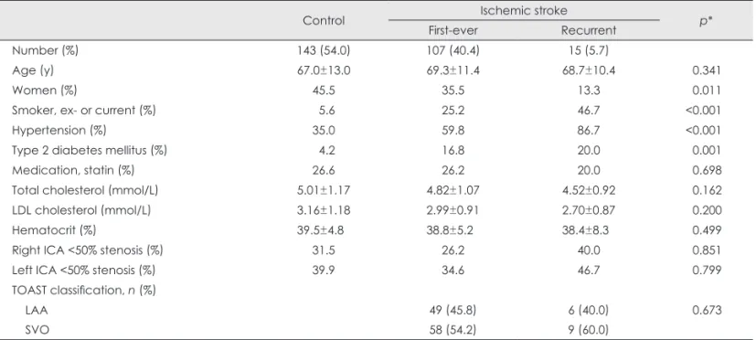
ICA shear stress was calculated in 143 (54.0%) control sub- jects and 122 (46.0%) patients with ischemic stroke of LAA or SVO, who had a normal ICA or an ICA with <50% steno- sis. Among the patients with ischemic stroke, 107 (87.7%), and 15 (12.3%) patients were categorized as first-ever and recurrent stroke, respectively, as indicated in Table 1. Male sex, smoking, hypertension, and type 2 diabetes were signifi- cantly and positively associated with stroke and its recurrence.
In contrast, age, use of statins, LDL cholesterol, and the he- matocrit did not differ across the three groups. ICA stenoses of <50% were evenly distributed in the groups. LAA and SVO appeared evenly in patients with first-ever and recurrent ischemic stroke.
Arterial diameters (ED) were significantly larger in the groups with ischemic stroke than the stroke-free group, espe- cially in the right ICA and CCA, as indicated in Table 2. PS and ED blood flow velocities were also significantly lower in patients with ischemic stroke than in the control group. Nei- ther plaque morphology (echogenicity and surface irregulari- ty) nor carotid IMT differed significantly between the groups (data not shown).
Mean values of PS and ED shear stress in both ICAs were significantly lower in patients with ischemic stroke, even af- ter adjusting for age, sex, and major cardiovascular risk fac-
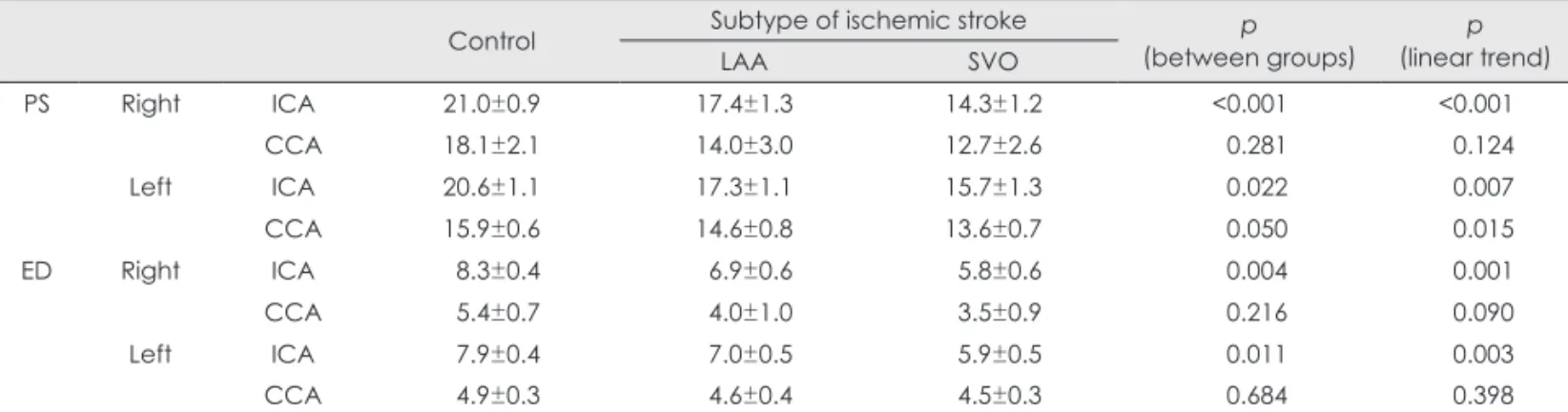
tors, as indicated in Table 3 and 4. The PS and ED shear stresses were lowest in patients with recurrent stroke (Table 3) and in those with SVO (Table 4). The shear stress was sig- nificantly lower in the left CCA. The PS and ED shear stresses in both ICAs showed independent and negative associations with ischemic stroke after adjusting for the potential con- founders, as indicated in Table 5. The interaction terms for carotid shear stress between the three study groups and inde- pendent variables including sex, smoking, hypertension, type 2 diabetes, and statin use were not significant (p>0.1).
Discussion
This study found that the vascular shear stress in carotid arte- rial segments was significantly lower in patients with isch- emic stroke than in the control group. Among the stroke pa- tients, the carotid arterial shear stress was lowest in those with recurrent stroke or SVO. Carotid arterial shear stress was measured in the extracranial portions of the ICA among sub- jects with normal findings or stenosis of <50%, as assessed ultrasonographically.
Our findings suggest that the milieu of low carotid artery Table 1. Characteristics of the included subjects
Control Ischemic stroke
First-ever Recurrent p*
Number (%) 143 (54.0) 107 (40.4) 15 (5.7)
Age (y) 67.0±13.0 69.3±11.4 68.7±10.4 0.341
Women (%) 45.5 35.5 13.3 0.011
Smoker, ex- or current (%) 5.6 25.2 46.7 <0.001
Hypertension (%) 35.0 59.8 86.7 <0.001
Type 2 diabetes mellitus (%) 4.2 16.8 20.0 0.001
Medication, statin (%) 26.6 26.2 20.0 0.698
Total cholesterol (mmol/L) 5.01±1.17 4.82±1.07 4.52±0.92 0.162
LDL cholesterol (mmol/L) 3.16±1.18 2.99±0.91 2.70±0.87 0.200
Hematocrit (%) 39.5±4.8 38.8±5.2 38.4±8.3 0.499
Right ICA <50% stenosis (%) 31.5 26.2 40.0 0.851
Left ICA <50% stenosis (%) 39.9 34.6 46.7 0.799
TOAST classification, n (%)
LAA 49 (45.8) 6 (40.0) 0.673
SVO 58 (54.2) 9 (60.0)
Data are mean±SD or percentage values.
*p values by analysis of variance or chi-square test for trend as appropriate.
ICA: internal carotid artery, LAA: large-artery atherosclerosis, LDL: low-density lipoprotein, SVO: small-vessel occlusion, TOAST: Trial of Org 10172 in Acute Stroke Treatment.
Table 2. Geometric and hemodynamic features in the carotid arteries of the study participants
Control Ischemic stroke
First-ever Recurrent p*
ED diameter (mm) Right ICA 4.6±1.1 5.2±1.0 5.8±0.9 <0.001
CCA 6.1±1.2 6.5±0.9 6.8±0.9 <0.001
Left ICA 5.2±1.2 5.2±1.1 6.0±0.7 0.035
CCA 6.1±0.9 6.3±0.8 6.6±0.7 0.025
PS velocity (cm/sec) Right ICA 61.7±22.3 55.2±20.3 52.1±22.3 0.034
CCA 60.1±19.4 54.6±13.8 53.3±13.5 0.027
Left ICA 63.5±21.3 58.3±20.0 56.0±14.7 0.093
CCA 66.4±20.2 57.2±17.2 56.3±11.0 <0.001
ED velocity (cm/sec) Right ICA 24.7±10.4 22.2±11.0 18.2±7.5 0.035
CCA 18.0±6.4 15.1±4.9 14.3±8.0 <0.001
Left ICA 25.5±9.4 22.0±7.8 20.2±7.6 0.003
CCA 20.8±7.7 18.3±9.3 16.3±6.1 0.022
Data are mean±SD values.
*p values by analysis of variance.
CCA: common carotid artery, ED: end-diastolic, ICA: internal carotid artery, PS: peak-systolic.
shear stress affects cerebral arteries adversely based on its as- sociation with both first-ever and recurrent ischemic strokes.
Among the subtypes of ischemic stroke, the carotid artery shear stress was much lower in SVO than LAA, as indicated in Table 4. We previously reported that the blood flow veloc- ities in cerebral arteries were lower in patients with LI (or SVO) than in patients with other ischemic stroke subtypes.19 A low shear stress along the carotid artery was previously re- ported to be associated with ischemic stroke of LAA, espe-
cially on the same side as the affected hemisphere.8 Howev- er, that study involved a relatively small number of patients with LAA (n=25), and did not include patients with SVO.
The cross-sectional design of the present study meant that we could not determine whether low carotid artery shear stress was causally related to ischemic stroke. Nevertheless, several mechanisms may contribute to an increased risk of ischemic stroke in regions of low wall shear stress,20 includ- ing the production of open gaps due to the endothelial cells Table 3. Adjusted values of carotid artery PS and ED shear stress (dyne/cm2)
Control Ischemic stroke p
(between groups)
p (linear trend) First-ever Recurrent
PS Right ICA 21.0±0.9 16.0±0.9 13.2±2.5 <0.001 <0.001
CCA 18.1±2.1 13.2±2.1 13.9±5.5 0.294 0.178
Left ICA 20.6±1.1 16.8±1.0 14.0±2.7 0.020 0.005
CCA 15.9±0.6 14.0±0.6 13.9±1.4 0.079 0.040
ED Right ICA 8.3±0.4 6.4±0.4 5.0±1.2 0.004 0.001
CCA 5.4±0.7 3.7±0.7 4.3±1.8 0.219 0.166
Left ICA 7.9±0.4 6.5±0.4 5.6±1.0 0.025 0.007
CCA 4.9±0.3 4.5±0.3 4.3±0.7 0.669 0.374
Data are mean±SE values adjusted for age, sex, smoking, hypertension, type 2 diabetes, total cholesterol, LDL cholesterol, hemato- crit, and statin medication.
CCA: common carotid artery, ED: end-diastolic, ICA: internal carotid artery, LDL: low-density lipoprotein, PS: peak-systolic.
Table 4. Adjusted values of carotid artery shear stress (dyne/cm2) according to subtypes of ischemic stroke
Control Subtype of ischemic stroke p
(between groups) p
(linear trend)
LAA SVO
PS Right ICA 21.0±0.9 17.4±1.3 14.3±1.2 <0.001 <0.001
CCA 18.1±2.1 14.0±3.0 12.7±2.6 0.281 0.124
Left ICA 20.6±1.1 17.3±1.1 15.7±1.3 0.022 0.007
CCA 15.9±0.6 14.6±0.8 13.6±0.7 0.050 0.015
ED Right ICA 8.3±0.4 6.9±0.6 5.8±0.6 0.004 0.001
CCA 5.4±0.7 4.0±1.0 3.5±0.9 0.216 0.090
Left ICA 7.9±0.4 7.0±0.5 5.9±0.5 0.011 0.003
CCA 4.9±0.3 4.6±0.4 4.5±0.3 0.684 0.398
Data are mean±SE values adjusted for age, sex, smoking, hypertension, type 2 diabetes, total cholesterol, LDL cholesterol, hemato- crit, and statin medication.
CCA: common carotid artery, ED: end-diastolic, ICA: internal carotid artery, LAA: large-artery atherosclerosis, LDL: low-density lipo- protein, SVO: small-vessel occlusion, PS: peak-systolic.
Table 5. Multivariate association of carotid shear stress with ischemic stroke
Carotid shear stress (dyne/cm2) Ischemic stroke
Odds ratio 95% confidence interval p
PS Right ICA 0.93 0.89–0.97 0.002
CCA 0.96 0.91–1.02 0.174
Left ICA 0.95 0.92–0.99 0.011
CCA 0.93 0.87–0.99 0.027
ED Right ICA 0.89 0.82–0.97 0.011
CCA 0.83 0.65–1.05 0.122
Left ICA 0.89 0.81–0.99 0.030
CCA 0.92 0.81–1.06 0.246
Data adjusted for age, sex, smoking, hypertension, type 2 diabetes, total cholesterol, LDL cholesterol, hematocrit, and statin medication.
CCA: common carotid artery, ED: end-diastolic, ICA: internal carotid artery, LDL: low-density lipoprotein, PS: peak-systolic.
becoming more spherical.21 The low shear stress may result in altered physiological responses that include a decrease in endothelial nitric oxide synthase mRNA and protein expres- sion,22 increased release of proinflammatory mediators such as macrophage chemoattractant-1,23 increased uptake of oxi- dized LDL,24 altered redox state of the vasculature, and al- tered smooth-muscle-cell gene expression.25 Regions of low arterial shear stress are considered to be vulnerable to future atherosclerotic growth26 and thrombosis evolution.2,3 However, the exact mechanisms underlying how low arterial shear stress affects neurovascular units and cerebrovascular networks need further evaluation.27
Wall shear stress occurs immediately adjacent to the arte- rial wall. A limitation of the current method includes the use of flow velocities that were obtained in the center of the artery instead of along the vessel wall, and the use of the estimated blood viscosity. Further investigations are required to deter- mine if the arterial shear stress measured with the centerline flow velocity is a parameter for which there are critical thresh- old values for eliciting vascular diseases or pathologic condi- tions. Our study involving 122 patients with ischemic stroke showed that the ICA PS shear stress was 16–17 dyne/cm2, which is consistent with a previous report of low carotid arte- rial shear stresses in 126 subjects with high-risk vascular pro- files.20
Therapeutic implications might be considered for improv- ing the carotid and cerebral arterial shear stress when a low arterial shear stress is diagnosed. However, very few experi- mental trials have investigated the possible beneficial effects of shear-stress-modifying therapy.28 Considering the unstable cerebral and carotid hemodynamics in the acute phase of isch- emic stroke, shear-stress-modifying therapy might be more suitable for the subacute or chronic phases of ischemic stroke in order to prevent recurrent attacks. The variability or persis- tency of subphysiologic arterial shear stress in the transition from the acute to chronic phases requires further evaluation in each subtype of ischemic stroke.
The present study was subject to several limitations. First, the cross-sectional methodology with an interval between the examination of carotid arterial shear stress and ischemic stroke might have hindered the accurate determination of temporal changes in carotid shear stress according to the subtypes of ischemic stroke. Second, the internal diameters of arteries were measured only in the ED phase, so the calculated PS shear stresses might be higher than the true values. Third, the application of the Poiseuille equation in ICA was not consis- tent with the basic assumption of some patients having mild stenosis (<50%). However, excluding those subjects with mild stenosis did not change the present results markedly (data not shown). The Poiseuille equation can only be applied to a
human artery if certain basic assumptions are met. The ICA has no branches along its intracranial course, and if mild ste- nosis (<50%) affects the local hemodynamics, the present study might have included ICAs with higher shear stresses and thereby have underestimated the true difference. Finally, the blood in the present study was assumed to be Newtonian, even though each carotid shear stress was calculated using patient-specific blood viscosity derived from the hematocrit.
Blood actually behaves as a non-Newtonian fluid, in that its viscosity changes with the shear rate.29 Future studies that di- rectly measure the non-Newtonian characteristics of the blood should enhance the accuracy of vascular shear stress mea- surements and potentially also the clinical utility of carotid ar- terial shear stress measurements.
ICA shear stresses were significantly lower in patients with ischemic stroke than in the control subjects. Further study is needed to define the causal relationship between ca- rotid arterial shear stress and ischemic stroke.
Conflicts of Interest
The authors have no financial conflicts of interest.
Acknowledgements
This study was supported by the Research Institute of Clinical Medicine, Chonbuk National University, and the Biomedical Research Institute of Chonbuk National University Hospital.
REFERENCES
1. Cunningham KS, Gotlieb AI. The role of shear stress in the patho- genesis of atherosclerosis. Lab Invest 2005;85:9-23.
2. Giannoglou GD, Soulis JV, Farmakis TM, Farmakis DM, Louridas GE. Haemodynamic factors and the important role of local low static pressure in coronary wall thickening. Int J Cardiol 2002;86:27-40.
3. Nesbitt WS, Westein E, Tovar-Lopez FJ, Tolouei E, Mitchell A, Fu J, et al. A shear gradient-dependent platelet aggregation mechanism drives thrombus formation. Nat Med 2009;15:665-673.
4. Malek AM, Alper SL, Izumo S. Hemodynamic shear stress and its role in atherosclerosis. JAMA 1999;282:2035-2042.
5. Lee SW, Antiga L, Spence JD, Steinman DA. Geometry of the carotid bifurcation predicts its exposure to disturbed flow. Stroke 2008;39:
2341-2347.
6. Groen HC, Gijsen FJ, van der Lugt A, Ferguson MS, Hatsukami TS, van der Steen AF, et al. Plaque rupture in the carotid artery is localized at the high shear stress region: a case report. Stroke 2007;38:2379-2381.
7. Palm-Meinders IH, Box FM, de Craen AJ, Blauw GJ, van Buchem MA, van der Grond J. Diastolic wall shear stress in the internal carotid artery is associated with different cardiovascular risk factors than sys- tolic wall shear stress. Cerebrovasc Dis 2009;28:185-190.
8. Carallo C, Lucca LF, Ciamei M, Tucci S, de Franceschi MS. Wall shear stress is lower in the carotid artery responsible for a unilateral ischemic stroke. Atherosclerosis 2006;185:108-113.
9. Rothwell PM, Warlow CP. Low risk of ischemic stroke in patients with reduced internal carotid artery lumen diameter distal to severe symp- tomatic carotid stenosis: cerebral protection due to low poststenotic flow? On behalf of the European Carotid Surgery Trialists’ Collabor- ative Group. Stroke 2000;31:622-630.
10. Adams HP Jr, Bendixen BH, Kappelle LJ, Biller J, Love BB, Gordon DL, et al. Classification of subtype of acute ischemic stroke. Defini-
tions for use in a multicenter clinical trial. TOAST. Trial of Org 10172 in Acute Stroke Treatment. Stroke 1993;24:35-41.
11. Hardie K, Hankey GJ, Jamrozik K, Broadhurst RJ, Anderson C. Ten- year risk of first recurrent stroke and disability after first-ever stroke in the Perth Community Stroke Study. Stroke 2004;35:731-735.
12. Touboul PJ, Hennerici MG, Meairs S, Adams H, Amarenco P, Born- stein N, et al. Mannheim carotid intima-media thickness consensus (2004-2006). An update on behalf of the Advisory Board of the 3rd and 4th Watching the Risk Symposium, 13th and 15th European Stroke Conferences, Mannheim, Germany, 2004, and Brussels, Bel- gium, 2006. Cerebrovasc Dis 2007;23:75-80.
13. Petersen C, Peçanha PB, Venneri L, Pasanisi E, Pratali L, Picano E.
The impact of carotid plaque presence and morphology on mortality outcome in cardiological patients. Cardiovasc Ultrasound 2006;4:16.
14. Grant EG, Benson CB, Moneta GL, Alexandrov AV, Baker JD, Bluth EI, et al. Carotid artery stenosis: gray-scale and Doppler US diagnosis- -Society of Radiologists in Ultrasound Consensus Conference. Radi- ology 2003;229:340-346.
15. Jiang Y, Kohara K, Hiwada K. Association between risk factors for atherosclerosis and mechanical forces in carotid artery. Stroke 2000;31:
2319-2324.
16. Guyton AC, Hall JE. Textbook of Medical Physiology. 10th ed. Phila- delphia, PA: W.B. Saunders, 2000.
17. Cho YI, Kensey KR. Effects of the non-Newtonian viscosity of blood on flows in a diseased arterial vessel. Part 1: Steady flows. Biorheology 1991;28:241-262.
18. Schulz UG, Rothwell PM. Major variation in carotid bifurcation anat- omy: a possible risk factor for plaque development? Stroke 2001;32:
2522-2529.
19. Kim JT, Lee SH, Hur N, Jeong SK. Blood flow velocities of cerebral arteries in lacunar infarction and other ischemic strokes. J Neurol Sci 2011;308:57-61.
20. Irace C, Cortese C, Fiaschi E, Carallo C, Farinaro E, Gnasso A. Wall shear stress is associated with intima-media thickness and carotid ath-
erosclerosis in subjects at low coronary heart disease risk. Stroke 2004;
35:464-468.
21. Levesque MJ, Liepsch D, Moravec S, Nerem RM. Correlation of en- dothelial cell shape and wall shear stress in a stenosed dog aorta. Ar- teriosclerosis 1986;6:220-229.
22. Wilcox JN, Subramanian RR, Sundell CL, Tracey WR, Pollock JS, Harrison DG, et al. Expression of multiple isoforms of nitric oxide syn- thase in normal and atherosclerotic vessels. Arterioscler Thromb Vasc Biol 1997;17:2479-2488.
23. Sheikh S, Rainger GE, Gale Z, Rahman M, Nash GB. Exposure to flu- id shear stress modulates the ability of endothelial cells to recruit neu- trophils in response to tumor necrosis factor-alpha: a basis for local variations in vascular sensitivity to inflammation. Blood 2003;102:
2828-2834.
24. Zhu CH, Ying DJ, Mi JH, Zhu XH, Sun JS, Cui XP. Low shear stress regulates monocyte adhesion to oxidized lipid-induced endothelial cells via an IkappaBalpha dependent pathway. Biorheology 2004;41:
127-137.
25. Khatri JJ, Johnson C, Magid R, Lessner SM, Laude KM, Dikalov SI, et al. Vascular oxidant stress enhances progression and angiogenesis of experimental atheroma. Circulation 2004;109:520-525.
26. Lee SH, Hur N, Jeong SK. Geometric analysis and blood flow simu- lation of basilar artery. J Atheroscler Thromb 2012;19:397-401.
27. del Zoppo GJ. Virchow’s triad: the vascular basis of cerebral injury.
Rev Neurol Dis 2008;5 Suppl 1:S12-S21.
28. Zhang Y, He X, Liu D, Wu G, Chen X, Ma H, et al. Enhanced external counterpulsation attenuates atherosclerosis progression through mod- ulation of proinflammatory signal pathway. Arterioscler Thromb Vasc Biol 2010;30:773-780.
29. Jeong SK, Cho YI, Duey M, Rosenson RS. Cardiovascular risks of anemia correction with erythrocyte stimulating agents: should blood viscosity be monitored for risk assessment? Cardiovasc Drugs Ther 2010;24:151-160.