Copyright ⓒ 2018 by Korean Society for Surgery of the Hand, Korean Society for Microsurgery, and Korean Society for Surgery of the Peripheral Nerve. All Rights reserved.
This is an Open Access article distributed under the terms of the Creative Commons Attribution Non-Commercial License (http://creativecommons.org/licenses/by-nc/4.0/) which permits unrestricted non-commercial use, distribution, and reproduction in any medium, provided the original work is properly cited.
Fibroma of the tendon sheath is a rare benign soft tis- sue tumor that typically occurs in the upper extremities, especially the fingers. The most common symptom is a slow-growing mass with no pain or swelling. When it occurs in a finger, finger movement may be limited due to interference with tendon excursion through the flexor pulley system. Moreover, depending on where the tumor occurs, fibroma may also cause localized symptoms such as carpal tunnel syndrome1 or trigger-finger2. Fibroma of
the tendon sheath is almost always found adhering to the tendon sheath or tendon3, whereas it is extremely rare to find this tumor within the tendon. Grenga4 reported a case of intratendinous fibroma in a 10-year-old boy present- ing with a slow-growing, non-tender mass in the palmar aspect of a finger, which limited finger movement. The mass was 1.5 cm in size and was located within the flexor digitorum profundus (FDP) tendon, interfering with the excursion of this tendon. Closed tears of the FDP tendon Hand and
Microsurgery
야구 선수의 수지에서 심수지굴곡건 파열을 동반한 건내 섬유종:
증례 보고
김규진
1ㆍ이재훈
21가천대 길병원 정형외과, 2경희대학교 의과대학 강동경희대학교병원 정형외과
Intratendinous Fibroma with a Flexor Profundus Tendon Tear in the Finger of an Adolescent Baseball Player: A Case Report
Kyu Jin Kim
1, Jae Hoon Lee
21Department of Orthopedic Surgery, Gachon University Gil Medical Center, Incheon, Korea
2Department of Orthopaedic Surgery, Kyung Hee University Hospital at Gangdong, School of Medicine, Kyung Hee University, Seoul, Korea
Flexor digitorum profundus (FDP) tears in adolescents appear as avulsion tears in the FDP tendon, whereas longitudinal tears are very rare. Moreover, there has been only one reported case of intratendinous fibroma occurring in the flexor ten- don of a finger. A longitudinal tear of the flexor profundus tendon associated with an intratendinous fibroma has not been previously reported. We report one case of a longitudinal partial tear accompanied by an intratendinous fibroma at the FDP tendon of the left middle finger after a hyperextension injury caused by the impact of a baseball. Given the rarity of longitudinal flexor tendon tears in adolescents, in such cases, the possibility of an underlying pathology should be consid- ered.
Key Words: Finger, Flexor digitorum profundus, Intratendinous fibroma
Received August 6, 2018, Revised [1] August 29, 2018, [2] September 4, 2018, Accepted September 4, 2018 Corresponding author: Jae Hoon Lee
Department of Orthopaedic Surgery, Kyung Hee University Hospital at Gangdong, 892 Dongnam-ro, Gangdong-gu, Seoul 05278, Korea TEL: +82-2-440-6153, FAX: +82-2-440-7498, E-mail: [email protected]
Case Report
in pediatric patients are very rare, whereas cases involv- ing avulsion tear of the FDP tendon are more common5. However, reports of a closed longitudinal tear of the FDP tendon have not been found in the English literature to date. We report the case of a 12-year-old baseball player with a longitudinal partial tear at the FDP finally diag- nosed as an intratendinous fibroma.
CASE REPORT
A 12-year-old male baseball player complained of pain in the proximal interphalangeal joint (PIPJ) of the middle finger after a hyperextension injury caused by a baseball impact six weeks previously, which was managed with conservative treatment. Physical examination revealed
tenderness and swelling at the palmar side of the PIPJ of the left middle finger. Although passive range of motion showed no limitation, active range of motion was limited (range, 0-70 degrees). There was no triggering.
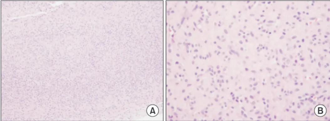
Preoperative radiography revealed no abnormalities, whereas magnetic resonance imaging (MRI) revealed a longitudinal partial tear at the FDP tendon of the left middle finger (Fig. 1). Surgical treatment was planned to allow the patient to return to his sports activity. After making a zigzag skin incision on the palmar side of the middle PIPJ under general anesthesia, the cruciate pulley and parts of the A2 pulley were incised. A longitudinal tear was found in the FDP tendon. Further splitting of the tear revealed a 0.8×0.5×0.5 cm mass (Fig. 2). The mass was well demarcated and was excised easily, followed by tenorrhaphy with continuous suture. Histopathologi- cal examination confirmed the mass to be a fibroma (Fig.
3). In the one-month postoperative follow-up, there was no pain in the PIPJ of the middle finger, and the range
Fig. 1. Magnetic resonance image showing a longitudinal partial tear of the middle flexor digitorum profundus tendon and an intratendinous tumor (arrow).
FDP
FDP
Fig. 2.Intraoperative photograph showing a longitudinal split of the flexor digitorum profundus (FDP) tendon and the intratendinous tumor (arrow).
A B
Fig. 3. Histopathologic finding of intratendinous fibroma. (A) Low- power view (H&E stain, ×100) showing numerous hyalinized fibrous tissues. (B) High-power view (H&E stain, ×400) showing scattered fibro blasts with large
of motion in the joint returned to normal. In the one- year postoperative follow-up, the patient was able to play baseball without pain. Written informed consent was ob- tained from the patient for publication of this case report and any accompanying images.
DISCUSSION
The most common form of benign soft tissue tumor in the hand is ganglion, followed by giant cell tumor. Fi- broma is a very rare form of benign soft tissue tumor. It occurs mostly in patients aged 20-49 years, and its preva- lence is twice as high among male than female patients.
The diagnosis of fibroma is based on the medical history of the patient and clinical tests; however, radiological examinations such as MRI or ultrasonography may be useful. Plain radiography typically shows negative re- sults; however, positive results may be found when a large mass is compressing the surrounding muscles or fat or when bony erosion is present. Differential diagnoses include epidermal cyst, neurofibroma, nodular fasciitis, leiomyoma, and giant cell tumor. In particular, the clini- cal features of giant cell tumor and fibroma are very simi- lar to each other. Both types of tumor commonly occur in the fingers, attached to the tendon sheath, and they show similar MRI signal intensity and form a grayish-white multiseptated mass that is hard and well encapsulated.
However, a fibroma can be differentiated from a giant cell tumor on the basis of histopathological findings. Fi- brotic changes in the matrix of a fibroma are more severe than in a giant cell tumor. Moreover, in fibroma, spindle cells are more abundant, the frequency of giant cells is low, and foam cells and siderophages are rare. Satti6 de- scribed fibroma in the tendon sheath as an end or harden- ing stage of a giant cell tumor that may have been the re- sult of progressive vascular damage. Fibroma may occur anywhere on the limbs; however, it is especially common in the finger, hand, and wrist areas. Millon et al.7 reported that, of 208 hand tumor excision cases in soft tissue over a 15-year period, fibromas of the tendon sheath of the finger flexor tendon accounted for 1%. The relapse rate after tumor excision was ≥24%8. Fibroma that occurs in
a finger may interfere with tendon excursion, causing limited movement and triggering9. There has been only one previously reported case of fibroma found within a tendon4; it is typically found as an indolent mass adhering directly to the tendon sheath or the tendon itself3,7. In our case, a fibroma with a size of 0.8×0.5×0.5 cm, which was too small to be detected by visual inspection alone, was found, and it remained asymptomatic until several finger injuries occurred. It is believed that the finger injury led to the underlying intratendinous fibroma, causing a lon- gitudinal tear of the FDP tendon, which in turn caused limited movement and pain in the finger.
Most cases of closed flexor tendon injury following finger injury in children involve avulsion rupture of the FDP tendon10, which is rare5. Mid-substance rupture of the flexor tendon in adults often occurs when an underly- ing pathologic condition is present, but it rarely occurs without any underlying pathology5. Traumatic closed mid-substance flexor tendon rupture in children is ex- tremely rare, although Badur et al.5 reported a case of mid-substance flexor tendon rupture that occurred in a 12-year-old child with no underlying pathology. In our case, the underlying pathology of the child was intraten- dinous fibroma, and this was the first case in which mid- substance longitudinal partial tear was caused by this tumor.
We report one case of a longitudinal partial tear accom- panied by an intratendinous fibroma at the FDP tendon of the left middle finger after a hyperextension injury caused by the impact of a baseball. Longitudinal flexor tendon tear in adolescents is extremely rare; when it oc- curs, the possibility of an underlying pathology should be considered.
CONFLICTS OF INTEREST
The authors have nothing to disclose.
REFERENCES
1. Garrido A, Lam WL, Stanley PR. Fibroma of a tendon sheath at the wrist: a rare cause of compression of the
median nerve. Scand J Plast Reconstr Surg Hand Surg.
2004;38:314-16.
2. Oni OO. A tendon sheath tumour presenting as trigger fin- ger. J Hand Surg Br. 1984;9:340.
3. Cooper PH. Fibroma of tendon sheath. J Am Acad Derma- tol. 1984;11:625-8.
4. Grenga TE. Intratendinous fibroma of flexor tendon. J Hand Surg Am. 1990;15:92-3.
5. Badur N, Gutierrez Monclus R, Ferreres I Claramunt A, Leclère FM. Spontaneous rupture of a flexor digitorum profundus tendon at two levels in zones II and III in a child. Hand (NY). 2013;8:97-101.
6. Satti MB. Tendon sheath tumours: a pathological study of the relationship between giant cell tumour and fibroma of tendon sheath. Histopathology. 1992;20:213-20.
7. Millon SJ, Bush DC, Garbes AD. Fibroma of tendon sheath in the hand. J Hand Surg Am. 1994;19:788-93.
8. Chung EB, Enzinger FM. Fibroma of tendon sheath. Can- cer. 1979;44:1945-54.
9. Feinberg MS. Fibroma of a tendon causing limited finger motion: a case report. J Hand Surg Am. 1979;4:386.
10. Lourie GM, Hamby Z, Raasch WG, Chandler JB, Porter JL. Annular flexor pulley injuries in professional baseball pitchers: a case series. Am J Sports Med. 2011;39:421-4.
야구 선수의 수지에서 심수지굴곡건 파열을 동반한 건내 섬유종:
증례 보고
김규진
1ㆍ이재훈
21가천대 길병원 정형외과, 2경희대학교 의과대학 강동경희대학교병원 정형외과
청소년에서 심수지굴곡건 파열은 주로 견열 파열로 나타나며 종적 파열은 매우 드물다. 수지의 굴곡건에 발생하는 건내 섬유종의 보고는 지금까지 1예가 있었지만, 건내 섬유종과 관련된 심수지굴곡건의 종파열은 아직까지 보고된 바 없었다. 저자들은 야구 중 충격에 의한 과신전 손상을 받은 이후 좌측 3수지의 건내 섬유종과 동반된 심수지굴 곡건의 종 파열을 보고한다. 청소년에서 굴곡건의 종 파열은 드물지만 발생한 경우 병적 질환에 의한 파열일 수 있 음을 고려하여야 하겠다.
색인단어: 수지, 심수지굴건, 건내 섬유종
접수일 2018년 8월 6일 수정일 1차: 2018년 8월 29일, 2차: 2018년 9월 4일 게재확정일 2018년 9월 4일 교신저자 이재훈
05278, 서울시 강동구 동남로 892, 강동경희대학교병원 정형외과 TEL 02-440-6153 FAX 02-440-7498 E-mail [email protected]