Ann Hepatobiliary Pancreat Surg 2018;22:150-155
https://doi.org/10.14701/ahbps.2018.22.2.150
Case Report
A huge intraductal papillary neoplasm of the bile duct treated by right trisectionectomy after right portal vein embolization
Vusal Aliyev1, Kentaro Yasuchika1, Ahmed Hammad1,2, Tetsuya Tajima1, Ken Fukumitsu1, Koichiro Hata1, Hideaki Okajima1, and Shinji Uemoto1
1Division of Hepato-Biliary-Pancreatic and Transplant Surgery, Department of Surgery, Graduate School of Medicine, Kyoto University, Kyoto, Japan, 2Department of Surgery, Mansoura University, Mansoura, Egypt
Intraductal papillary neoplasm of the bile duct (IPNB) is a rare variant of bile duct tumors characterized by papillary growth within the bile duct lumen and recognized precursor of invasive carcinoma. IPNB was detected incidentally in a 60-year-old woman during check up. Radiologic images revealed a huge cystic mass with papillary projection and markedly dilated bile ducts. Biopsies revealed high-grade IPNB. Cholangioscopy detected a connection between the right posterior bile duct and cyst lumen with epithelial dysplasia of the bile duct. Right posterior sectional duct opened in the left hepatic duct. Consequently, right trisectionectomy and extrahepatic bile duct resection were conducted. Histological studies revealed intraductal papillary neoplasm with high-grade intraepithelial neoplasia (carcinoma in situ). IPNB patients without distant metastases are candidates for surgery and complete resection should be conducted to achieve long-term survival. (Ann Hepatobiliary Pancreat Surg 2018;22:150-155)
Key Words: Intraductal papillary neoplasm of the bile duct; Portal vein embolization; Right trisectionectomy
Received: October 23, 2017; Revised: October 28, 2017; Accepted: October 29, 2017 Corresponding author: Kentaro Yasuchika
Division of Hepato-Biliary-Pancreatic and Transplant Surgery, Department of Surgery, Graduate School of Medicine, Kyoto University, 54 Kawahara-cho, Shogoin, Sakyo-ku, Kyoto 606-8507, Japan
Tel: +81-75-751-4323, Fax: +81-75-751-4348, E-mail: [email protected]
Copyright Ⓒ 2018 by The Korean Association of Hepato-Biliary-Pancreatic Surgery
This is an Open Access article distributed under the terms of the Creative Commons Attribution Non-Commercial License (http://creativecommons.org/
licenses/by-nc/4.0) which permits unrestricted non-commercial use, distribution, and reproduction in any medium, provided the original work is properly cited.
Annals of Hepato-Biliary-Pancreatic Surgery ∙ pISSN: 2508-5778ㆍeISSN: 2508-5859
INTRODUCTION
Intraductal papillary neoplasm of the bile duct (IPNB) is a rare variant of bile duct tumors, characterized by pap- illary or villous growth within the bile duct lumen and is regarded as a biliary counterpart of intraductal papillary mucinous neoplasm (IPMN) of the pancreas.1 IPNB was recently defined in 2010 WHO classification as a distinct clinical and pathologic entity.2 IPNB is mostly found in the Far East nations where hepatolithiasis and clonorch- iasis infections are endemic.3 Most patients are age 50-70.4 Common clinical signs of IPNB are abdominal pain, jaundice and cholangitis. The most common radio- logic findings for IPNB are bile duct dilatation and intra- ductal masses.5 Here, we report surgical treatment of a huge asymptomatic IPNB.
CASE
A 60-year-old Japanese woman was admitted to our hospital due to a huge liver mass detected incidentally during the patient’s routine respiratory examination diag- nosed with bronchial asthma and obstructive pulmonary disease. She had no symptoms or past medical history of liver disease. She smoked 15 cigarettes per day and quit 2 months prior the operation. Laboratory values on admis- sion were as follows: Total leucocytic count 5.17×10³/cc, hemoglobin 13.4 g/dl, platelets count 274×109/L, aspartate aminotransferase 25 IU/L, alanine aminotransferase 28 IU/L, alkaline phosphatase 271 IU/L, gamma-glutamyl transpeptidase 38 IU/L, total and direct bilirubin 0.5 and 0.1 mg/dl, albumin 3.8 g/dl, -fetoprotein (AFP) 4.1 ng/ml, carcinoembryonic antigen (CEA) 6.8 ng/ml and cancer antigen 19-9 (CA19-9) 0.6 U/ml.
Abdominal ultrasonography (US) and enhanced com- puted tomography (CT) revealed a large cystic mass with
Fig. 1. Preoperatoive imaging findings. (A) Abdominal ultrasonography (US) revealed multiple hyper-echoic solid lesions (arrows). (B) Enhanced computed tomography (CT) revealed a huge (10×7 cm) cystic tumor with surrounding high-density com- ponents (arrows) involving the right hemiliver and the left medial section.
Fig. 2. Non-enhanced magnetic resonance imaging (A) revealed huge solid mass in the right hemiliver and the left medial segment of the liver. Bile ducts were markly dilated. Suspicious communication between right posterior bile duct (arrow) and the cystic lesion. Magnetic resonance cholangiography (B) revealed a marked aneurysmal dilatation of the bile duct.
multiple internal papillary projections involving the right- hemiliver and left medialsection (Fig. 1). Magnetic reso- nance imaging (MRI) and magnetic resonance cholangiog- raphy (MRC) detected a large cystic lesion involving the right posterior bile ducts (Fig. 2).
Endoscopic retrograde cholangiogram (ERC) confirmed contrast filling of the cyst lumen (Fig. 3). On peroral chol- angioscopy (POCS), a large amount of mucus and papil- lary proliferating epithelium was observed in the cyst cav- ity while the orifice of the posterior duct was invaded by
the tumor. From the lower bile duct up to the hepatic hi- lum, right anterior segment bile duct and left hepatic duct endo-lumen had no abnormal findings (Fig. 4). Multiple biopsies were conducted from the cyst cavity. Based on above mentioned radiological and endovisual findings, a diagnosis of IPNB was reached. Biopsy results confirmed our diagnosis, as a high-grade IPNB. Positron emission to- mography (PET) demonstrated malignant lesions with marginal dominance of FDG accumulation in the right lobe. Intra- and extra-hepatic metastases were not detected
152 Ann Hepatobiliary Pancreat Surg Vol. 22, No. 2, May 2018
Fig. 4. Peroral cholangioscopy findings. Papilla Vater canula- tion (A). No abnormal finding on hepatic hilum (B). Tumor in- vading the right posterior bile duct (C). Biliary papillomatosis inside of the cyst (D).
Fig. 3. Endoscopic retrograde cholangiogram revealed com- munication between the proximal right hepatic duct and cyst.
Fig. 5. Positron emission tomography-computed tomography scan revealed tumorous lesions with abnormal uptake and marginal dominance of FDG accumulation in the right lobe.
Intrahepatic and extrahepatic metastasis were not detected.
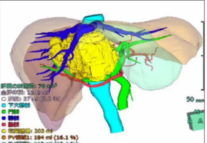
(Fig. 5). Three-dimensional liver CT scan demonstrated right, middle hepatic veins and right portal vein in contact with the tumor (Fig. 6).
Based on findings, right trisectionectomy of liver with extrahepatic bile duct resection and reconstruction were
planned. Liver volumetry was undertaken, total liver and the functional volume of the left lateral section were esti- mated at 1,143 ml (100%) and 303 ml (26.5%), respectively. So, we first conducted percutaneous trans- hepatic portal vein embolization (PTPE) to induce hyper-
Fig. 8. Sagittal computed tomography before (A) and after (B) percutaneous transhepatic portal vein embolization.
Fig. 7. Right portal vein emboli- zation.
Fig. 6. Three-dimensional computed tomography image be- fore percutaneous transhepatic portal vein embolization.
trophy of the remnant liver. After 4 weeks, the estimated remnant liver volume increased to 434 ml (38%) (Figs.
7 and 8).
Four weeks after PTPE, the patient underwent surgery.
Right trisectionectomy with extrahepatic bile duct re- section and Roux-en-Y cholangiojejunostomy were conducted. Intraoperative frozen section confirmed can- cer-free surgical margins, including segment II and III bile duct stumps. Segment II and III bile ducts were separately anastomosed to the ascending jejunal limb. Macroscopic examination of the resected specimen revealed a large mixed cystic-solid lesion with fibrous capsule and multi- ple papillary projections (Fig. 9). Histological studies re- vealed intraductal papillary neoplasm with high-grade in- traepithelial neoplasia (carcinoma in situ), and no regional lymph node metastases (pTis pN0). Microscopic findings
154 Ann Hepatobiliary Pancreat Surg Vol. 22, No. 2, May 2018
Fig. 10. Gross appearance of the resected section. (A) Well defined cystic mass with multiple polypoid mural nodules (black arrows). Communication between the right posterior bile duct and the cyst cavity (yellow arrow). (B) Segment II and III bile ducts stump (B2 and B3, yellow arrows).
Fig. 9. Microscopic finding showing prominent papillary pro- liferation with fibrovascular cores.
include prominent papillary proliferation with fi- brovascular cores, and epithelial subtypes was a pan- creatobiliary (Fig. 10). Immunohistochemical staining re- sults were positive for CK7, MUC4, MUC5AC, MUC6, and negative for MUC1, MUC1, MUC2, CK20, p53.
Ki-67 level was 20%. The patient’s post-operative course was uneventful and was discharged from our institution on the 15th post-operative day. So far, 12 months post-op- eratively, the patient has no recurrence and appears healthy.
DISCUSSION
The INPB is a rare neoplasm of the liver, adopted as
a distinct entity in the 2010 World Health Organization (WHO) classification.6 According to the WHO classi- fication system, IPNB is defined as a cystic lesion lined with biliary, mucinous or oncocytic epithelium in papil- lary configurations without ovarian-like stroma.7 Recently, IPNB is a biliary counterpart of IPMN, because clin- icopathologic and morphologic similarities.8
The most common presenting symptom of IPNB is in- termittent abdominal pain, jaundice and acute cholangitis, however, up to 5% of patients may be asymptomatic.9 Approximately 30% of patients have history of hep- atolithiasis and clonorchiasis infection.10 In our patient, the tumor was discovered incidentally with no history of hepatolithiasis or clonorchiasis infection. It is critical to diagnose IPNB correctly, because this tumor has malig- nant potential and shares similar radiological and clinical characteristics with other cystic liver lesions.2 IPNB can be detected in the extrahepatic and intrahepatic bile ducts.11 Radiologic findings of IPNB include bile duct di- latation and intraductal masses.12 The US, CT, MRI, ERC, POC and IDUS are useful for diagnosis of tumor ex- tension, involvement of the bile duct and presence of mu- cin (Figs. 1-4). Solid, irregular, thickened lesions in the cystic or dilated bile duct are radiologic findings strongly considered as evidence of malignancy.13 The POCS is available in a few centers. In our case, we conducted POCS to approach the bile duct directly, assess the extent of the tumor and conduct a biopsy for histological exami- nation (Fig. 4). The POCS is useful in detecting super-
ficial tumors spreading along the biliary epithelium, that is crucial to reach the right decision before surgery.14
Patients with IPNB should be treated, even if not malignant. Because, papillary tumors and mucin may lead to obstructive jaundice and cholangitis.10 In patients with- out distant metastasis, surgical intervention is the first choice of treatment including pancreaticoduodenectomy (31%), hemihepatectomy (28%), bile duct resection (18%), segmental liver resection (15%) or liver transplant (5%).15 It has been proposed that the curative resection of IPNB with a malignant potential has a favorable prog- nosis of 47.0-82.0% 5-year overall survival rate.16-18 However, microscopic positive margin (R1 resection), in- vasive carcinoma, extraductal invasion, lymphovascular invasion, lymph node metastasis, tumor positivity for Mucin-1, and positivity for carcinoembryonic antigen (CEA) are poor prognostic factors with higher incidence of recurrence after surgical resection of IPNB.18,19 Mucus production can occur after surgical resection, even if no malignant IPNB tissue remains. Therefore, biliary tract af- ter management after surgical resection is paramount.
We report a rare case of IPNB in an asymptomatic patient. The US, CT, MR, MRCP, IDUS and POCS are useful in pre-operative diagnosis to assess tumor invasion depth and extension along the bile duct. IPNB patients without distant metastases are candidates for surgery.
Complete resection should be conducted to achieve long-term survival.
REFERENCES
1. Shibahara H, Tamada S, Goto M, Oda K, Nagino M, Nagasaka T, et al. Pathologic features of mucin-producing bile duct tu- mors: two histopathologic categories as counterparts of pancre- atic intraductal papillary-mucinous neoplasms. Am J Surg Pathol 2004;28:327-338.
2. Nakanuma Y, Curabo MP, Franceschi S, Gores G, Paradis V, Sripa B. WHO classification of tumours of the digestive system.
Lyon: IARC, 2010:217-224.
3. Ohtsuka M, Shimizu H, Kato A, Yoshitomi H, Furukawa K, Tsuyuguchi T, et al. Intraductal papillary neoplasms of the bile duct. Int J Hepatol 2014;2014:459091.
4. Barton JG, Barrett DA, Maricevich MA, Schnelldorfer T, Wood CM, Smyrk TC, et al. Intraductal papillary mucinous neoplasm of the biliary tract: a real disease? HPB (Oxford) 2009;11:
684-691.
5. Kim KM, Lee JK, Shin JU, Lee KH, Lee KT, Sung JY, et al.
Clinicopathologic features of intraductal papillary neoplasm of the bile duct according to histologic subtype. Am J Gastroenterol 2012;107:118-125.
6. Aoki S, Okayama Y, Kitajima Y, Hayashi K, Imai H, Okamoto T, et al. Intrahepatic biliary papilloma morphologically similar to biliary cystadenoma. J Gastroenterol Hepatol 2005;20:321-324.
7. Bosman FT, Carneiro F, Hruban RH, Theise ND. WHO classi- fication of tumours of the digestive system. Lyon: IARC, 2010:3-8.
8. Zen Y, Fujii T, Itatsu K, Nakamura K, Minato H, Kasashima S, et al. Biliary papillary tumors share pathological features with intraductal papillary mucinous neoplasm of the pancreas.
Hepatology 2006;44:1333-1343.
9. Wan XS, Xu YY, Qian JY, Yang XB, Wang AQ, He L, et al.
Intraductal papillary neoplasm of the bile duct. World J Gastroenterol 2013;19:8595-8604.
10. Ohtsuka M, Kimura F, Shimizu H, Yoshidome H, Kato A, Yoshitomi H, et al. Similarities and differences between intra- ductal papillary tumors of the bile duct with and without macro- scopically visible mucin secretion. Am J Surg Pathol 2011;35:512-521.
11. Tan Y, Milikowski C, Toribio Y, Singer A, Rojas CP, Garcia-Buitrago MT. Intraductal papillary neoplasm of the bile ducts: a case report and literature review. World J Gastroenterol 2015;21:12498-12504.
12. Lee SS, Kim MH, Lee SK, Jang SJ, Song MH, Kim KP, et al.
Clinicopathologic review of 58 patients with biliary papillomatosis. Cancer 2004;100:783-793.
13. Yoon KH, Ha HK, Kim CG, Roh BS, Yun KJ, Chae KM, et al. Malignant papillary neoplasms of the intrahepatic bile ducts:
CT and histopathologic features. AJR Am J Roentgenol 2000;
175:1135-1139.
14. Sakai Y, Tsuyuguchi T, Ishihara T, Sugiyama H, Miyakawa K, Yasui S, et al. Usefulness of peroral cholangioscopy in pre- operative diagnosis of intraductal papillary neoplasm of the bile duct. Hepatogastroenterology 2010;57:691-693.
15. Schlitter AM, Born D, Bettstetter M, Specht K, Kim-Fuchs C, Riener MO, et al. Intraductal papillary neoplasms of the bile duct: stepwise progression to carcinoma involves common mo- lecular pathways. Mod Pathol 2014;27:73-86.
16. Paik KY, Heo JS, Choi SH, Choi DW. Intraductal papillary neo- plasm of the bile ducts: the clinical features and surgical out- come of 25 cases. J Surg Oncol 2008;97:508-512.
17. Hokuto D, Nomi T, Yasuda S, Yoshikawa T, Ishioka K, Yamada T, et al. Long-term observation and treatment of a widespread intraductal papillary neoplasm of the bile duct extending from the intrapancreatic bile duct to the bilateral intrahepatic bile duct:
A case report. Int J Surg Case Rep 2017;38:166-171.
18. Kubota K, Nakanuma Y, Kondo F, Hachiya H, Miyazaki M, Nagino M, et al. Clinicopathological features and prognosis of mucin-producing bile duct tumor and mucinous cystic tumor of the liver: a multi-institutional study by the Japan Biliary Association. J Hepatobiliary Pancreat Sci 2014;21:176-185.
19. Narita M, Endo B, Mizumoto Y, Matsusue R, Hata H, Yamaguchi T, et al. Multicentric recurrence of intraductal papil- lary neoplasms of bile duct in the remnant intrahepatic bile duct after curative resection. Int J Surg Case Rep 2015;12:123-127.