INTRODUCTION
Tumefactive multiple sclerosis (MS) is an uncommon manifes- tation of multiple sclerosis (1). The characteristic MRI findings for this demyelinating disease are a large isolated mass in the white matter with incomplete rim enhancement, slight perilesional ede- ma and a mass effect (2, 3). The clinical features of tumefactive MS are variable and polysymptomatic, depending on the location and size of the mass (4). Patients with this lesion commonly pres- ent with headache, cognitive abnormalities, and sub-acute pro- gressive motor and/or sensory symptoms (5). Most patients with this type of MS manifest only a single acute clinical presentation, whereas those with typical MS present with recurrent episodes of neurological symptoms (4, 6). For these reasons tumefactive MS is indistinguishable from glial neoplasm, lymphoma, abscess or other similar types of lesions (2). Thus, awareness of the charac-
teristic MRI appearance of these lesions can be helpful for correct- ly diagnosing this condition in a noninvasive manner (6).
We report herein a case of pathologically confirmed tumefac- tive MS in an uncommon location, which showed atypical mag- netic resonance (MR) imaging findings and a good response to steroid therapy. We also summarize the typical MR imaging findings for tumefactive MS that can be used to differentiate this lesion from others.
CASE REPORT
A 43-year-old woman presented with paresthesia of the right leg, a limping gait, blurred vision, and motor weakness of the right arm and leg. These symptoms had progressed for three weeks without any prior neurological symptoms. She showed ataxia on the right side on neurological examination.
J Korean Soc Radiol 2013;69(5):337-341 http://dx.doi.org/10.3348/jksr.2013.69.5.337
Received May 20, 2013; Accepted August 14, 2013 Corresponding author: Hyun Sook Kim, MD Department of Radiology, Eulji Hospital, Eulji University, 68 Hangeulbiseok-ro, Nowon-gu, Seoul 139-711, Korea.
Tel. 82-2-970-8290 Fax. 82-2-970-8346 E-mail: khs46359@eulji.ac.kr
This is an Open Access article distributed under the terms of the Creative Commons Attribution Non-Commercial License (http://creativecommons.org/licenses/by-nc/3.0) which permits unrestricted non-commercial use, distri- bution, and reproduction in any medium, provided the original work is properly cited.
Tumefactive multiple sclerosis (MS) is a rare type of demyelinating disease. Typical magnetic resonance (MR) image findings show incomplete ring enhancement with a mild mass effect. This lesion is otherwise indistinguishable from other mass-like lesions in the brain. Knowledge of the MR imaging findings for tumefactive MS is thus helpful for correct diagnosis and appropriate therapy. In this report we de- scribe the MR image findings for pathology-confirmed tumefactive MS in an un- common location, alongside a discussion of its aggressive features.
Index terms
Tumefactive Multiple Sclerosis Tumoral Multiple Sclerosis Tumefactive Demyelinating Lesion Incomplete Rim Enhancement Multiple Sclerosis
A Tumefactive Multiple Sclerosis Lesion in the Brain: An Uncommon Site with Atypical Magnetic Resonance Image Findings
1종양유사성 다발성 경화증의 뇌병변: 병변의 비전형적인 위치 및 자기공명영상 소견1
Min Sun Jeong, MD
1, Hyun Sook Kim, MD
1, Jae Hoon Kim, MD
2, Eun Kyung Kim, MD
3, Yun Sun Choi, MD
1Departments of 1Radiology, 2Neurosurgery, 3Pathology, Eulji Hospital, Eulji University, Seoul, Korea
Our first diagnostic impression was of a brain tumor such as a high-grade glioma. The other potential diagnoses were primary lymphoma, tumefactive MS, or brain abscess.
Proton MR spectroscopy was obtained at the center of the mass. An increased concentration of choline and lactate and a decreased concentration of N-acetylaspartate (NAA) was ob- served (Fig. 1D). These findings were more compatible with brain tumor and tumefactive MS than with abscess. Stereotactic biopsy was then performed. The lesion showed normal brain tissue, and was composed of proliferated astrocytes, histiocytes and perivascular lymphocytes. Immunohistochemical staining for CD68 revealed numerous histiocytes and microglial cells in the lesion, although these findings are not detailed in the pres- ent report. The microscopic findings suggested benign reactive gliosis rather than an astrocytic tumor (Fig. 2). Finally, a brain tumor was ruled out and tumefactive MS was confirmed.
The patient was treated with oral methylprednisolone 12 mg/
day for two months. Follow-up MRI revealed a marked decrease in the size of the irregular hyperintense lesion in the left tempo- ral white matter, midbrain, internal capsule, external capsule, and posterior thalamus on T2-weighted image (Fig. 3). The neurologic symptoms also showed improvement.
DISCUSSION
The occurrence of solitary tumor-like multiple sclerosis, which is considered as a fulminant acute demyelinating plaque or con- glomeration of acute plaques forming a mass in the brain, is rare The laboratory tests were negative, including examination of
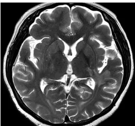
the cerebrospinal fluid. MRI of the brain showed a 5 cm irregu- lar shaped hyperintense mass-like lesion with a bizarre margin and perilesional edema involving the white matter in the left temporal lobe on T2-weighted imaging. The lesion also involved the left posterior thalamus, posterior limb of the internal cap- sule, external capsule, and midbrain crus, with extension to the periventricular white matter of the left lateral ventricular atrium (Fig. 1A, B). Thick irregular and open rim enhancement with a central nonenhancing portion was noted on the post-gadolini- um enhancement image (Fig. 1B). The left retrolenticular white matter was postulated to be the epicenter of the lesion. The le- sion extended along the course of the left internal capsule fiber with incomplete rim enhancement of the left midbrain crus (Fig. 1B, C).
Fig. 1. A 43-year-old female with tumefactive multiple sclerosis.
A. An irregularly shaped mass-like lesion approximately 5 cm in size involves the white matter of the left temporal lobe. The lesion shows high signal intensity on axial T2-weighted image and extends to the left posterior thalamus, posterior limb of the internal capsule, and the external capsule. Also this hyperintensity extends to the periventricular white matter near the left lateral ventricular atrium.
B, C. After gadolinium enhancement, the lesion demonstrates thick and irregular rim-like enhancement with extension along the course of the left internal capsule on coronal image (B) and the left midbrain crus, with incomplete rim enhancement on axial image (C).
D. Magnetic resonance spectroscopy of the mass in left temporal lobe demonstrates elevated choline and lactate, and decreased N-acetylaspartate.
A B C D
Fig. 2. Photomicrography of the center of the mass. Section shows proliferation of reactive astrocyte and perivascular infiltration of lym- phocytes and histiocytes (H&E, ×200).
oma, primary lymphoma, and solitary metastasis, as well as oth- er pseudotumoral brain lesions such as abscess, acute dissemi- nated encephalomyelitis (ADEM) (2).
High-grade glioma mostly demonstrates heterogeneous sig- nal intensity, irregular edges, extensive peritumoral edema or a mass effect, and there will commonly be an irregularly enhanc- ing solid portion of the tumor present on gadolinium enhanced images (2, 4, 8-10). Brain lymphoma is easily confused with tu- mefactive MS owing to its unifocal features on MRI and its good response to steroid therapy (11). However, primary lymphoma in the brain shows marked homogeneous enhancement on T1- weighted post-contrast images and iso- or mild hyperintensity on T2-weighted images (2). A solitary cerebral metastasis often demonstrates central heterogeneous signal intensity with pe- ripheral hypointensity on T2-weighted images and complete rim enhancement on gadolinium administration. Brain abscess- es commonly have a complete hypointense rim on T2-weighted images, hyperintensity on diffusion weighted imaging, and reg- ular complete rim enhancement on gadolinium enhanced im- ages (2, 10). ADEM usually appears as small and bilateral le- sions and the large isolated form is rare. Vaccination or infection commonly precedes ADEM, whereas tumefactive MS is rarely accompanied by this type of preceding history (2).
In our case, central homogeneous signal intensity with rim enhancement of the lesion was compatible with tumefactive multiple sclerosis rather than with intracranial neoplasm. How- ever, the lesion revealed irregular and thick rim-enhancement (1). According to recent study, the prevalence of tumefactive MS
has been reported to be 0.09% (7). However, it is likely that the true incidence rate is unknown, despite attempts in the literature to define it (5, 8). This type of MS commonly occurs in women with an average age of 37 years and usually features a single neu- rologic episode, whereas in classic MS there are repeated sub- acute episodes. Although the neurologic symptoms are depen- dent upon the location of the lesion, the common clinical features are motor, cognitive, sensory, cerebellar, and brainstem symptoms, in that order of frequency (5, 6). Visual disturbances, seizure, the appearance of a sudden cognitive deficit, and bowel dysfunction are relatively uncommon clinical presentations. The frontal lobe and parietal lobe are the most common locations of tumefactive MS (4, 5). Rare locations of the lesion, reported in less than 10% of cases, are the temporal lobe, cerebellum, deep gray matter, and brain stem. Corticosterioid therapy is impor- tant for improving the clinical symptoms and for decreasing the size of the lesion (2, 5).
Many researches have reported specific MRI findings in tume- factive MS. First, tumefactive MS lesions present as large isolated mass-like lesions that are round in form, and have a well-circum- scribed margin. These lesions usually show homogeneous hy- pointensity on T1-weighted images and hyperintensity on T2- weighted images (2). Tumefactive MS is typically located in the supratentorial brain and is centered within the white matter (9).
Second, tumefactive MS reveals a proportionally minor mass ef- fect and perilesional vasogenic edema relative to the size of the lesion, compared to other mass-like lesions in the brain (1-3).
Third, rim-like smooth enhancement, either complete or incom- plete, or open rim or arc-like enhancement, is noted in approxi- mately half of patients. This enhancement pattern consists of an enhanced leading edge of demyelination and a central nonen- hancing portion representing a more chronic inflammatory state (8, 9). The open part of the ring enhancement is generally oriented towards the basal ganglia and the cortex (2). In addi- tion, a multiplicity of the lesion can be helpful in the diagnosis of tumefactive MS. The lesion is typically located in the periven- tricular and subcortical white matter, or cervical spine (4, 5). In addition, MR spectroscopy usually shows similar spectrum to that of glioma, presenting as elevated choline and suppressed N- acetylaspartate (9).
Tumefactive MS can mimic tumors including high-grade gli-
Fig. 3. After two months of steroid therapy, the irregular hyperintense lesion markedly decreases in size; the lesion have been located in the left temporal lobe, midbrain, internal capsule, external capsule, and posterior thalamus on the axial T2-weighted image.
mefactive multiple sclerosis mimicking neoplasm. Acta Chirurgica Latviensis 2010;10:91-97
5. Lucchinetti CF, Gavrilova RH, Metz I, Parisi JE, Scheithauer BW, Weigand S, et al. Clinical and radiographic spectrum of pathologically confirmed tumefactive multiple sclerosis.
Brain 2008;131(Pt 7):1759-1775
6. Mandrioli J, Ficarra G, Callari G, Sola P, Merelli E. Monofo- cal acute large demyelinating lesion mimicking brain glio- ma. Neurol Sci 2004;25 Suppl 4:S386-S388
7. Annesley-Williams D, Farrell MA, Staunton H, Brett FM.
Acute demyelination, neuropathological diagnosis, and clini- cal evolution. J Neuropathol Exp Neurol 2000;59:477-489 8. Law M, Yang S, Wang H, Babb JS, Johnson G, Cha S, et al.
Glioma grading: sensitivity, specificity, and predictive values of perfusion MR imaging and proton MR spectroscopic im- aging compared with conventional MR imaging. AJNR Am J Neuroradiol 2003;24:1989-1998
9. Given CA 2nd, Stevens BS, Lee C. The MRI appearance of tumefactive demyelinating lesions. AJR Am J Roentgenol 2004;182:195-199
10. Schwartz KM, Erickson BJ, Lucchinetti C. Pattern of T2 hy- pointensity associated with ring-enhancing brain lesions can help to differentiate pathology. Neuroradiology 2006;48:
143-149
11. Kantarci OH, Weinshenker BG. Natural history of multiple sclerosis. Neurol Clin 2005;23:17-38, v
with a bizarre margin, corresponding to an intracranial neo- plasm. In addition, the location of the lesion was an uncommon site for multiple sclerosis. For these reasons, this patient under- went brain biopsy to assist in correct diagnosis. She could then be diagnosed with tumefactive multiple sclerosis and underwent corticosteroid therapy. After two months, the neurologic deficits improved, with a decrease in the size of the brain lesion on MRI.
This case suggests that when a patient who has an isolated large mass in the white matter of the brain with incomplete rim enhancement on MRI shows transient neurological dysfunc- tion, consideration of tumefactive MS may be helpful for avoid- ing unnecessary procedures and is also likely to be useful in se- lection of the appropriate treatment.
REFERENCES
1. Rusin JA, Vezina LG, Chadduck WM, Chandra RS. Tumoral multiple sclerosis of the cerebellum in a child. AJNR Am J Neuroradiol 1995;16:1164-1166
2. Comi G. Multiple sclerosis: pseudotumoral forms. Neurol Sci 2004;25 Suppl 4:S374-S379
3. Kim DS, Na DG, Kim KH, Kim JH, Kim E, Yun BL, et al. Dis- tinguishing tumefactive demyelinating lesions from glio- ma or central nervous system lymphoma: added value of unenhanced CT compared with conventional contrast-en- hanced MR imaging. Radiology 2009;251:467-475 4. Elsone L, Platkajis A, Karelis G, Dzelzite S, Murzina M. Tu-
종양유사성 다발성 경화증의 뇌병변: 병변의 비전형적인 위치 및 자기공명영상 소견1
정민선
1· 김현숙
1· 김재훈
2· 김은경
3· 최윤선
1종양유사성 다발성 경화증은 탈수초화 병변 중 드문 질환이다. 이 병변의 전형적인 자기공명영상 소견은 불완전한 고리형 조영증강 및 경미한 종괴효과인데 뇌의 다른 종괴양 병변들과의 구별이 어려운 경우가 많다. 종양유사성 다발성 경화증의 다양한 자기공명영상 소견을 숙지하여야 적합한 치료를 하는 데 도움을 줄 수 있다. 이에 저자들은 비전형적인 위치에서 공격적인 자기공명영상 소견을 보이는 종양유사성 다발성 경화증 1예를 보고하고자 한다.
을지대학교 의과대학 을지병원 1영상의학과, 2신경외과, 3병리과