Address reprint requests to Weon Wook Park, M.D.
Department of Orthopaedic Surgery, College of Medicine, Pusan National University 1-10 Ami-Dong, Seo-Gu, Pusan 602-739, Korea
Tel : 82-51-240-7248, Fax : 82-51-247-8395, E-mail : pww@scoliosis.co.kr
변연척추의 전산화 단층 촬영 혹은 자기 공명 영상 소견
박원욱・최영준・안성준*・김정태*
부산대학교 의과대학 정형외과학교실, 부산메리놀병원 정형외과*
Limbus Vertebra Demonstrated by Computed Tomography or Magnetic Resonance Image
Weon-Wook Park, M.D., Young-Jun Choi, M.D., Sung-Jun Ahn, M.D.*, Jung-Tae Kim, M.D.*
Department of Orthopaedic Surgery, College of Medicine, Pusan National University, Pusan, Korea Department of Orthopaedic Surgery, Maryknoll Hospital, Pusan, Korea*
– Abstract –
Study Design : We studied retrospectively the limbus vertebra by computed tomography or magnetic resonance image.
Objectives : To analyze the clinical and radiologic characteristics of the limbus vertebra and to distinguish it from a fracture, infection or tumor.
Summary of Literature Review : The limbus vertebra is common. However, the clinical manifestations including the level, symptoms and radiologic characteristics of the limbus vertebra are not understood exactly in the literatures.
Materials and Methods : We presented 25 cases of the limbus vertebra that were confirmed by plain roentgenogram combined with computed tomography (CT) or magnetic resonance imaging (MRI). Of the 25 patients, 18 were males and 7 females.
Results : The levels of the limbus vertebra were L3 (2 cases), L4 (13 cases), and L5 (8 cases). There were two cases of 2 level involvement (L3/4 and L4/5). All cases showed the lower lumbar lesion and complained of the lower back pain. The accompa- nying diseases included 10 cases of herniated intervertebral discs, 2 cases of ankylosing spondylitis, 2 cases of spinal stenosis and one spondylolisthesis. Three patients were first diagnosed as tuberculous spondylitis and 2 patient as spine fracture on plain roentgenograms. But they can be confirmed by demonstrating the herniation of disc material between the anterosuperior bony fragment and the rest of the body in CT or MRI.
Conclusions : The CT or MRI could be great diagnostic modalities. The pathogenesis is thought to be the herniation of disc material into the vertebral body such as Schmorl’s node and disc degeneration. Most limbus vertebra was found at the lower lumbar region and accompanied with disc bulging and degeneration. The correlation between the limbus vertebra and lower back pain is not certain.
Key Words : Lumbar, Limbus vertebra, MRI, CT
Journal of Korean Spine Surg.
Vol. 8, No. 4, pp 475~481, 2001
등 임상 양상에 대해 연구 분석하였다.
연구 대상 및 방법
1 9 9 4년부터 2 0 0 0년까지 본원 정형외과를 방문한 2 5례
의 변연척추 환자를 대상으로 하였다. 전 환자에 대하여 증상, 과거력 등 임상 양상을 조사하였다. 모든 예에서 요추 전후면 및 측면 단순 방사선 촬영을 하였고 특수검 사로 전산화 단층촬영( C T )을 8례, 자기공명영상( M R I )을1 4례에서 시행하였으며, 두 가지 모두 다 검사한 경우가 3례였다. 전산화 단층촬영(Siemens Somatom plus)은 횡
단면(axial view)과 시상 재건면(sagittal reformat view)이 촬영되었고 자기공명영상 (Siemens Magnetom Vision1 . 5 T )은 T1, T2 영상의 횡단면과 시상면이 촬영되었다.
단순 방사선 측면 사진에서 상부 척추 종판과 결손 부위 가 이루는 각을 limbus angle이라고 정의하고 전 예에 대 해 limbus angle을 측정하였다. 또한 상부 종판에서 변연 척추가 시작되는 부위가 어디인지를 조사하였다.
결 과
총 25례의 환자 중 남자가 16례였고 여자는 9례였다.
연령은 8세부터 63세까지 광범위하였고 평균연령은 35 세였다(Table 1). 25례 중 초기진단이 변연척추였던 경 우는 20례로 80%였고 척추 결핵이 2례, 척추 골절이 3례 로 오진율은 20%였으나(Table 2) 대개 증례 수집 초기의 경우였다. 변연척추가 발생한 부위는 제 3요추 2례, 제 4 요추 13례, 제 5요추 8례였고(Fig. 1) 제 3요추와 제 4요 추가 동반된 예와 제 4요추와 제 5요추가 동반된 경우가 각각 1례가 있었다(Table 3). 모든 환자는 하부요통을 동 반하였지만 변연척추와의 연관성은 알 수 없었다. 과거
31.9°
였다(Fig. 2). 변연척추의 시작 부위는 상부 종판의 전방 1/3 근처 혹은 그보다 더 전방이었다.고 찰
변연척추라는 말은 추간판이 추체 내로 전방 탈출된 것 을 뜻하기도 하고5 , 1 9 )위치에 관계없이 척추체의 모서리에 있는 골편을 지칭하기도 한다. 동의어는 여러 가지가 있 으며 그 중에 persistent epiphysis1 7 )
, ossified vertebral rim
1 4 ), intercalary bone
1 2 )등이 있는데 이들은 외상과는 무관하다 는 것을 뜻하고, posterior bony avulsion(PBA)1 1 ), limbus frac- t u r e
1 8 ), end plate fracture
1 3 )라는 말은 원인이 외상이라는 것 을 뜻한다. 변연척추의 발생 원인은 아직 정립되지 않았 다. Hellstradius9 ), Resnick과 N i w a y a m a
1 5 )는 ring apophysis의Table 1. Age distribution
M F
05~10 1
11~20 3 1
21~30 7
31~40 4 1
41~50 2 2
51~60 3
60~00 1
Total 16 (64%) 9 (36%)
Table 2. Initial diagnosis by plain radiographic examination
Disease No. of patients
Spinal Tuberculosis 02
Spine fracture 03
Limbus vertebra 20
Misdiagnosis rate 5/25 = 20%
일부가 척추체에 유합되지 않아 발생한다고 하였으나 만 약 그렇다면 골결손 부위에 추간판 조직이 아닌 다른 조 직이 있어야 할 것이다. 그러나 결손 부위에는 추간판 조 직인 수핵이 있다는 것이 자기공명영상이나 추간판조영 술에서 확인되었으며 이는 1 9 7 1년 S c h m o r l1 6 )이 언급한 바 있다. 한편 Swischuk 등1 7 )은 변연척추가 발생한 분절의 추 간판은 모두 퇴행성 변화를 보였다고 하였고 저자의 경 우도 자기공명영상을 촬영한 1 7례에서 변연척추가 있는 분절의 추간판은 정도의 차이는 있지만 모두 퇴행성 변 화를 보였다. 추간판탈출증의 근본 원인이 추간판의 변 성임을 감안한다면 변연척추의 원인이 추간판의 퇴행성 변화라는 것을 추정해볼 수 있다.
변연척추의 다른 발생원인으로 외상으로 인해 골편이
분리되고 그곳으로 추간판이 탈출될 수 있다는 것이다
1 , 6 , 1 8 )
. 저자의 경우 환자가 기억하지 못하는 경미한 외상이
있었을 가능성을 배제할 수는 없지만 2 5례 중 6례에서만 가벼운 외상이 있었으며 이 사실로 외상으로 인해 변연척 추가 발생했다는 가설을 받아들이기는 어렵다고 생각된 다. 따라서 가장 가능성 있는 설명은 외상이나 ring apoph-
y s i s의 불유합이 아니라 어떤 이유로든지 추간판 퇴행이
발생하여 추간판 물질이 추체내로 탈출하여 골편이 분리 되어 변연척추가 발생한다는 것이다. 이런 이유로 저자도Schmorl`s node나 변연척추가 같은 원인에 의해 발생한다
Fig. 1-A. Lateral radiography of the lumbar spine. The bony defect with sclerotic contour at the anterosuperior margin of L4 bodyand a small detached triangular bony fragment was seen.
Fig. 1-B. CT sagittal reformat view. The bony defect at the anterior edge of the vertebral body with irregular and increased density was seen.
Fig. 1-C. Sagittal view of MRI showed that the erosion of the anterior part of the vertebral end plates, presence of an anterior bone fragment and herniation of disc material between this fragment and the vertebral body.
A B C
Table 3. Level of the limbus vertebra
Vertebra Cases
L3 2 (8%)
L4 13 (52%)
L5 8 (32%)
L3 & L4 1 (4%)
L4 & L5 1 (4%)
Total 25 (100%)
Table 4. Accompanied spinal disease
Disease Cases
HIVD 10 (40%)
same level 4(
upper level 3(
lower level 2(
combined 1(
Ankylosing spondylitis 2 (8%)
Spinal stenosis 2 (8%)
Spondylolisthesis 1 (4%)
None 10 (40%)
Total 25 (100%)
는 Swischuk 등1 7 )과 Henales 등1 0 )의 생각에 동의한다.
이와는 반대로 모두 같은 뜻인 척추 종판 골절 ( e n d
plate fracture), 변연골절(rim injury), 혹은 후방 변연골절 (posterior limbus fracture)은 외상과 연관된 경우가 많다
6,13,18)
. 이들은 골편의 위치와 그 위치로 인해 신경학적 증
상을 동반할 수 있다는 점만 다를 뿐 변연척추와 그 모 양은 대칭이다. 즉 추간판의 전방 척추내 탈출은 외상과 연관이 적고 후방 탈출은 외상과 연관이 많다는 뜻이 된 다. 추간판 탈출의 방향과 외상 유무와의 관계는 문헌상 에는 거의 보고가 없다.
변연척추의 진단은 대개 단순 방사선 촬영으로 가능하 나 골절(Fig. 3), 감염, 및 종양과의 감별이 필요하다5 )
. 저
자의 경우 2 5례 중 비록 증례를 수집한 초기의 진단이었 지만 진단이 결핵성 척추염이었던 경우가 2례, 골절이었 던 경우가 3례로 오진율은 2 0 %였다. 그러나 이들은 컴퓨 터 단층촬영이나 자기공명영상 촬영에서 변연척추로 확 인되었다. 변연척추의 단순방사선 소견은 골절이나 종 양, 감염과는 달리 척추체에 압박(compression, collapse)체 전상방 골결손과 상부 척추 종판과의 각도는 평균
3 1 . 9도였는데 추간판 섬유륜의 섬유 주행 각도 3 0도와
비슷하다는 것이 흥미롭다. 변연척추의 특징적 단순방 사선 소견으로는 골편과 인접한 척추체 면의 골경화상 인데 골절, 감염 및 종양과 구분되는 소견이다(Fig. 1A).그러나 골경화상은 오랜 기간 지속된 변연척추의 방사 선 소견이며 발생한지 얼마 되지 않은 변연척추는 아직 보고된 바가 없지만 발견된다면 골경화상은 없을 가능 성이 크다고 생각된다. 추간판 간격 협소는 Y a g a n1 9 )은 없 다고 하였으나 Swischuk 등1 7 )과 Goldman 등6 )은 있다고 하 였다. 저자의 경우에는 없는 경우( 1 8례, 72%)가 있는 경 우( 7례, 28%)보다 더 많았다.
Y a g a n
1 9 )은 변연척추의 컴퓨터 단층촬영 소견으로 첫 째 척추체의 전상방에 전위가 없는 골편의 확인, 둘째 골편과 추체 사이의 아주 일정한 모양의 결손, 셋째 골 결손 후면의 골경화와 척추체 전면의 골경화, 넷째 컴퓨 터 단층촬영의 Scout view에서 이환된 추간판에 인접된 척추체 높이가 감소되지 않은 것을 언급하였는데, 저자 도 이와 비슷한 소견을 보였다.한편 Yagan은 축성 면(axial view)과 Scout view에 대해 서만 언급하였으나 저자의 경우 시상면 재구성 화면
(sagittal reformat view) 에서는 골편이 더욱 잘 보였다 (Fig. 1B). 자기공명영상 소견은 컴퓨터 단층촬영과 큰
차이는 없었지만 이환된 분절의 추간판에 퇴행성 변화 가 있으면서 골결손 부위에도 추간판이 있다는 것을 확 인시켜 주었다(Fig. 1C). 또한 자기공명영상 촬영을 시행 한 17례 중 7례(41.2%)에서 척추체의 퇴행성 변화가 관 찰되었으며 이는 골편과 척추체 사이에 운동(motion)이 있다는 것을 의미하며 단순방사선에서 골경화상이 보 이는 것도 이런 운동 때문일 것으로 생각된다. 골결손 부위에 차있는 것은 추간판의 수핵이며 자기공명영상 촬영에서는 추간판이 퇴행성 변화를 보여 확인하기 어 렵지만 추간판 조영술에서 알 수 있다5).
Fig. 2. Lateral radiography of the lumbar spine. The length of detached bony fragment usually did not exceed 1/3 of endplate length and the angle between endplate and bony defect line was under 40 degrees.
Swischuk 등
17)은 12례의 변연척추 중 흉추 1례, 흉요추 부 1례에 발생한 경우를 보고하였으나 주로 요추에 발 생한다고 하였다1,8,17). 저자의 경우도 제 4요추 전상방이 13례(52%), 제 5요추 전상방이 8례(32%)로 하요추가 대
부분이었고 상부 요추와 천추에 발생한 예는 없었다. 한 편 후방 변연골절의 발생 부위는 하요추부에 많이 발생 하고, Schmorl’s node는 제 3 - 4요추에 많으며 제 5요추- 천추 간은 비교적 적다4,8). 즉 변연척추, 후방 변연 골절
은 추간판 탈출증과 함께 Schmorl`s node보다 하부인 하 요추부에 많이 발생한다.변연척추가 있는 경우 요통이 동반될 수 있다3 , 7 , 8 )
. 저자
의 경우 전 환자가 모두 요통이 있었지만 변연척추가 있 으면서 요통이 없다면 병원을 내원하지 않을 것이므로 변연척추를 가진 모든 환자가 요통이 있다고 할 수는 없 을 것이다. 저자의 경우 변연척추의 발생원인이라고 하 기 어려운 외상이 있었던 경우가 6례 있었으며 이것이 요 통의 원인이 될 수도 있었을 것이다. 또한 동반 질환으로 요추 추간판 탈출증 1 0례, 강직성 척추염 2례, 척추관협 착증 2례, 척추전방전위증이 1례가 있었으나 변연척추와 의 연관성은 없다고 생각되며, 변연척추만 있었던 경우 가 1 0례 있었다. 즉 변연척추로 인해 요통이 발생되었다 고는 보기 어렵다는 것이다. 동반질환으로는 추간판탈출 증이 가장 많았으며 변연척추와 같은 분절에 발생한 경 우가 두 분절에 발생한 경우를 포함하면 5례였으며 이는 추간판의 퇴행성 변화와 관계가 있을 것으로 생각된다.결 론
변연척추는 하부 요추에 많이 발생하며 전 연령층에 서 발견되지만 10대와 20대의 젊은 연령에서 흔히 발견 된다. 전산화 단층촬영과 자기공명영상은 추체와 골편 사이로 수핵탈출을 증명함으로 변연척추의 진단에 도 움이 되며, Schmorl’s node와 같이 추체내로의 수핵탈출 과 추간판의 퇴행성 변화가 변연척추의 발생기전으로 사료된다. 요통은 전례에서 동반되었지만 변연 척추가 요통의 원인이라고 단정지을 수는 없다고 생각된다.
REFERENCES
01) Begg AC : Nuclear herniations of the intervertebral disc:
Their radiological manifestations and significance. J Bone Joint Surg, 36-B:180-193, 1954.
02) Brocher JEW : Spine. In: Schinz HR, Baensch WE, From- hold W, Glauner R, Uehlinger E, Wellauer J, etc. Roentgen diagnosis. 2nd ed. New York: Grune and Stratton, 32-38, 1 9 6 9 .
03) Epstein JA and Lavin LS : Herniated lumbar disc in teen- age children, J Neurosurg, 21:1070, 1964.
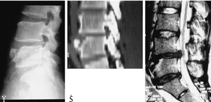
Fig. 3-A. This case was differential diagnosis. The bony defect at the anterosuperior margin of the L4 body implies the vertebral body fracture.
Fig. 3-B. CT sagittal reformat view. Fracture line is well visible. No sclerosis and no disc material is in the fracture site.
Fig. 3-C. Sagittal view of MRI. The findings of no herniation of disc material between this fragment and the vertebral body implies vertebral body fracture. In addition, bony contusion of vertebral body is another evidence of vertebral body fracture.
A B C
Juvenile discogenic disease. Spine, 19:335-340, 1994.
09) Hellstadius A : A contribution to the question of the origin of anterior paradiscal defects and so-called persisting apo- physes in the vertebral bodies. Acta Orthop Scand, 18:377, 1 9 4 9 .
10) Henales V, Hervas JA, Lopez P, Martinez JM, Ramos R and Herrera M : Intervertebral disc herniations(limbus vertebrae) in pediatric patients: report of 15 cases. Pediatr Radiol, 23:608-610, 1993.
11) Laredo JD, Bard M, Chretien J and Kahn MF : Lum - bar posteior marginal intra-oseous cartilaginous node.
health and disease. 2nd ed. New York: Grune and Stratton, 2-14, 158-172, 1971.
17) Swischuk LE, John SD and Allbery S : Disc degenerative disease in childhood: Scheuermann’s disease, Schmorl’s nodes, and limbus vetebra: MRI findings in 12 patients.
Pediatr Radiol, 28:334-338, 1998.
18) Takata K, Inoue SI, Takahashi K and Ohtsuka Y : F r a c - ture of the posterior margin of a lumbar vertebral body. J Bone Joint Surg, 70-A:589-594, 1988.
19) Yagan R : CT diagnosis of limbus vertebra. J Comput Assist Tomogr, 8:148-151, 1984.
연구계획 : 컴퓨터 단층촬영 또는 자기공명영상 촬영을 통해 변연척추를 후향적으로 연구하였다.
연구목적 : 변연척추의 임상적 및 방사선학적인 특징의 분석을 통해 골절, 감염 또는 종양 등과의 감별을 위하여 연 구 분석하였다.
대상 및 방법 : 컴퓨터 단층촬영 또는 자기공명영상 촬영을 단순 방사선 사진과 함께 촬영하여 확진된 25례의 변연 척추를 대상으로 하였으며 남자가 18례, 여자가 7례이었다.
결과 : 변연척추의 분절은 각각 제 3요추 2례, 제 4요추 13례, 제 5요추 8례이었고, 두 분절(제 3-4, 제 4-5요추)에 걸쳐 나타난 경우가 2례에서 있었다. 25례의 환자는 모두 하부 요추에 변연척추를 보였고 공통적으로 요통을 호소하였다.
동반 질환으로 10례에서 탈출된 추간판, 2례에서 강직성 척추염, 2례에서 척추관 협착증 및 1례의 척추전방전위증이 보였다. 초기 단순방사선 사진 상 3례에서는 결핵성 척추염으로 2례에서는 척추 골절로 진단되었지만 컴퓨터 단층 촬영과 자기공명영상 촬영에서 추체와 골편 사이로 수핵 탈출을 증명함으로 변연척추를 확진할 수 있었다.
결론 : 컴퓨터 단층촬영 또는 자기공명영상 촬영은 변연척추의 진단에 도움을 주며 Schmorl’s node와 같이 추체내로 의 수핵 탈출과 추간판의 퇴행성 변화가 변연척추의 발생기전으로 사료된다. 변연척추는 대부분 하부 요추에 발생 되었고, 추간판의 팽윤과 변성 및 하부 요통을 동반하였다.
색인단어 : 요추, 변연척추, 자기공명 영상장치, 컴퓨터 단층촬영 국 문 초 록
※ 통신저자 : 박 원 욱
부산광역시 서구 아미동 1가 10 부산대학교 의과대학 정형외과학교실
Tel : 82-51-240-7248, Fax : 82-51-247-8395, E-mail : pww@scoliosis.co.kr