INTRODUCTION
The gastrointestinal tract is the most commonly involved extranodal site in non-Hodgkin’s lymphoma (1, 2). However, the esophageal involvement by lymphoma is the rarest event, accounting for less than 1% of patients with lymphoma (1-3).
Since Isaacson and Wright introduced the concept of mucosa- associated lymphoid tissue (MALT) lymphoma in 1984 (4), there have been many reports regarding primary gastrointesti- nal MALT lymphoma, especially primary gastric MALT lym- phoma. To our knowledge, only one case of primary esophageal MALT lymphoma has been reported (5). Recent advancement in endoscopic ultrasonography (EUS) provides detailed struc- tural abnormalities and depth of invasion in various gastroin- testinal diseases including gastric lymphoma (6). Usually, gas- tric MALT lymphoma presents itself as a thickening of the mucosa and submucosa layer in the gastric wall, or discrete tumor masses; however, nearly all cases show infiltration of the mucosa (7, 8). We report a case of primary esophageal MALT lymphoma presenting itself as a large submucosal tum- or (SMT), which was examined by endoscopic ultrasonogra- phy.
CASE REPORT
A 61-yr-old man was referred to Soon Chun Hyang Uni-
versity hospital for evaluation of an esophageal SMT that had been detected incidentally by endoscopy. The patient denied dysphagia and any systemic symptoms. He had a history of pulmonary tuberculosis, which had been completely healed after a long-term medication (isoniazid, rifampin, ethambu- tol, and pyrazinamide for 9 months) 15 yr before. He also had a history of syphilis, which had been completely treated one year before. He had been taking a Comthyroid�(a combina- tion of synthetic hormones; levothyroxine 0.05 mg and lio- thyronine 0.0125 mg) daily for the past 10 months because of an autoimmune thyroiditis. His blood sugar level was slight- ly high, but had been well controlled by diet control and exer- cise. He had a history of alcohol abuse for over 20 yr. He had a 20-pack-year smoking history. Physical examination revealed no abnormal finding except slightly pale conjunctivae. Lab- oratory data included white blood cell count 7,100/ L (normal range, 4,000-10,000/ L), hemoglobin value 10.3 g/dL (normal range, 13-17 g/dL), MCV 79.9 fL (normal range, 81-96 fL), and platelet count 307×103/ L (normal range, 130-450× 103/ L). Fasting blood sugar level was 93 mg/dL (normal range, 70-110 mg/dL) with HbA1cof 7.0% (normal range, 3.8-5.4%). Thyroid function test revealed TSH 1.89 U/mL (normal range, 0.25-4.0 U/mL), T3 uptake 27.9% (normal range, 23-34%), T3 116.2 ng/dL (normal range, 60-190 ng/
dL), and T4 7.73 g/dL (normal range, 4.5-11 g/dL). Both thyroglobulin antibodies and microsomal antibodies were pos- itive (1.49 U/mL and 2.2 U/mL, respectively; normal range,
120 J Korean Med Sci 2003; 18: 120-4
ISSN 1011-8934
Copyright � The Korean Academy of Medical Sciences
A Case of Primary Esophageal B-cell Lymphoma of MALT type, Presenting as a Submucosal Tumor
The primary esophageal lymphoma is extremely rare, and shows various mor- phologic characteristics. Only a single case of mucosa-associated lymphoid tissue (MALT) type lymphoma confined to the esophagus has been reported in the lit- erature. A 61-yr-old man was referred to our hospital for evaluation of an esophageal submucosal tumor (SMT) that had been detected incidentally by endoscopy. He had a history of pulmonary tuberculosis with long-term anti-tuberculosis medica- tion 15 yr before, and also had a history of syphilis, which had been treated one year before. He had been taking a synthetic thyroid hormones for the past 10 months because of an autoimmune thyroiditis. Endoscopy showed a longitudinal round and tubular shaped smooth elevated lesion, which was covered with intact mucosa and located at the mid to distal esophagus, 31 cm to 39 cm from the incisor teeth. Endoscopic ultrasonography (EUS) showed a huge longitudinal growing intermediate- to hypo-echoic mass located in the submucosal layer with internal small, various sized honeycomb-like anechoic lesions suggesting germinal centers. Subsequently, he underwent a surgery, which confirmed the mass as a primary esophageal low-grade B-cell lymphoma of MALT type.
Key Words : MALT; Lymphoma; Esophagus; Endosonography
Chan Sup Shim, Joon Seong Lee, Jin Oh Kim, Joo Young Cho, Moon Sung Lee, So Young Jin*, Wook Youm�
Institute for Digestive Research, Department of Pathology*, Department of Chest Surgery�, College of Medicine, Soon Chun Hyang University, Seoul, Korea
Address for correspondence Chan-Sup Shim, M.D.
Institute for Digestive Research, Digestive Disease Center College of Medicine, Soon Chun Hyang University, 657 Hannam-dong, Yongsan-gu, Seoul 140-743, Korea
Tel : +82-2-709-9681, Fax : +82-2-749-1968 E-mail : [email protected]
Received : 9 March 2002 Accepted : 25 April 2002
<0.3 U/mL for both).
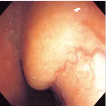
Initial endoscopy showed a longitudinal round and tubu- lar shaped smooth elevated lesion, which was covered with intact normal mucosa and located at the mid to distal esoph- agus, 31 cm to 39 cm (2 cm from the Z-line) from the incisor teeth. There was no such finding as reflux esophagitis or Bar- rett’s esophagus (Fig. 1). EUS showed an intermediate echoic mass, which was located in the deep mucosal and submucos-
al layer and measured 2×8 cm. The mass was well demarcat- ed, lobulated, and had multiple small anechoic lesions (honey- comb in appearance) within its solid portion (Fig. 2). A small round hypoechoic mass of 1.0 cm in diameter was also shown around the esophageal wall from the aorticopulmonary (AP) window, suggesting regional lymph node hyperplasia. These findings suggested the possibility of the esophageal stromal cell tumor or cavernous hemangioma. He was discharged with- out further evaluation including EUS-guided aspiration cytol- ogy for the proper tissue diagnosis.
There was no change in EUS findings 6 months after dis- charge. Barium esophagogram showed a smooth elongated protruded mass lesion in the distal esophagus, 2 cm above the esophagogastric junction, and this finding was compatible with a stromal tumor (Fig. 3). Dynamic chest computed tomo- graphy showed a well-defined, poorly enhanced mass in the distal esophagus, suggesting an esophageal SMT. Two small lymph nodes were also detected in the paraseptal region and AP window. There was a centrilobular and paraseptal emphy- sema. Abdominal ultrasonography showed normal abdomi- nal organs, except for a mild fatty liver.
Surgical resection was performed under the impression of esophageal stromal cell tumor. After exposure of distal esoph- agus and confirmation of the exact tumor site, longitudinal myotomy was done along the tumor and we tried to separate the tumor mass from the mucosa. However, since the tumor was unusual for stromal cell tumor and had different patho- logic characteristics, it was impossible to separate the tumor
Fig. 1.Endoscopic view of the large longitudinal round mass with normal overlying mucosa in mid and distal esophagus.
Fig. 2.Endoscopic ultrasonogram showing a well-demarcated, 2
×1.5×8 cm-sized, lobulated mass with intermediate echoic pat- tern and honeycomb-like multiple small anechoic lesions, origi- nating from the third hyperechoic layer (submucosa), consistent with a submucosal tumor such as esophageal stromal cell tumor or cavernous hemangioma.
Fig. 3.Esophagogram showing a large submucosal tumor (arrows) with luminal narrowing, about 10 cm in length, in mid and distal esophagus.
122 C.S. Shim, J.S. Lee, J.O. Kim, et al.
mass from the mucosa. We removed the elongated tumor mass with underlying mucosa and closed the remaining mucosa and muscle separately layer by layer, and then covered the myo-
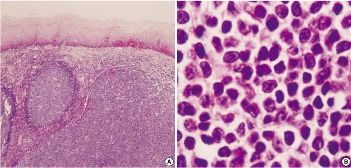
tomy site with a pleural flap. The gross examination of the surgical specimen revealed a well-circumscribed multinodu- lar grayish white mass (6×2×1.5 cm), which was covered by normal esophageal mucosa on one side. The cut surface was creamy yellow and lobulated in appearance. Histological- ly the esophageal mucosa was unremarkable. The lamina pro- pria and the submucosa were replaced by dense infiltration of lymphoid cells. These lymphoid cells were arranged in large follicles with occasional germinal centers. Proliferating lym- phoid cells were relatively monotonous centrocyte-like cells with irregular nuclear membrane and abundant clear cyto- plasm (Fig. 4). Immunohistochemistry disclosed positive stain- ing for CD20, Bcl 2, and CD10 and negative staining for Bcl 6, CD3, CD5, and cycline D1. These cells showed a monoclon- al restriction pattern for the kappa light chain on immunos- taining and a positive monoclonal band for immunoglobulin heavy chain gene rearrangement (Fig. 5). These findings were consistent with a low-grade B-cell lymphoma of MALT type.
Regional lymph nodes showed reactive hyperplasia.
Follow-up endoscopy with rapid urease test after surgery showed mild atrophic gastritis with Helicobacter pylori infection, so he received the modified triple therapy (clarithromycin, amoxicillin, and proton pump inhibitor for 1 week) and erad- ication of H. pylori was confirmed one month later.
DISCUSSION
MALT lymphoma is a distinct subgroup of non-Hodgkin’s lymphoma with particular clinicopathologic characteristics.
Fig. 4.(A) Histologic section disclosed proliferation of lymphoid cells in a large follicular pattern with occasional germinal center formation (H&E, ×40). (B) Proliferating lymphoid cells had central nuclei with irregular nuclear membrane and abundant clear cytoplasm (H&E,×
1,000).
A B
Fig. 5. Molecular analysis of immunoglobulin heavy chain gene rearrangement by polymerase chain reaction using FR3A-LJH as a primary PCR (Lane A) and using FR3A-VLJH as a secondary PCR (Lane B). A monoclonal dominant band corresponding to a monoallelic clonal rearrangement was noted on 90-100 bp (Lane B). Lane C, beta-globin control; Lane D, molecular markers.
A B C D
90-100 bp
The gastrointestinal tract is the most commonly involved site, but it may be observed in the lungs, breast, bladder, conjuncti- va, kidney, liver, skin, salivary glands, thyroid, and thymus (9).
Organized MALT within the gastrointestinal tract is dis- tributed throughout the small intestine, appendix, and colo- rectum, forming Payer’s patches, but is normally not found within the stomach mucosa, esophageal mucosa, salivary gland, or thyroid. However, MALT lymphoma usually develops from acquired MALT rather than the primary organized MALT.
These acquired MALT tend to appear in patients with a his- tory of autoimmune diseases or chronic inflammatory disor- ders. In the stomach, this lesion is induced by long-standing gastric infection with H. pylori and gastric MALT lymphoma may eventually develop as a result of persistent immunolog- ical stimulation. Acquired MALT also develops within the salivary glands, thyroid or lungs as a result of myoepithelial sialoadenoiditis, due to Sjogren’s syndrome, Hashimoto thy- roiditis, and interstitial lymphoid pneumonia (9, 10).
In our patient, the MALT lymphoma developed in the sub- mucosal layer and was found only in the mucosa and submu- cosal layer of the esophagus definitely apart from the cardia although he had the H. pylori infection in his stomach. Atyp- ical hyperplastic lymphoproliferative responses are known to occur due to Epstein-Barr virus, human immunodeficiency virus (HIV), human T-cell leukemia/lymphoma virus type I (HTLV-I), and certain drugs, and in post-transplant organ reci- pients, in autoimmune disorders, and in acquired or genetic hypogammaglobulinemias (11). In our patient, we could not detect the exact cause of esophageal MALT lymphoma, how- ever, there were potential risk factors, such as an autoimmune disorder, drugs, and chronic periesophageal inflammation, which were supported by the presence of autoimmune thy- roiditis, history of long-term anti-tuberculosis medication, and treatment of syphilis, respectively.
Usually CD10 expression has been described as the sensi- tive and specific marker of follicular center cell lymphoma.
Recently the specificity of CD10 expression was challenged due to observation of CD10 expression in the neoplastic T cells of angioimmunoblastic T-cell lymphoma (12). There- fore CD10/Bcl-6 coexpression is regarded as a reliable mark- er for follicular center B cell differentiation (13). Though CD10 was positive in our patient, the possibility of malignant lym- phoma of follicular center cell origin is less likely because CD 10 expression is not accompanied by Bcl-6.
The major finding of low-grade gastric MALT lymphoma on EUS, as has been suggested in many reports, is the thick- ening of the wall layer, especially the first to the third layer (8, 9, 14, 15). Some cases show discrete tumor masses destroy- ing the mucosal layer. However, our case presented itself as a well-demarcated SMT-like intermediate- to hypo-echoic mass located in the submucosal layer, which misled us to an impres- sion of esophageal stromal cell tumor or cavernous heman- gioma. Internal, small, various sized, honeycomb-like anechoic lesions may suggest germinal centers. In general, the lym-
phomatous involvement of the gastric wall proceeds mainly by longitudinal (or horizontal) growth within the wall, while the growth of cancer is predominantly vertical with early inva- sion of the deep wall. Mucosal involvement in lymphoma is often less extensive than the infiltration of underlying layers (16). From these observations, we considered the EUS findings of our case to suggest a low-grade lymphoma, because the lymphoma of the patient had developed in the submucosal layer and was found only in the mucosa and submucosal layer of the esophagus, representing a longitudinal SMT.
Another case of primary esophageal MALT lymphoma re- ported also presented itself as a SMT-like lesion, which was very similar to our case, despite the lack of description of patho- logic findings (5). To our knowledge, these two cases are the only reported cases of primary esophageal MALT lymphoma, which showed a predominant esophageal involvement with- out peripheral or mediastinal lymph node involvement, as well as splenic or hepatic involvement, and a normal white cell count (17). This SMT-like growth might be a character- istic of primary esophageal MALT lymphoma.
REFERENCES
1. Amer MH, El-Akkad S. Gastrointestinal lymphoma in adults: clini- cal features and management of 300 cases. Gastroenterology 1994;
106: 846-58.
2. Herrmann R, Panahon A, Barcos MP, Walsh D, Stutzman L. Gas- trointestinal involvement in non-Hodgkin’s lymphoma. Cancer 1980;
46: 215-22.
3. Taal BG, Van Heerde P, Somers R. Isolated primary oesophageal involvement by lymphoma: a rare cause of dysphagia: two case his- tories and a review of other published data. Gut 1993; 34: 994-8.
4. Isaacson P, Wright DH. Extranodal malignant lymphoma arising from mucosa-associated lymphoid tissue. Cancer 1984; 53: 2515-24.
5. Nishiyama Y, Yamamoto Y, Ono Y, Satoh K, Ohkawa M, Yamauchi A, Tanabe M. Visualization of esophageal non-Hodgkin’s lymphoma with Ga-67 scintigraphy. Ann Nucl Med 1999; 13: 419-21.
6. Shim CS. Role of endoscopic ultrasonography for gastric lesions.
Endoscopy 1998; 30 (Suppl 1): A55-9.
7. Pavlick AC, Gerdes H, Portlock CS. Endoscopic ultrasound in the evaluation of gastric small lymphocytic mucosa-associated lymphoid tumors. J Clin Oncol 1997; 15: 1761-6.
8. Sackmann M, Morgner A, Rudolph B, Neubauer A, Thiede C, Schulz H, Kraemer W, Boersch G, Rohde P, Seifert E, Stolte M, Bayerdoerf- fer E, MALT Lymphoma stady group. Regression of gastric MALT lymphoma after eradication of Helicobacter pylori is predicted by endosonographic staging. Gastroenterology 1997; 113: 1087-90.
9. Thieblemont C, Berger F, Coiffier B. Mucosa-associated lymphoid tissue lymphomas. Curr Opin Oncol 1995; 7: 415-20.
10. Weston AP, Cherian R, Horvat RT, Lawrinenko V, Dixon A, McGre- gor D. Mucosa-associated lymphoid tissue (MALT) in Barrett’s esoph- agus: prospective evaluation and association with gastric MALT, MALT lymphoma, and Helicobacter pylori. Am J Gastroenterol 1997;
. .
124 C.S. Shim, J.S. Lee, J.O. Kim, et al.
92: 800-4.
11. Harrington DS, Masih AS, Purtilo DT. Atypical immune prolifera- tions. In: Hoffman R, Benz EJ, Shattil SJ, Furie B, Cohen HJ, Siber- stein LE, editors. Hematology. Basic principles and practice. 2rd ed.
New York: Churchill-Livingstone; 1995: 1387-99.
12. Attygalle A, Al-Jehani R, Diss TC, Munson P, Liu H, Du MQ, Issac- son PG, Dogan A. Neoplastic T cells in angioimmunoblastic T-cell lymphoma express CD10. Blood 2002; 99: 627-33.
13. Ree HJ, Yang WI, Kim CW, Huh J, Lee SS, Cho EY, Ko YH, Cha- rney D. Coexpression of Bcl-6 and CD10 in diffuse large B-cell lym- phomas: significance of Bcl-6 expression patterns in identifying ger- minal center B-cell lymphoma. Hum Pathol 2001; 32: 954-62.
14. Nobre-Leitao C, Lage P, Cravo M, Cabeçadas J, Chaves P, Alberto-
Santos A, Correia J, Soares J, Costa-Mira F. Treatment of gastric MALT lymphoma by Helicobacter pylori eradication: a study con- trolled by endoscopic ultrasonography. Am J Gastroenterol 1998;
93: 732-6.
15. Cheng PN, Sheu BS. Application of endoscopic sonography to moni- tor the resolution of H. pylori-related gastric MALToma. Endoscopy 2000; 32: A45 (P119).
16. Caletti G, Bocus P, Togliani T, Roda E. Gastric lymphoma, infiltra- tive disorders, and large gastric folds. In: van Dam J, Sivak MV edi- tors. Gastrointestinal Endoscopy. 1st ed. Philadelphia; WB Saun- ders Co; 1999: 175-83.
17. Dawson IMP, Cornes JS, Morson BC. Primary malignant lymphoid tumours of the intestinal tract. Br J Surg 1961; 49: 31-84.