Korean J Thorac Cardiovasc Surg 2011;44:189-192 □ Case Report □ DOI:10.5090/kjtcs.2011.44.2.189 ISSN: 2233-601X (Print) ISSN: 2093-6516 (Online)
− 189 −
*Division of Cardiovascular Surgery, Yonsei Cardiovascular Hospital, Yonsei University College of Medicine Received: September 30, 2010, Revised: November 18, 2010, Accepted: February 9, 2011
Corresponding author: Young-Nam Youn, Division of Cardiovascular Surgery, Yonsei Cardiovascular Hospital, Yonsei University College of Medicine, 134, Sinchon-dong, Seodaemun-gu, Seoul 120-752, Korea
(Tel) 82-2-2228-8487 (Fax) 82-2-313-2992 (E-mail) [email protected] C The Korean Society for Thoracic and Cardiovascular Surgery. 2011. All right reserved.
CC This is an open access article distributed under the terms of the Creative Commons Attribution Non-Commercial License (http://creative-commons.org/licenses/by-nc/3.0) which permits unrestricted non-commercial use, distribution, and reproduction in any medium, provided the original work is properly cited.
Extra-anatomic Bypass Grafting after Endovascular Embolization
for the Treatment of Mycotic Aneurysm
−
2 case reports −
Kwan-wook Kim, M.D.*, Jung Hwan Kim, M.D.*, Young-Nam Youn, M.D., Ph.D.*
Mycotic aneurysm is a disease requiring immediate treatment because of the high risk of rupture. A difficult surgi-cal approach, especially in the case of occurrence on the iliac artery, involving endovascular embolization and ex-tra-anatomic bypass grafting, is known to be a suitable treatment. We performed exex-tra-anatomic bypass grafting af-ter endovascular embolization successfully in two patients. The postoperative computed tomography of both patients showed complete exclusion of the mycotic aneurysm.
Key words: 1. Mycotic aneurysm 2. Endovascular stent 3. Extra-anatomic bypass
CASE REPORT 1) Case 1
A 64-year-old male patient who had been on diabetes med-ication with no other particular medical history visited the clinic for symptoms of chillness, and his temperature was measured to be 38.8oC. An abdomical CT revealed a 6-cm abscess surrounding a pseudoaneurysm of about 4 cm near the right internal iliac artery, and mycotic aneurysm was diagnosed. Although no particular strain was identified from the blood culture test conducted at the time of the clinic visit, based on the week-old abdominal CT results, which clearly showed the growing aspect of the aneurysm, risk of death due to rupture was judged to be high. Before conducting a femoral artery bypass, a distal part of the right internal iliac artery was occluded using a 10-mm Amplatzer vascular plug. The right common iliac artery and external iliac artery were
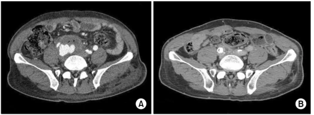
also occluded in succession using 14-mm and 12-mm Amplatzer vascular plugs, respectively. Post-procedure angiog-raphy showed complete disappearance of the mycotic aneur-ysm sac. The patient was immediately moved to the OR and a left fermoral artery-right femoral artery bypass was per-formed using a 10-mm ringed Gore-Tex graft. On the third day post-operation, a pigtail catheter was inserted into the ab-scess near the mycotic aneurysm of the mycotic aneurysm of the right iliac artery and the surrounding abscess was drained. Postoperative abdominal CT showed complete occlusion of the right iliac artery and internal and external iliac arteries. When compared to the preoperative abdominal CT, the my-cotic aneurysm and abscess were found to be completely re-moved (Fig. 1). The patient was discharged after 4 weeks of antibiotic treatment with no symptomatic exacerbation found. Currently, he has been off antibiotics for one year, and no complications have been detected in outpatient monitoring.
Kwan-wook Kim, et al
− 190 −
Fig. 1. (A) Preoperative CT scan, mycotic aneurysm was enclosed by abscess. (B) Postoperative CT scan shows the resolved mycotic aneur-ysm and abscess.
2) Case 2
A 56-year-old female visited the clinic with symptoms of fever, coughing, and chillness for 5 days. Her temperature at the time of the visit was 38.2oC. She complained of pain in her left leg. Two years and 5 months before her visit to the clinic, the patient had been diagnosed with perforated appen-dicitis and had received an appendectomy. However, post-operative complications including intraperitoneal abscess and enterocutaneous fistula resulted in treatments including right hemicolectomy, oophorectomy, and colostomy. In addition, the histopathology tests of the patient at the time resulted in the diagnosis of stage IIA cecal cancer, and she received ad-juvant concurrent chemo-radiotherapy for 12 months there-after. Since then, a PET scan for post-treatment evaluation showed a lump on the soft tissue of the right anterior ab-dominal wall. Upon removal, it was diagnosed as metastatic adenocarcinoma. The patient received chemotherapy for an additional 11 months and was discharged. A CT scan to de-termine the cause of pain in the left lower limb at the time of the visit revealed a deep vein thrombosis. Inferior vena cava filter insertion was performed. In addition, the part of small intestine creating an enterocutaneous fistula was re-moved following the previous operation. When detaching a severe adhesion on the abdominal wall, the right external iliac artery was damaged and sutured. On the tenth day post operation, lower right abdominal pain and wound infection were found. In a CT scan conducted during conservative treatment, an 8-cm lump suggestive of acute hematoma in the right external iliac fossa and a false aneurysm connected to the right external iliac fossa were found. An S&G stent graft of 10-mm diameter and 40-mm length was promptly inserted
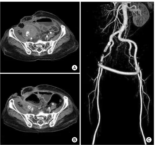
into the right external iliac fossa. Thereafter, the false aneur-ysm was found to have disappeared. However, a profound amount of hematoma was present in the affected area. Although no strains were identified from blood culture test-ing, a joining of the infected area in the lower right abdomen and the false aneurysm was observed, resulting in the diag-nosis of mycotic aneurysm. A 10-mm long Amplatzer vas-cular plug was inserted into the right proximal external iliac artery. The patient was then immediately moved to the OR and the right distal external iliac artery was ligated. A 12-mm ringed Gore-Tex graft was used to perform a left femoral ar-tery-right femoral artery bypass. A postopeative lower ex-tremity vascular CT scan showed complete withdrawal of the mycotic aneurysm previously present at the right external iliac artery (Fig. 2). The patient was discharged 18 days after the operation without specific symptoms, but remote meta-stasis of the cecal cancer aggravated and she died after 4 months.
DISCUSSION
Mycotic aneurysm constitutes about 1% of all aneurysms, and its high risk of rupture requires immediate treatment. It tends to occur in a wide range of arteries, including in the lesser curve of the aortic arch and on the opposite side of the visceral branch vessel of the abdominal aorta. Although the condition is often thought to be related to structural character-istics of the artery or the turbulent blood flow that occurs in the opposite side of abdominal branching blood vessels, the actual cause of the condition is still unknown. The major bacterial strains known to cause mycotic aneurysms are E. coli, Staphylococcus, Salmonella, and Streptococcal species.
Extra-anatomic Bypass Grafting for Mycotic Aneurysm
− 191 −
Fig. 2. (A) Preoperative CT scan, mycotic aneurysm connected with Rt. External iliac artery. Large amo-unt of hematoma was observed. (B) Postoperative CT scan shows the resolved mycotic aneurysm and de-creased hematoma. (C) Postoperat-ive 3-dimensional CT angiography.
These strains are related to atherosclerotic ulcers in the inner arterial membrane, and they act as a nidus for invading a secondarily infected arterial wall, triggering a false aneurysm or rupture of the artery [1]. Although the onset rate of my-cotic aneurysm is comparatively low, the difficulty of diag-nosis often results in late discovery of the disease after seri-ous advancement of the condition. The mortality rate is fairly high, since it often develops in patients with lowered im-munity or other underlying diseases. Diagnosis of mycotic aneurysm can be done by identifying three typical symptoms: abdominal pain, fever, and a pulsating lump. However, it is unusual to find all the symptoms at once and symptoms are often nonspecific [2]. A CT scan of a subject with a sus-pected mycotic aneurysm can reveal detailed information of the lesion and diagnosis can be done through blood culture testing. However, only 10∼27% of postoperative mycotic aneurysms are found to be benign in tissue culture testing, and there have been no reports of any relationship to
prog-nosis, such as a postoperative infection of the graft [3]. Treatment of mycotic aneurysm includes early diagnosis as well as broad removal of the lesion surgically following arti-ficial blood vessel placement and long-term use of antibiotics. Although treatment plays a critical role in a patient’s prog-nosis, several studies have reported high postoperative death rates (16∼44%) despite ongoing technical advancement. Unlike the general aneurysm, mycotic aneurysm is often lo-cated near the upper portion of renal arteries. In that case, in
situ graft placement is preferred, since the range of the lesion
is broad and reconstruction of blood vessels that reach sur-rounding organs is required. On the other hand, in cases of mycotic aneurysm in the lower portion of renal arteries, ex-tra-anatomic artificial blood vessel bypass has been recog-nized as the appropriate treatment. In both cases, if staph-ylococcus aureus, salmonella species, or other pyogenic in-fection is grossly suspected, in situ graft placement is for-bidden, since the chance of a postoperative graft infection is
Kwan-wook Kim, et al
− 192 − high [4,5]. A variety of vascular embolizations have been re-ported recently for extra-anatomic artificial vessel bypass on mycotic aneurysms in the inferior renal artery. Vascular em-bolization is known to be suitable for the treatment of aneur-ysms in blood vessels that have below-average size, since the operational approach can be difficult. However, it has been controversial to place a foreign material in the infected artery in treatment of mycotic aneurysm and the risk of post-treat-ment complications such as rupture can increase due to a weakening of the arterial wall. Therefore, these treatments can only be applied in patients with a low chance of con-tinuous exacerbation of infection [6]. In this case report, the authors have planned treatments after comprehensive consid-eration of the patients’ clinical symptoms as well as the loca-tion of lesions. We would like to introduce the bypass sur-gery in between bilateral femoral arteries following vascular embolization as a successful treatment option for mycotic aneurysm.
REFERENCES
1. Svensson LG. Descending thoracic and thoracoabdominal
aortic surgery. In: Silke FW, del Nido PJ, Swanson SJ. Sabiston & spencer surgery of the chest. 7th ed.
Philadel-phia: Elsevier Saunders. 2005;1165-94.
2. Oderich GS, Panneton JM, Bower TC, et al. Infected aortic
aneurysm: aggressive presentation, complicated early out-come, but durable results. J Vasc Surg 2001;34:900-8.
3. Cordero JA Jr, Darling RC 3rd, Chang BB, et al. In situ
prosthetic graft replacement for mycotic thracoabdominal aneurysms. Am Surg 1996;62:35-9.
4. Hsu RB, Tsay YG, Wang SS, et al. Surgical treatment for
primary infected aneurysm of the descending thoracic aorta, abdominal aorta and iliac arteries. J Vasc Surg 2002;36:
746-50.
5. Iida Y, Obitsu Y, Yokoi Y, et al. Successful treatment of
multiple mycotic aortic aneurysms, using a hybrid procedure.
J Vasc Surg 2010;51:1521-4.
6. Kische S, Ince H, Peuster M. Coil occlusion of a subclavian
mycotic aneurysm. Catheter Cardiovasc Interv 2010;75:1116-