변형된 장 족무지 굴건 이전술을 이용한 진구성 아킬레스 건 파열의 치료
한림대학교 강남성심병원 정형외과학교실 김형년⋅서일우⋅박용욱
Treatment of Old Achilles Tendon Rupture using Modified Flexor Hallucis Longus Tendon Transfer
Hyong-Nyun Kim, M.D., Il-Woo Suh, M.D., Yong-Wook Park, M.D.
Department of Orthopedic Surgery, Kangnam Sacred Heart Hospital, Hallym University College of Medicine, Seoul, Korea
=Abstract=
Purpose: The purpose of this study was to evaluate the clinical results of the old Achilles tendon rupture treated with modified flexor hallucis longus (FHL) tendon transfer.
Materials and Methods: Seventeen patients with old Achilles tendon rupture treated with modified FHL tendon transfer between March 2004 and February 2008 were enrolled in this study. Technically FHL was pass through the distal portion of the ruptured tendon instead of the drilled hole made on the calcaneus. The mean age of the patients was 37 years (range, 22~67 years), mean follow-up period was 28 months (range, 12~30 months). Patients' subjective satisfaction, calf circumferential diameter, range of motion of ankle and AOFAS ankle-hind foot score and Arner-Lidholm score was evaluated.
Results: The average gap between the ruptured tendon was 52 mm (range, 47~56 mm). The AOFAS score improved from 47 pre-operatively to 91 points at the last follow-up. Sixteen patients were satisfied with the result free from discomfort, a patient had mild discomfort who had DM. fourteen patients had decreased range of motion less than 5 degrees while 2 patients had more than 7 degrees decrease compared to the intact side but had no discomfort in daily activities. Nine patients had less than 1 cm calf circumferential diameter difference and 7 patients had 1 to 3 cm diameter difference compared to the intact side. One who had more than 3 cm diameter difference had deteriorated muscle strength.
Conclusion: Modified FHL tendon transfer can be a useful technique for the treatment of old Achilles tendon rupture when the gap is with large gap placed too proximal.
Key Words: Achilles tendon, Old Achilles tendon rupture, Modified FHL tendon transfer
∙Address for correspondence Yong-Wook Park, M.D.
Department of Orthopedic Surgery, Kangnam Sacred Heart Hospital, 948-1 Dalim-1dong, Youngdeungpo-gu, Seoul, 150-719, Korea Tel: +82-2-829-5165 Fax: +82-2-2634-1908
E-mail: [email protected]
서 론
아킬레스 건 급성 파열은 대개 30~40대의 연령에서 스 포츠와 관련하여 발생하는데 최근 스포츠 및 여가 활동의 증가로 발생 빈도가 증가하는 추세에 있다13). 급성 아킬레 스 건 파열을 치료하지 않고 방치하거나 보존적 치료가 실 패한 경우 파열 단 사이에 반흔 조직이 채워지고 파열된 건 은 수축, 위축되며 결국 족저 굴곡력이 약화되어 보행에 지
Figure 1. Pre-operative T1-weighted MRI shows a chronic Achilles tendon ruptured with retracted tendon ends and the fibrous scar tissue filled between the tendon stumps.
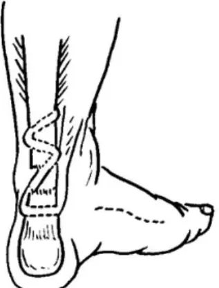
Figure 2. Diagram of modified FHL tendon transfer showing FHL tendon passing through the distal portion of the ruptured tendon instead of the drilled hole made on the calcaneus.
장을 주게 된다1,9,13,17). 진구성 아킬레스 건 파열의 수술적 치료 시 파열 단 사이에 채워진 반흔 조직을 제거하고 나서 생기는 결손 부위는 파열단 사이의 단단 봉합이 어려워 급 성 아킬레스 건 파열의 수술적 치료보다 더 복잡하고 어렵 다. 결손 부위가 클 경우 장 족무지 굴근건 이전술을 통한 치료로 좋은 결과들이 보고되고 있는데14,15,20,22-24) 파열 부 위가 근위부이거나 결손 부위가 커서 장 족무지 굴근건을 충분하게 이전, 보강하지 못하는 경우를 경험하게 된다. 이 에 저자들은 장 족무지 굴근건을 아킬레스 건 부착부 종골 을 통과시키지 않고 파열 원위단 아킬레스 건을 통과시키는 변형된 장 족무지 굴근건 이전술을 이용하여 만족할 만한 결과를 얻었기에 문헌 고찰과 함께 보고하는 바이다
대상 및 방법
2004년 3월부터 2008년 2월까지 본 교실에 내원한 28예 의 진구성 아킬레스 건 파열 환자 중 변형된 장 족무지 굴근 건 이전술을 통해 치료하였으며 추시가 가능하였던 17예를 대상으로 후향적 연구를 하였다. 평균 나이는 37세(범위, 22~67세)이였으며 환자 중 당뇨가 있던 환자는 5예, 아킬 레스 건 부착 부위에 스테로이드 주사를 맞은 기왕력이 있 는 환자가 1예가 있었으며 아킬레스 건 손상 후 진단하기까 지는 평균 4.5개월(범위, 4주~14개월)이었다. 파열 부위는 아킬레스 건의 종골 부착 부위에서부터 평균 53 mm(범위, 44~65 mm) 상방이었으며 파열단 사이의 결손 간격은 평 균 52 mm(범위, 47~56 mm)이었다. 최종 추시 관찰까지는 평균 28개월(범위, 12~30개월) 이었으며. 최종 추시 때 환
자의 임상적 결과 판정은 미국정형외과족부족관절학회의 족관절-후족부 평가와 환자의 만족도, 종아리 근력 정도, 종아리 둘레, 족근 관절 운동 범위에 근거한 Arner- Lidholm 평가 기준을 이용하여 우수, 양호, 불량으로 판정 하였다2). 환자의 만족도에서는 보행 시 불편감 정도, 보행 력, 발뒤꿈치 들기 유무와 종아리 근력 정도를 보았고 객관 적 평가로 양측 종아리 둘레 차이와 양측 족근 관절 운동범 위 차이를 측정하였다.
수술 방법은 환자를 전신 마취 또는 척추 마취 후 앙와위 에 두고 환측 하지를 건측 하지 위에 4자 모양을 이루게 올 려놓은 후 중족부 내측에 주상골에서부터 중족골 두에 이르 는 종 절개를 가한다. 족무지 외전근과 단 족무지 굴근을 족 저부로 도수 견인하여 중족부 심부를 노출시키면 장 족무지 굴근건과 장 족지 굴근건이 나타나며 장 족무지 굴근건을 장 족지 굴근과 봉합할 정도를 남겨둔 채 최대한 원위부에 서 절개하여 충분한 길이의 이식건을 얻는다(Fig. 3A). 장 족무지 굴근건과 장 족지 굴근건을 5개의 족지가 모두 중립 위인 상태에서 봉합을 한다. 다음 후족부에 종골의 아킬레 스 건 부착부 1.0 cm 내측에서 건의 내측을 따라 약 10 cm 정도 종 절개를 가한 후 아킬레스 건을 노출시켰다. 파열 부 위의 반흔 조직을 신선 건이 나타날 때까지 제거한 후 슬관 절 굴곡 30도 족관절 중립위에서 단단 간의 결손 간격을 측 정하였다. 하퇴의 후방 구획의 근막을 종 절개하여 장 족무 지 굴근을 노출시키고 중족부에서 절개하였던 이식건을 후 방 절개 부위로 도수 견인하여 얻는다. 파열 부위가 근위부 이거나 결손부위가 커서 이식건의 길이가 충분하지 못할 때 족근 관절 족저 굴곡 10도의 상태에서 아킬레스 건의 파열 원위단 부착부에서 1 cm 근위부에 절개를 가하여 내측에서 외측으로 이식건을 통과시킨 후 2.0 EthibondⓇ (Ethicon
A B
Figure 3. Intra-operative photograph shows (A) harvested FHL tendon from the midfoot, (B) recontructed Achilles tendon with FHL passing through the distal stump and woven through the proximal stump.
Inc, U.K.)로 아킬레스 건 통과 부위과 이식건을 봉합하였 다. 이식건은 결손 부위를 건너 아킬레스 건에 지그재그 형 태를 이루도록 통과시킨 후 봉합하였다. 또한 장 족무지 굴 근을 아킬레스 건의 내측에 봉합하여 보강하고 혈류 개선을 도왔다(Fig. 2, 3B). 환자는 2주간의 단하지 석고 고정 후 봉합사의 제거와 함께 단하지 보조기(walking boots)를 착 용하여 체중부하를 허용하였다.
결 과
환자의 주관적 만족도는 보행 시 불편감 없음이 16예였 고 당뇨가 동반되었던 1예에서 가벼운 불편감을 호소하였 다. 또한 보행 시 정상 보행력을 보인다가 15예였고 보행 능 력이 다소 떨어진다가 2예였고, 여기에는 당뇨가 동반되었 던 1예가 속하였다. 당뇨가 동반되었던 1예를 제외한 16예 에서 한 발뒤꿈치 들림이 가능하였다. 미국정형외과족부족 관절학회의 족관절 후족부 평가 점수는 수술 전 평균 47점 에서 수술 후 최종 추시에서 평균 91점으로 향상되었다.
Arner-Lidholm 평가 기준에 따르면 우수가 15예, 양호 2 예로 전 예에서 양호 이상의 주관적 만족을 보였다. 총 17명 의 환자 중 시행한 환측과 건측의 종아리 둘레 차이가 1 cm 이하를 보인 경우가 9예, 1~3 cm 차이를 보인 경우가 7예, 3 cm 이상의 차이를 보인예가 1예에서 있었으며 1예에서 이 로 인한 근력 약화를 호소하였다. 환측과 건측의 족근 관절 운동 범위의 차이가 5도 이하를 보이는 경우가 14예, 2예에 서는 7도 이상의 차이를 보이고 있었으나 이로 인한 일상 생활에 불편을 호소하지 않았다. 수술 후 재파열이나 심부 감염 등의 합병증은 없었다.
고 찰
진구성 아킬레스 건 파열은 급성 손상을 진단하지 못하 였거나 치료하지 않고 방치된 경우, 잘못 진단되었던 경우 또는 급성 손상의 보존적 치료가 실패한 경우를 포함하며 손상 후 4주 이상이 경과한 경우를 말한다9). 파열 후 10일 이 지나면서부터 파열 단 사이에 반흔 조직이 채워지고 파 열된 건은 수축, 위축되며 결국 족저 굴곡력이 약화되어 보 행에 지장을 주게 된다1,17). 급성 아킬레스 건 파열은 문진과 이학적 검사만으로도 진단이 비교적 용이하지만 Ballas 등3) 은 전체 손상의 5분의 1에서 진단을 놓칠 수 있다고 보고하 였고 Boyden 등4)은 25%에서 진단을 놓칠 수 있다고 보고 하였다. Nestorson 등18)은 25명의 고령 환자에서 진단이 늦어지거나 이뤄지지 못하여 치료가 1주일 이상 지연되었던 경우가 9건(36%)이었다고 보고하였다. 급성 파열의 진단과 치료가 이루어지지 않을 경우 통증과 부종은 가라앉으며 결 손 부위는 반흔 조직으로 채워져 이학적 검사에서 결손 부 위가 촉지 되지 않는 경우도 있고 후 경골근과 장 족무지 굴 근 등 족저 굴근에 의해 족근 관절의 굴곡 운동이 가능한 경 우가 있어 주의 깊은 문진과 이학적 검사가 필요하다13). 진 구성 아킬레스 건 파열의 경우 장 족지 굴근 및 장 족무지 굴근이 하퇴 삼두근의 기능을 대신하며 강화되어 갈퀴 족지 변형이 나타나거나 내측 종아치가 높아지기도 하며 또한 하 퇴 삼두근의 기능 약화로 환측 하지만으로 뒤꿈치를 들지 못하는 경우가 많다. 정확한 진단과 치료 방법의 결정을 위 하여 자기 공명 염상 검사가 도움이 되며 정상의 아킬레스 건이 T1과 T2 강조 영상 모두에서 저신호 강도를 보이는 반 면 파열 부위는 T1 강조 영상에서 저신호 강도를 T2 강조 영상에서 비연속성의 변화된 신호를 보인다10). 진구성 아킬 레스 건의 수술적 치료는 파열 단 사이에 채워진 반흔 조직
을 제거하고 나서 생기는 결손 부위의 단단 봉합이 어려워 급성 아킬레스 건 파열의 수술적 치료보다 더 복잡하고 어 렵다. 수술적 방법은 크게 단단봉합(end-to-end repair), 아킬레스 건 전진술 및 건판을 이용한 재건술(tendo-Achilles advancement or flap reconstruction),국소 건 이전술 (lcoal tendon transfer), 건 이식술(autograft, allograft or synthetic)로 나눌 수 있다9,11,13,17). 여러 가지 방법들 중 Myerson17)은 파열 단단 간 결손 간격과 환자의 나이 및 활 동 정도, 손상 후 경과한 기간 등으로 수술 방법을 결정할 것을 제안하고 진구성 아킬레스 건 파열을 결손 간격 정도 에 따라 치료 방법을 제시하였다. 결손 간격이 1~2 cm 이 하인 경우는 단단 봉합(end-to-end repair) 및 후방 구획 의 근막 절개술을, 간격이 2~5 cm인 경우는 V-Y 건판 전 진술을 건 이전술과 함께 혹은 단독으로 시행할 것을 제안 하였고 간격이 5 cm 보다 큰 경우는 장 족무지 굴근건을 이 용한 건 이전술을 V-Y 건판 전진술과 함께 혹은 단독으로 시행할 것을 제안하였다. 한편 Kuwada12)는 Type I은 부분 파열의 경우로 보존적 치료를, Type II는 결손 간격이 3 cm 이하의 완전 파열로 단단 봉합을, Type III는 결손 간격이 3~6 cm으로 건 이식술을 인조 건 보강술(synthetic graft augmentation)과 함께 혹은 단독으로 할 것을 제안하였다.
Type IV는 결손 간격이 6 cm 이상이 경우로 하퇴 삼두근의 전진술이나 유리 건 이전술, 인조 건 이전술을 제안하였다.
V-Y 건판 전진술은 Abraham과 Pankovich1)에 의해 처 음 소개되었는데 건판을 5 cm 이상 전진시켰을 때 근단위 (muscle unit)에 약화를 가져올 가능성이 많아 큰 결손 간 격을 갖는 진구성 아킬레스 건 파열에서는 사용에 제한이 있고 비복근 근막 젖혀내림 피판술의 경우 절개 부위가 크 다는 단점이 있다8,17). 본 연구에서는 술 전에 촬영한 자기 공명 영상 검사를 통해 단단 간 결손 간격을 측정하여 Myerson의 기준에 따라 수술을 계획하였고 실제 수술장에 서 반흔 조직을 제거한 후 측정하였을 경우 5 cm 보다 컸던 경우 모두 장 족무지 굴근건 이전술을 통해 아킬레스 건을 재건하였다. 장 족무지 굴근건은 다른 이식건보다 길고 내 구성이 강하며 수축의 축이 아킬레스 건과 같고 하퇴 삼두 근과 같은 자극에 수축하며 해부학적으로 인접하여 접근이 쉽다. 또한 족저 굴근에서 족저 굴근에 이식하므로 다른 근 육간의 균형을 유지할 수 있고 중족부에서 채취하는 경우 10~12 cm의 긴 이식건을 얻을 수 있는 장점이 있다14,22). 장 족무지 굴근의 근육 부위를 아킬레스 건의 파열 부위에 함 께 봉합하면 아킬레스 건의 혈액 공급 및 순환에 도움이 된 다5). 하지만 장 족무지 굴근의 이식으로 무지 지간 관절의 능동적 굴곡이 불가능하게 되지만 이것으로 인한 보행의 불
편을 호소하는 경우는 없었다. Coull 등6)은 족저 압력 분석 검사(pedobarography)에서 보행 시 족무지 원위 지골의 최 대 압력 지수가 저하되었지만 통계학적으로 유의하지 않았 으며 기능적으로 이상이 없음을 보고하였다. Wapner 등22) 이 처음 소개한 장 족무지 굴근건 이전술의 경우 파열된 부 위가 하퇴 1/3 이상 근위부이거나 결손 부위가 커서 이식건 의 길이가 짧을 경우 충분하게 결손 부위에 이식하기가 어 렵다. 본 연구에서는 변형된 장 족무지 굴근건 이전술을 시 행하여 만족할 만한 결과를 얻었기에 문헌 고찰과 함께 보 고하는 바이다.
결 론
진구성 아킬레스 건 파열의 수술적 치료로 변형된 장 족 무지 굴근건 이전술은 파열된 부위가 근위부이거나 결손 부 위가 크고 이식건의 길이가 짧아 충분하게 결손 부위에 이 식하기가 어려운 경우 사용할 수 있는 유용한 술식으로 사 료된다.
REFERENCES
1. Abraham E and Pankovich AM: Neglected rupture of the achilles tendon. treatment by V-Y tendinous flap. J Bone Joint Surg, 57-A: 253-255, 1975.
2. Arner O and Lindholm A: Subcutaneous rupture of the achilles tendon; a study of 92 cases. Acta Chir Scand Suppl, 116: 1-51, 1959.
3. Ballas MT, Tytko J and Mannarino F: Commonly missed orthopedic problems. Am Fam Physician, 57: 267-274, 1998.
4. Boyden EM, Kitaoka HB, Cahalan TD and An KN: Late versus early repair of Achilles tendon rupture. Clinical and biomechanical evaluation. Clin Orthop Relat Res, 317: 150- 158, 1995.
5. Carr AJ and Norris SH: The blood supply of the calcaneal tendon. J Bone Joint Surg, 71-B: 100-101, 1989.
6. Coull R, Flavin R and Stephens MM: Flexor hallucis longus tendon transfer: Evaluation of postoperative morbidity. Foot Ankle Int, 24: 931-934, 2003.
7. Den Hartog BD: Flexor hallucis longus transfer for chronic achilles tendonosis. Foot Ankle Int, 24: 233-237, 2003.
8. Elias I, Besser M, Nazarian LN and Raikin SM: Reconstruction for missed or neglected achilles tendon rupture with V-Y lengthening and flexor hallucis longus tendon transfer through one incision. Foot Ankle Int, 28: 1238-1248, 2007.
9. Gabel S and Manoli A 2nd: Neglected rupture of the achilles tendon. Foot Ankle Int, 15: 512-517, 1994.
10. Hahn F, Meyer P, Maiwald C, Zanetti M and Vienne P:
Treatment of chronic achilles tendinopathy and ruptures with flexor hallucis tendon transfer: Clinical outcome and MRI findings. Foot Ankle Int, 29: 794-802, 2008.
11. Kissel CG, Blacklidge DK and Crowley DL: Repair of neglected achilles tendon ruptures--procedure and functional results. J Foot Ankle Surg, 33: 46-52, 1994.
12. Kuwada GT: Classification of tendo-Achilles rupture with consideration of surgical repair techniques. J Foot Surg, 29:
361-365, 1990
13. Maffulli N and Ajis A: Management of chronic ruptures of the achilles tendon. J Bone Joint Surg, 90-A: 1348-1360, 2008.
14. Mann RA, Holmes GB,Jr, Seale KS and Collins DN: Chronic rupture of the achilles tendon: A new technique of repair. J Bone Joint Surg, 73-A: 214-219, 1991.
15. Martin RL, Manning CM, Carcia CR and Conti SF: An outcome study of chronic achilles tendinosis after excision of the achilles tendon and flexor hallucis longus tendon transfer.
Foot Ankle Int, 26: 691-697, 2005.
16. Mulier T, Rummens E and Dereymaeker G: Risk of neuro- vascular injuries in flexor hallucis longus tendon transfers:
An anatomic cadaver study. Foot Ankle Int, 28: 910-915, 2007.
17. Myerson MS: Achilles tendon ruptures. Instr Course Lect, 48:
219-230, 1999.
18. Nestorson J, Movin T, Möller M and Karisson J: Funcion after Achilles tendon rupture in the elderly: 25 patients older than 65 years followed for 3 years. Act Orthop Scan, 71: 64-68, 2000.
19. Reddy SS, Pedowitz DI, Parekh SG, Omar IM and Wapner KL: Surgical treatment for chronic disease and disorders of the achilles tendon. J Am Acad Orthop Surg, 17: 3-14, 2009.
20. Tashjian RZ, Hur J, Sullivan RJ, Campbell JT and DiGiovanni CW: Flexor hallucis longus transfer for repair of chronic achilles tendinopathy. Foot Ankle Int, 24: 673-676, 2003.
21. Us AK, Bilgin SS, Aydin T and Mergen E: Repair of neglected achilles tendon ruptures: Procedures and functional results.
Arch Orthop Trauma Surg, 116: 408-411, 1997.
22. Wapner KL, Pavlock GS, Hecht PJ, Naselli F and Walther R:
Repair of chronic achilles tendon rupture with flexor hallucis longus tendon transfer. Foot Ankle, 14: 443-449, 1993.
23. Wegrzyn J, Luciani JF, Philippot R, Brunet-Guedj E, Moyen B and Besse JL: Chronic achilles tendon rupture reconstruction using a modified flexor hallucis longus transfer. Int Orthop, 2009.
24. Wilcox DK, Bohay DR and Anderson JG: Treatment of chronic achilles tendon disorders with flexor hallucis longus tendon transfer/augmentation. Foot Ankle Int, 21: 1004-1010, 2000.