• 검색 결과가 없습니다.

Silymarin-Mediated Degradation of c-Myc Contributes to the Inhibition of Cell Proliferation in Human Colorectal Cancer Cells

N/A
N/A
Protected

Academic year: 2021

Share "Silymarin-Mediated Degradation of c-Myc Contributes to the Inhibition of Cell Proliferation in Human Colorectal Cancer Cells"

Copied!
7
0
0

로드 중.... (전체 텍스트 보기)

전체 글

(1)

Introduction

Silymarin from the Silybum marianum (milk thistle) has received a tremendous attention over the last decade (Abenavoli et al., 2010). It has been reported that silymarin has antioxidant activity (Draz et al., 2015), anti-diabetes (Kazazis et al., 2014), anti-obesity (Gu et al., 2016), anti- inflammatory activity (Guo et al., 2016) and hepatoprotective effect (Mereish et al., 1991). In previous study, we reported that silymarin suppressed the growth of human colorectal cancer cells through cyclin D1 proteasomal degradation (Eo et al., 2015) and induced apoptosis via activating ATF3 (Eo et al., 2016). In addition, the effect of silymarin on cell cycle arrest and apoptosis has been reported in ovarian cancer (Fan et al., 2014) and lung cancer (Wu et al., 2016). These studies for anti-proliferative effect of silymarin have been focused on cyclin D1 associated with the cell cycle regulation. However,

cancer cell growth has been controlled by a number of the cell cycle regulators.

Among the cell cycle regulators, c-Myc is overexpressed in various human cancers, including lung carcinoma (Little et al., 1983), breast carcinoma (Mariani-Costantini et al., 1988) and colon carcinoma (Augenlicht et al., 1997). c-Myc regulates the expression of various genes involved in controlling cell proliferation and apoptosis (Bretones et al., 2015). Thus, it has been accepted that c-Myc may be the potential target for cancer chemoprevention and therapy.

In this study, we elucidated the molecular mechanism of silymarin by which silymarin may inhibits cell proliferation in human colorectal cancer cells in order to search the new potential anti-cancer target associated with the cell growth arrest.

Materials and Methods

Reagents

Cell culture media, Dulbecco's Modified Eagle medium

Silymarin-Mediated Degradation of c-Myc Contributes to the Inhibition of Cell Proliferation in Human Colorectal Cancer Cells

Hyun Ji Eo

1

, Jin Boo Jeong

1,2

, Jin Suk Koo

1,2

and Hyung Jin Jeong

1,2

*

1

Department of Medicinal Plant Resources, Andong National University, Andong 36729, Korea

2

Agricultural Science and Technology Research Institute, Andong 36729, Korea

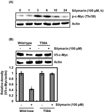
Abstract - In this study, we elucidated the molecular mechanism of silymarin by which silymarin may inhibits cell proliferation in human colorectal cancer cells in order to search the new potential anti-cancer target associated with the cell growth arrest. Silymarin reduced the level of c-Myc protein but not mRNA level indicating that silymarin-mediated downregulation of c-Myc may result from the proteasomal degradation. In the confirmation of silymarin-mediated c-Myc degradation, MG132 as a proteasome inhibitor attenuated c-Myc degradation by silymarin. In addition, silymarin phosphorylated the threonine-58 (Thr58) of c-Myc and the point mutation of Thr58 to alanine blocked its degradation by silymarin, which indicates that Thr58 phosphorylation may be an important modification for silymarin-mediated c-Myc degradation. We observed that the inhibition of ERK1/2, p38 and GSK3β blocked the Thr58 phosphorylation and subsequent c-Myc degradation by silymarin. Finally, the point mutation of Thr58 to alanine attenuated silymarin-mediated inhibition of the cell growth. The results suggest that silymarin induces the cell growth arrest through c-Myc proteasomal degradation via ERK1/2, p38 and GSK3β-dependent Thr58 phosphorylation.

Key words – Cancer chemoprevention, Cell growth arrest, c-Myc, Human colorectal cancer, Silymarin

*Corresponding author. E-mail : [email protected] Tel. +82-54-820-5464

These authors equally contributed to this study

ⓒ 2017 by The Plant Resources Society of Korea

Original Research Article

(2)

(DMEM)/F-12 1:1 Modified medium (DMEM/F-12) was purchased from Lonza (Walkersville, MD, USA). PD98059 (ERK1/2 inhibitor), SB203580 (p38 inhibitor), SP600125 (JNK inhibitor) and LiCl (GSK3β inhibitor) and MG132 were purchased from Sigma Aldrich (St. Louis, MO, USA).

Antibodies against c-Myc, phospho-c-Myc (Thr58), V5-tag and β-actin were purchased from Cell Signaling (Bervely, MA, USA). All chemicals were purchased from Fisher Scientific (Hampton, NH, USA), unless otherwise specified.

Cell culture and treatment

Human colorectal cancer cell lines such as HCT116, SW480, LoVo and HT-29 were purchased from Korean Cell Line Bank (Seoul, Korea) and cultured in DMEM/F-12 supplemented with 10% fetal bovine serum (FBS), 100 U/㎖

penicillin and 100 ㎍/㎖ streptomycin. The cells were main- tained at 37 ℃ under a humidified atmosphere of 5% CO

2

. Silymarin was purchased from Sigma Aldrich (St. Louis, MO, USA) and dissolved in dimethyl sulfoxide (DMSO).

DMSO was used as a vehicle and the final DMSO concent- ration did not exceed 0.1% (v/v).

Cell proliferation assay

Cell growth was measured using MTT assay system.

Briefly, cells were plated onto 96-well plated and grown overnight. The cells were transfected with wild type c-Myc and T58A c-Myc expression factor for 24 h and then treated with silymarin for the additional 24 h. Then, the cells were incubated with 50 ㎕ of MTT solution (1 ㎎/㎖) for an additional 2 h. The resulting crystals were dissolved in DMSO.

The formation of formazan was measured by reading absorbance at a wavelength of 570 ㎚.

SDS-PAGE and Western blot

After silymarin treatment, cells were washed with 1×

phosphate-buffered saline (PBS), and lysed in radioimmuno- precipitation assay (RIPA) buffer (Boston Bio Products, Ashland, MA, USA) supplemented with protease inhibitor cocktail (Sigma-Aldrich) and phosphatase inhibitor cocktail (Sigma-Aldrich) for 30 min at 4℃. Then, cell lysates were centrifuged at 15,000 rpm for 10 min at 4 ℃. Protein concentration was determined by the bicinchoninic acid

(BCA) protein assay (Pierce, Rockford, IL, USA). The equal proteins were separated on SDS-PAGE and transferred to PVDF membrane (Bio-Rad Laboratories, Inc., Hercules, CA, USA). The membranes were blocked for non-specific binding with 5% non-fat dry milk in Tris-buffered saline containing 0.05% Tween 20 (TBS-T) for 1h at room temperature and then incubated with specific primary antibodies in 5% non-fat dry milk at 4 ℃ overnight. After three washes with TBS-T, the membranes were incubated with horse radish peroxidase (HRP)-conjugated immunoglobulin G (IgG) for 1 h at room temperature and chemiluminescence was detected with ECL Western blotting substrate (Amersham Biosciences, Piscataway, NJ, USA) and visualized in Polaroid film.

Reverse transcriptase-polymerase chain reaction (RT-PCR) After silymarin treatment, total RNA was prepared using a RNeasy Mini Kit (Qiagen, Valencia, CA, USA) and total RNA (1 ㎍) was reverse-transcribed using a Verso cDNA Kit (Thermo Scientific, Pittsburgh, PA, USA) according to the manufacturer’s protocol for cDNA synthesis. PCR was carried out using PCR Master Mix Kit (Promega, Madison, WI, USA) with human primers for c-Myc and GAPDH as followed : c-Myc: forward 5’- cgcgctgagtataaaagccg -3’ and reverse 5’- ctattcgctccggatctccc-3’, GAPDH: forward 5’- acccagaagactgtggatgg-3’ and reverse 5’-ttctagacggcaggtcaggt-3’.

Expression vectors

Wild type V5-tagged c-Myc and point mutation of T58A of V5-tagged c-Myc were provided from Addgene (Cambridge, MA, USA). Each vector was transfected to HCT116 cells for 24 h using the PolyJet DNA transfection reagent (SignaGen Laboratories, Ijamsville, MD, USA) according to the manu- facturers’ instruction.

Statistical analysis

All the data are shown as mean ± SEM (standard error of mean). Statistical analysis was performed with one-way ANOVA followed by Dunnett's test. Differences with *P

<0.05 were considered statistically significant.

(3)

Results and Discussion

Effect of silymarin on c-Myc protein level in human colorectal cancer cells

As one of the transcription factors, c-Myc has been reported to regulate the expression of various genes involved in controlling cell proliferation and apoptosis (Bretones et al., 2015), which is associated with cancer progression and metastasis (Dang, 2012). Thus, c-Myc has been regarded as the potential molecular target for cancer therapy (Fletcher and Prochownik, 2015). Recently, c-Myc overexpression has been observed in human colorectal cancer (Wright et al., 2010). In our previous study of silymarin, we demonstrated that silymarin suppresses the growth of human colorectal cancer cells through cyclin D1 proteasomal degradation (Eo et al., 2015) and induces apoptosis through activating ATF3 expression (Eo et al., 2016). In this study, we investigated whether silymarin regulates c-Myc level, which affects the growth of human colorectal cancer cells. As shown in Fig.

1A, silymarin treatment dose-dependently decreased c-Myc protein level in HCT116 cells. In addition, the attenuation of c-Myc protein level was observed in SW480, LoVo and HT-29 cells treated with silymarin (Fig. 1B). In time-course experiment, c-Myc protein level started to be decreased at 10 h after silymarin treatment.

Contribution of proteasomal degradation to silymarin- mediated downregulation of c-Myc protein level

c-Myc expression has been reported to be regulated through its transcription via NF-κB signaling pathway (Liu et al., 2016). We have reported that silymarin induces NF-κB activation (Eo et al., 2015). Thus, we investigated that silymarin-mediated downregulation of c-Myc protein level is attributed to transcriptional regulation, mRNA level of c-Myc was determined in human colorectal cancer cells treated with silymarin. As shown in Fig. 2A and 2B, mRNA level of c-Myc was not changed by silymarin treatment. These data imply that silymarin-mediated downregulation of c-Myc may be independent on transcriptional regulation.

Fig. 1. Effect of silymarin on the protein level of c-Myc in human colorectal cancer cells. (A, B) The cells were plated onto 6-well

plate for 24 h. Then the cells were treated with silymarin at the indicated concentrations for the additional 24 h. (C) HCT116 cells

were plated onto 6-well plate for 24 h. Then the cells were treated with 100 μM of silymarin for the indicated times. Each cell lysate

was subjected to SDS-PAGE and Western blot was performed using antibodies c-Myc and actin. Actin was used as internal control

for Western blot analysis. *P<0.05 compared to cell without silymarin treatment.

(4)

There is growing evidence that c-Myc protein level can be regulated through its degradation (Guo et al., 2012; Jing et al., 2016). Thus, we investigated whether silymarin induces c-Myc degradation using MG132 as a proteasome inhibitor.

As shown in Fig. 2C, the inhibition of proteasome activity by MG132 blocked silymarin-mediated attenuation of c-Myc protein level. This finding suggests that silymarin-mediated downregulation of c-Myc may result from the proteasomal degradation.

Involvement of the phosphorylation on c-Myc threonine-58 (Thr58) in silymarin-mediated degradation of c-Myc

The phosphorylation site, Thr58 is associated with c-Myc degradation through the ubiquitin-proteasome pathway (Yeh et al., 2004). De-phosphorylation of Thr58 results in the increase of c-Myc stabilization and subsequently induces accumulation of c-Myc, which contributes to human cell oncogenesis (Yeh et al., 2004). In this study, we observed that silymarin treatment increased the phosphorylation status of

c-Myc Thr58 at 3 and 6 h after silymarin treatment (Fig. 3A).

To elucidate the contribution of silymarin-mediated phos- phorylation of Thr58 to c-Myc degradation, we applied to Wild type c-Myc expression vector and T58A c-Myc expression vector. As shown in Fig. 3B, silymarin treatment reduced V5-c-Myc in the cell transfected with Wild type c-Myc expression vector. However, T58A transfection abolished silymarin-mediated decrease of V5-c-Myc. These findings indicate that silymarin may phosphorylate Thr58 of c-Myc protein, which may contribute to c-Myc degradation.

Elucidation of upstream kinases involved in silymarin- mediated degradation of c-Myc

The posttranslational modification by various kinases such as MAPK (ERK1/2, p38, JNK) and GSK3 β has been reported to be associated with protein degradation (Diehl et al., 1997).

However, the upstream kinases attributed to c-Myc degradation have been not elucidated. Thus, we investigated whether MAPK (ERK1/2, p38, JNK) and GSK3 β affects silymarin- Fig. 2. Proteasomal degradation contributes to silymarin-mediated downregulation of c-Myc protein level. (A, B) The cells were plated onto 6-well plate for 24 h. Then the cells were treated with silymarin at the indicated concentrations for the additional 24 h. For RT-PCR analysis of c-Myc gene expression, total RNA was prepared after silymarin treatment. GAPDH was used as internal control for RT-PCR. (C) HCT116 cells were pretreated with 20 μM of MG132 for 2 h and then co-treated with 100 μM of silymarin for the additional 10 h. Each cell lysate was subjected to SDS-PAGE and Western blot was performed using antibodies c-Myc and actin.

Actin was used as internal control for Western blot analysis. *P<0.05 compared to cell without silymarin treatment.

(5)

mediated c-Myc degradation in order to search the upstream kinases related to c-Myc degradation. As shown in Fig. 4A, the inhibition of ERK1/2 by PD98059, p38 by SB203580 and GSK3 β by LiCl attenuated c-Myc degradation by silymarin, but not JNK inhibition. Next, we investigated the effect of the kinases such as ERK1/2, p38 and GSK3β on silymarin- mediated Thr58 phosphorylation in order to determine whether these kinases affect the protein modification of c-Myc. As shown in Fig. 4B, inhibitions of these kinases abolished Thr58 phosphorylation of c-Myc. These findings imply that ERK1/2, p38 and GSK3β may be the potential upstream kinases for silymarin-mediated Thr58 phosphorylation and subsequent degradation of c-Myc. Lastly, we investigated whether silymarin-mediated degradation of c-Myc contributes to the cell proliferation, MTT assay was performed. As shown in Fig. 4C, silymarin inhibited the proliferation of HCT116 cells transfected with Wild type c-Myc expression vector by 42%. Although silymarin suppressed the cell

proliferation in T58A c-Myc expression vector by 15%, T58A c-Myc expression vector attenuated silymarin-mediated inhibition of the cell proliferation compared to the cells transfected with Wild type c-Myc expression vector. This finding suggests that silymarin-mediated c-Myc degradation may contribute partially to the inhibition of cell growth.

Taken together, our findings indicate that silymarin induces c-myc degradation through Thr58 phosphorylation dependent on ERK1/2, p38 and GSK3β, which results in the cell growth arrest. Therefore, these findings can provide information on the anti-proliferative effect and the potential molecular mechanism of silymarin.

Acknowledgment

This work was supported by a grant from 2015 Research Funds of Andong National University (H.J. Jeong), Republic of Korea.

Fig. 3. Effect of silymarin on Thr58 phosphorylation of c-Myc and its contribution to c-Myc degradation. (A) HCT116 cells were

plated onto 6-well plate for 24 h. Then, the cells were treated with 100 μM of silymarin for the indicated times. (B) HCT116 cells

were plated onto 6-well plate for 24 h. Then, the cells were transfected with V5-tagged Wild type c-Myc or V5-tagged T58A c-Myc

expression vector for 24h. After transfection, the cells were treated with 100 μM of silymarin for 24 h. Each cell lysate was subjected

to SDS-PAGE and Western blot was performed using antibodies p-c-Myc (Thr58), V5-tag and actin. Actin was used as internal

control for Western blot analysis. *P<0.05 compared to cell without silymarin treatment.

(6)

References

Abenavoli, L., R. Capasso, N. Milic and F. Capasso. 2010. Milk thistle in liver diseases: past, present, future. Phytother. Res.

24:1423-1432.

Augenlicht, L.H., S. Wadler, G. Corner, C. Richards, L. Ryan, A.S. Multani, S. Pathak, A. Benson, D. Haller and B.G.

Heerdt. 1997. Low-level c-myc amplification in human colonic carcinoma cell lines and tumors: a frequent, p53-independent mutation associated with improved outcome in a randomized multi-institutional trial. Cancer Res. 57:1769-1775.

Bretones, G., M.D. Delgado and J. Leon. 2015. Myc and cell cycle control. Biochim. Biophys. Acta 1849:506-516.

Dang, C.V. 2012. MYC on the path to cancer. Cell 149:22-35.

Diehl, J.A., F. Zindy and C.J. Sherr. 1997. Inhibition of cyclin D1 phosphorylation on threonine-286 prevents its rapid degradation via the ubiquitin-proteasome pathway. Genes Dev. 11:957-972.

Draz, E.I., A.A. Abdin, N.I. Sarhan and T.A. Gabr. 2015.

Neurotrophic and antioxidant effects of silymarin comparable to 4-methylcatechol in protection against gentamicin-induced ototoxicity in guinea pigs. Pharmacol. Rep. 67:317-325.

Eo, H.J., G.H. Park and J.B. Jeong. 2016. Activating transcription factor 3 is a molecular target for apoptotic effect of silymarin in human colorectal cancer cells. Kor. J. Plant Res. 29:

281-288.

Eo, H.J., G.H. Park, H.M. Song, J.W. Lee, M.K. Kim, M.H.

Fig. 4. Effect of ERK1/2, p38, JNK and GSK3 β on silymarin-mediated Thr58 phosphorylation and subsequent degradation of

c-Myc, and contribution of silymarin-mediated c-Myc degradation to the cell proliferation. (A) HCT116 cells were pretreated with

PD98059 (40 μM, ERK1/2 inhibitor), SB203580 (40 μM, p38 inhibitor), SP900125 (40 μM, JNK inhibitor) or LiCl (20 mM, GSK3β

inhibitor) for 2 h and then co-treated with 100 μM of silymarin for the additional 10 h. (B) HCT116 cells were pretreated with

PD98059 (40 μM, ERK1/2 inhibitor), SB203580 (40 μM, p38 inhibitor) or LiCl (20 mM, GSK3β inhibitor) for 2 h and then

co-treated with 100 μM of silymarin for the additional 6 h. Each cell lysate was subjected to SDS-PAGE and Western blot was

performed using antibodies c-Myc, p-c-Myc (Thr58) and actin. Actin was used as internal control for Western blot analysis. (C)

HCT116 cells were plated onto 6-well plate for 24 h. Then, the cells were transfected with V5-tagged Wild type c-Myc or V5-tagged

T58A c-Myc expression vector for 24h. After transfection, MTT assay was performed. *P<0.05 compared to cell without silymarin

treatment.

(7)

Lee, J.R. Lee, J.S. Koo and J.B. Jeong. 2015. Silymarin induces cyclin D1 proteasomal degradation via its phos- phorylation of threonine-286 in human colorectal cancer cells. Int. Immunopharmacol. 24:1-6.

Fan, L., Y. Ma, Y. Liu, D. Zheng and G. Huang. 2014. Silymarin induces cell cycle arrest and apoptosis in ovarian cancer cells. Eur. J. Pharmacol. 743:79-88.

Fletcher, S. and E.V. Prochownik. 2015. Small-molecule inhibitors of the Myc oncoprotein. Biochim. Biophys. Acta 1849:

525-543.

Gu, M., P. Zhao, J. Huang, Y. Zhao, Y. Wang, Y. Li, S. Fan, Y.M. Ma, Q. Tong, L. Yang, G. Ji and C. Huang. 2016.

Silymarin ameliorates metabolic dysfunction associated with diet-induced obesity via activation of Farnesyl X receptor. Front. Pharmacol. 7:345.

Guo, Y., S. Wang, Y. Wang and T. Zhu. 2016. Silymarin improved diet-induced liver damage and insulin resistance by decreasing inflammation in mice. Pharm. Biol. 8:1-6.

Guo, Z., Y. Zhou, B.M. Evers and Q. Wang. 2012. Rictor regulates FBXW7-dependent c-Myc and cyclin E degradation in colorectal cancer cells. Biochem. Biophys. Res. Commun.

418:426-432.

Jing, H., J. Hu, B. He, Y.L. Negron Abril, J. Stupinski, K. Weiser, M. Carbonaro, Y.L. Chiang, T. Southard, P. Giannakakou, R.S. Weiss and H. Lin. 2016. A SIRT2-selective inhibitor promotes c-Myc oncoprotein degradation and exhibits broad anticancer activity. Cancer Cell 29:767-768.

Kazazis, C.E., A.A. Evangelopoulos, A. Kollas and N.G.

Vallianou. 2014. The therapeutic potential of milk thistle in diabetes. Rev. Diabet. Stud. 11:167-174.

Little, C.D., M.M. Nau, D.N. Carney, A.F. Gazdar and J.D.

Minna. 1983. Amplification and expression of the c-myc oncogene in human lung cancer cell lines. Nature 306:

194-196.

Liu, X., X.Y. Xiao, Q.Y. Shou, J.F. Yan, L. Chen, H.Y. Fu and J.C. Wang. 2016. Bufalin inhibits pancreatic cancer by inducing cell cycle arrest via the c-Myc/NF-kappaB pathway. J.

Ethnopharmacol. 193:538-545.

Mariani-Costantini, R., C. Escot, C. Theillet, A. Gentile, G.

Merlo, R. Lidereau and R. Callahan. 1988. In situ c-myc expression and genomic status of the c-myc locus in infiltrating ductal carcinomas of the breast. Cancer Res.

48:199-205.

Mereish, K.A., D.L. Bunner, D.R. Ragland and D.A. Creasia.

1991. Protection against microcystin-LR-induced hepato- toxicity by Silymarin: biochemistry, histopathology, and lethality. Pharm. Res. 8:273-277.

Wright, J.B., S.J. Brown and M.D. Cole. 2010. Upregulation of c-MYC in cis through a large chromatin loop linked to a cancer risk-associated single-nucleotide polymorphism in colorectal cancer cells. Mol. Cell. Biol. 30:1411-1420.

Wu, T., W. Liu, W. Guo and X. Zhu. 2016. Silymarin suppressed lung cancer growth in mice via inhibiting myeloid-derived suppressor cells. Biomed. Pharmacother. 81:460-467.

Yeh, E., M. Cunningham, H. Arnold, D. Chasse, T. Monteith, G. Ivaldi, W.C. Hahn, P.T. Stukenberg, S. Shenolikar, T.

Uchida, C.M. Counter, J.R. Nevins, A.R. Means and R.

Sears. 2004. A signalling pathway controlling c-Myc de- gradation that impacts oncogenic transformation of human cells. Nat. Cell Biol. 6:308-318.

(Received 15 November 2016 ; Revised 9 January 2017 ; Accepted 9 January 2017)

수치

Fig. 3. Effect of silymarin on Thr58 phosphorylation of c-Myc and its contribution to c-Myc degradation
Fig. 4. Effect of ERK1/2, p38, JNK and GSK3 β on silymarin-mediated Thr58 phosphorylation and subsequent degradation of  c-Myc, and contribution of silymarin-mediated c-Myc degradation to the cell proliferation

참조

관련 문서

Inhibitory effects of classified methanol extracts of Smilax china L on the COX-2 and iNOS expression of human colorectal cancer cell lines... Inhibitory

Activated H-Ras expression in human fibroblast cell lines increases the activity of Ku80 to bind injuried DNA, reduces γ-H2AX expression by UV irradiation,

4 &gt; Effects of Taro on COX-2 expression and iNOS expression(hot water) in human thyroid cancer cells. The cells were pretreated for 48hours with either

These results suggest that the bilobalide inhibits the cell proliferation and induces the apoptotic cell death in FaDu human pharyngeal squamous cell carcinoma via both

In the present study we investigated the proliferation effect of human oral cancer KB cell treated with pulsatilla koreana extract.. We analyzed the effects of this

They also showed significantly improved cell migration and proliferation with respect to negative control in the scratch - wound healing assay using confluent monolayers

Inhibitory effects of classified methanol extracts of Gastrodia elata Blume on the COX-2 and iNOS expression of human colorectal cancer cell lines...

Characterization of the glycoprotein D gene products of equine herpesvirus 1 using a prokaryotic cell expression vector.. Expression of equine herpesvirus-1 glycoprotein D by