Roles of TLR-4 and NF-κB in Interleukin-6 Expression Induced by Heat Shock Protein 90 in Vascular Smooth Muscle Cells
Byung-Yong Rhim, Kang-Seong Kim and Koanhoi Kim*
Department of Pharmacology, School of Medicine - Pusan National University, Busan, Korea Received September 5, 2008 /Accepted December 11, 2008
This study has investigated whether extracellular HSP90 predisposes vascular smooth muscle cells (VSMCs) to pro-inflammatory phenotype. Exposure of rat aortic smooth muscle cells to HSP90 not on- ly enhanced IL-6 release but also profoundly induced IL-6 transcript via promoter activation.
HSP90-induced IL-6 promoter activation was suppressed by dominant-negative forms of Toll-like re- ceptor (TLR)-4 and myeloid differentiation factor 88 (MyD88), but not by dominant-negative-forms of TLR-3 and TIR-domain-containing adapter-inducing interferon-β (TRIF). Curcumin, which inhibits di- merization of TLR-4, also attenuated the IL-6 induction by HSP90. Mutation at the NF-κB- or C/EBP-binding site in the IL-6 promoter region suppressed the promoter activation in response to HSP90. The gene delivery of IκB using recombinant adenoviruses and treatment with resveratrol, which inhibit NF-κB activity, attenuated the HSP90-induced IL-6 release from VSMCs. The present study proposes that extracellular HSP90 would contribute to inflammatory reaction in the stressed vasculature by inducing IL-6 in VSMCs, and that TLR-4 and NF-κB would play active roles in the process.
Key words : Interleukin 8, heat shock protein 90, toll-like receptor, vascular smooth muscle cell
*Corresponding author
*Tel:+82-51-240-7963, Fax:+82-51-244-1036
*E-mail : [email protected]
Introduction
Heat shock proteins (HSPs), the highly conserved mole- cules which participate in protein folding and assembly, are crucial for correct transportation of proteins through the cell [21]. Among the families of HSPs grouped according to their molecular weight, HSP60 and HSP70 have been the subject of extensive studies with regard to immunogenic and in- flammatory properties in association with atherosclerosis [16,19]. HSP60 localizes selectively in atherosclerotic lesions as opposed to non-atherosclerotic regions of the arterial wall.
In advanced atherosclerotic lesions, HSP70 is over-expressed in several cell types, including monocytes, macrophages, dendritic cells and smooth muscle cells [7,29]. HSPs are nor- mally intracellular whereas HSP90 is secreted from vascular smooth muscle cells (VSMCs) during oxidative stress and from necrotic, not apoptotic, macrophages [2,11]. However, it is not reported whether the secreted HSP90 affects VSMCs in terms of inflammatory property.
VSMCs reside mostly in the media of healthy arteries and regulate vascular tone. In atherosclerosis, VSMCs undergo phenotypic and cellular changes thought to be crucial in the
development of atherosclerotic plaque, and thus producing cytokines and excessive extracellular matrix proteins [12,13].
It is evident that interleukin-6 (IL-6) plays an active role in this process. IL-6 induces an acute phase response and stim- ulates lymphocyte proliferation as well as differentiation of B cells and their antibody production [9]. The effects of this cytokine also include growth- and differentiation-inducing activities of nonlymphoid cells, particularly of VSMCs and vascular endothelial cells [6,17]. Findings from previous clin- ical studies have shown that IL-6 can be a risk factor for the development of atherosclerosis and restenosis. Plasma levels of IL-6 are enhanced in patients with unstable angina and with restenosis independent of other risk factors [10,27].
Thus, understanding IL-6 regulation is important in vascular biology due to close association of this cytokine with vas- cular diseases.
VSMCs produce IL-6 and the production is enhanced in
response to platelet-derived growth factor-BB, thrombin, an-
giotensin II [14], and mechanical stress [28]. The presence
of IL-6 protein and mRNA has been observed in human nor-
mal and atherosclerotic arterial walls. When the cytokine has
been detected by immunohistochemical staining, the normal
intima presents only cellular deposits. In contrast, intimal
thickening and fibrous plaque show extended intracellular
and extracellular deposits of IL-6 [18,20]. These findings sug-
gest that some local factors induce synthesis of this cytokine
in the cells of the atherosclerotic arterial wall.
To explore the potential role of secreted HSP90 in vascular inflammation, we investigated whether extracellular HSP90 induces IL-6 in VSMCs, and found that aortic smooth muscle cells (AoSMCs) exhibited IL-6 up-regulation via transcrip- tional activation in response to HSP90. Furthermore, the in- volvement of Toll-like receptors (TLRs) and transcription el- ements in HSP90-induced IL-6 up-regulation in VSMCs was investigated.
Materials and Methods Cell culture and reagents
A7r5 rat AoSMCs (American Type Culture Collection, Manassas, VA) were cultured in Dulbecco's modified Eagle's medium-high glucose (DMEM) supplemented with 10% fetal bovine serum (FBS), 50 units/ml penicillin and 50 μg/ml streptomycin in a humidified atmosphere of 5% CO
2. Recombinant HSP90 was purchased from ProSpec-Tany Technogene Ltd (Rehovot, Israel). Resveratrol (3,4’,5-trihy- droxy-trans-stilbene) and curcumin were purchased from Sigma-Aldrich (St. Louis, MO). The reporter plasmid con- taining a 651-bp fragment of the IL-6 gene promoter region (pIL-6-Luc) located directly upstream of the transcriptional start site, as well as its mutant constructs were kindly pro- vided by Dr. Oliver Eickelberg [4]. Plasmids encoding domi- nant-negative forms of TLR pathway were purchased from Invivogen (San Diego, CA).
Enzyme linked immunosorbent assay (ELISA) of IL-6 The cytokine content of culture supernatant was de- termined through the use of commercially available micro- titer plate ELISA kit according to the manufacturer’s in- structions (Biotrak ELISA, Amersham, Buckinghamshire, UK). Cells were serum starved for 6 hr and exposed to HSP90 prior to isolation of culture medium. The isolated cul- ture medium and IL-6 standards were added to the micro- titer plate pre-coated with a monoclonal antibody against IL-6. After incubation for 1 hr, the plate was washed. Next, the plate was incubated with an enzyme-linked polyclonal antibody specific for IL-6. The substrate solution was added after several washes, and the color intensity was read.
Reverse transcription (RT) - polymerase chain reaction (PCR)
Total RNAs were reverse-transcribed for an hour at 42
oC
with Moloney Murine Leukemia Virus reverse transcriptase, followed by PCR analysis. For PCR analysis, primers for IL-6 were 5’- AGTTGCCTTCTTGGGACTGA-3’ (forward) and 5’- CAGAATTGCCATTGCACAAC-3’ (reverse). Products were size-separated by electrophoresis on 2% agarose gels and vi- sualized by ethidium bromide staining. To ensure that cor- rect sequences had been amplified, amplification products were sequenced.
Transient transfection and luciferase assay
Rat AoSMCs were seeded in 100-mm culture dishes 24 hr before transfection. Cells were transfected with 10 μg IL-6 reporter plasmid and 3 μg β-galactosidase plasmid using Lipofectamine. Cells were then re-fed DMEM containing 10% FBS 6 hr post-transfection and incubated overnight.
Transfected cells were exposed to 500 ng/ml HSP90 for the indicated time periods. Luciferase activity was measured us- ing a luciferase assay kit (Promega, Madison, WI) with signal detection in a luminometer and normalized to β-galactosi- dase activity.
Statistics
Statistical analyses were performed by ANOVA, and p<0.05 was considered statistically significant.
Results
The effects of HSP90 on IL-6 transcript and protein in VSMC
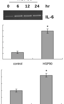
To investigate the effects of HSP90 on IL-6 expression in VSMCs, the level of IL-6 transcript was examined by RT-PCR after exposure of rat AoSMCs to recombinant HSP90. Figure 1A shows that IL-6 transcript was elevated in the presence HSP90. The elevation was observed as early as 6 hr post-treatment and persisted up to 24 hr after treatment. We also examined by ELISA whether VSMCs re- leased IL-6 protein, and found that secreted IL-6 increased profoundly in response to HSP90 (Fig. 1B). The amount of IL-6 in the medium increased from 90 pg/ml to 370 pg/ml in the presence of HSP90. Reporter gene assays were carried out to investigate whether HSP90 increased IL-6 transcript via promoter activation. Rat AoSMCs were transfected with the IL-6 reporter plasmid, and luciferase activity was as- sessed in the presence or absence of HSP90 (Fig. 1C).
Compared with control exposed to BSA, luciferase activity
increased by 2.1-fold in the presence of HSP90, indicating
0 6 12 24 hr IL-6 HSP90
(A)
0 75 150 225 300 375 450
control HSP90
IL -6 ( p g/ m l)
(B) *
0 50 100 150 200 250
control HSP90
% of contro l
*
0 50 100 150 200 250
control HSP90
% of contro l
(C) *
Fig. 1. The effects of HSP90 on IL-6 expression. (A) Up-regu- lation of IL-6 mRNA by HSP90. Rat AoSMCs were treat- ed for the indicated time periods with 500 ng/ml of HSP90, and IL-6 transcripts were amplified using RT-PCR. (B) Enhancement of IL-6 release by HSP90. Rat AoSMCs (1x10
6cells) cultured in growth media were in- cubated in the presence of BSA (control) or HSP90 (500 ng/ml, for 12 hr). IL-6 secreted into the culture medium was measured using ELISA. Data are expressed as means±SDs (n=3 replicates/group). * P<0.01 vs. control.
(C) Promoter activation of IL-6 gene by HSP90. Rat AoSMCs were transfected with the pIL-6-Luc construct and incubated in the presence of BSA (control) or HSP90 (500 ng/ml, for 6 hr). Luciferase activity was determined and normalized to β-galactosidase activity. Induction was calculated relative to the activity of control cells.
Data are expressed as means±SDs (n=3 replicates/
group). * P<0.01 vs. control.
that HSP90 promoted IL-6 transcriptional activity.
Involvement of TLR-4 pathways in IL-6 promoter activation
To investigate roles of TLRs in HSP90-induced promoter activation of IL-6 gene, a reporter gene assay was carried out after transfection of rat AoSMCs with the IL-6 reporter plasmid in combination with dominant-negative forms in TLR pathways. HSP90-induced luciferase activity driven by
IL-6 promoter was affected by co-transfection with domi- nant-negative forms of TLR-4 (Fig. 2A) and myeloid differ- entiation factor 88 (MyD88) (Fig. 2B). Of the two, domi- nant-negative MyD88 profoundly inhibited promoter acti- vation of IL-6 gene. Co-transfection with dominant negative forms of TLR-3 and TIR-domain-containing adapter-induc- ing interferon-β (TRIF), however, did not inhibit HSP90-in- duced IL-6 gene activation. The effects of phytochemicals known to modify TLR-4 signaling on HSP90-induced IL-6 up-regulation were investigated by reporter gene assays (Fig. 3A). Curcumin and resveratrol significantly sup- pressed the HSP90-induced IL-6 promoter activity. The sup- pression was more significant in the presence of curcumin.
Pre-treatment with curcumin suppressed the HSP90-in- duced IL-6 promoter activity approximately to that of control. When the effects of the two phytochemicals on IL-6
0 50 100 150 200 250 300
control pCM V dn-TLR4 dn-TLR3
% of control
*
**
- HSP90 + HSP90
(A)
0 50 100 150 200 250 300
control pCMV dn-MyD88 dn-TRIF
% of control
*
**
- HSP90 + HSP90
(B)
Fig. 2. Involvement of TLR-4 in the activation of IL-6 gene by HSP90 in VSMCs. The IL-6-Luc plasmid was co-trans- fected into rat AoSMCs with the indicated dominant- negative forms of TLRs (A) or with dominant negative forms of TLR-4 adaptor molecules (B). After stimulation with HSP90 (500 ng/ml, for 6 hr), luciferase activity was determined and normalized to β-galactosidase activity.
Induction was calculated relative to the activity of con-
trol cells. Data are expressed as means±SDs (n=3 repli-
cates/group). * P<0.01 vs. control. **P<0.01 vs. pCMV.
0 50 100 150 200 250
control DMSO Cur Res
% of control
*
** **
- H S P 90 + H S P 90
(A)
0 50 100 150 200 250 300 350 400
control DMSO Cur Res
IL -6 ( pg /m l)
*
** **
- HSP90 + H S P90
(B)
Fig. 3. Attenuation of IL-6 up-regulation induced by resveratrol and curcumin. (A) The effects of curcumin and resveratrol on the promoter activation of IL-6 gene. The IL-6-Luc plasmid was transfected into rat AoSMCs and incubated for 2 hr with 10 μM curcumin (Cur) or 25 μM resveratrol (Res). The transfected cells were processed for luciferase and β-galactosidase assays after stimulation with HSP90 (500 ng/ml, for 6 hr). Induction was calculated relative to the activity of the control cells that were incubated without HSP90. Data are expressed as means±SDs (n=3 replicates/ group). *P<0.01 vs. control.
**P<0.05 vs. DMSO. (B) The effects of curcumin and resveratrol on IL-6 release. Rat AoSMCs were stimulated with HSP90 (500 ng/ml, for 12 hr) in the absence or presence of the indicated phytochemicals. IL-6 in the culture medium was measured using ELISA. Data are expressed as means±SDs (n = 3 replicates/group). *P<0.01 vs. control. **P<0.01 vs. DMSO.
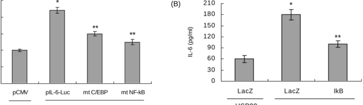
0 50 100 150 200 250
pCMV pIL-6-Luc mt C/EBP mt NF-kB
% of control
*
** **
- HSP90 + HSP90
(A)
0 30 60 90 120 150 180 210
LacZ LacZ IkB
IL-6 (pg/ml)