서 론
제1형 당뇨병(Insulin Dependent Diabetes Mellitus, IDDM) 은 췌장 islet에서 insulin을 분비하는 β세포의 파괴로 인하여 나 타나는 자가면역질환(autoimmune disease)으로1) 생성 초기에 췌 장 islet 주위로 면역세포들이 침습하여 interleukin(IL)-1β와 interferon(IFN)-γ와 같은 싸이토카인(cytokine)을 분비하게 된다
2,3). 분비된 싸이토카인은 췌장 β세포에 작용하여 iNOS(inducible nitric oxide synthase)의 발현을 증가시키고 이 로 인한 산화질소(nitric oxide, NO) 생성의 증가는 췌장 β세포 파괴의 주 매개체로서 알려져 있다3-6).
천화분은 葫蘆科(박과 ; Cucurbitaceae)에 속한 다년생 攀援 性草質藤本인 하늘타리 및 동속 근연식물의 괴근으로 淸熱生津, 淸肺化痰, 消腫排膿의 효능을 지닌 것으로 알려져 있으며7), 실험 적으로 alloxan으로 유발된 당뇨 흰쥐에 대한 천화분 추출물의 항당뇨 효과가 보고되었다8).
이에 저자는 천화분 추출물이 백서 췌장 β-세포인 RINm5F 세포에서 싸이토카인에 의해 유발된 독성에 대한 방어효과를 조 사하였으며 그 기전 조사를 위하여 NF-κB 등을 관찰한 결과 유 의한 변화를 얻었기에 보고하는 바이다.
재료 및 방법
1. 천화분 추출물의 조제
천화분 200 g에 3차 증류수 1.8 L를 환저류 플라스크에 넣고
싸이토카인 유발 췌장 β세포 독성에 대한 천화분 추출물의 방어효과
송미영․김은경․송제호1*
전북대학교 의과대학 생화학교실, 1 : 원광대학교 뷰티디자인학부․생활자원개발연구소
Protective Effect of Radix Trichosanthis Extracts on Cytotoxicity of Pancreatic β-Cells by Cytokines
Mi Young Song, Eun Kyung Kim, Je Ho Song1*
Department of Biochemistry, Medical School and Institute for Medical Sciences, Chonbuk National University, 1 : Division of Beauty Design & Institute for Better Living, Wonkwang University
In this study, the preventive effects of Radix Trichosanthis extracts (RTE) against cytokine-induced β-cell death were assessed. Cytokines generated by immune cells infiltrating pancreatic islets are crucial mediators of β-cell destruction in insulin-dependent diabetes mellitus. The treatment of RIN cells with interleukin-1β (IL-1β) and interferon- γ (IFN-γ) resulted in a reduction of cell viability. RTE protected IL-1β and IFN-γ-mediated viability reduction in a concentration-dependent manner. Incubation with RTE also induced a significant suppression of IL-1β and IFN-γ -induced inducible nitric oxide synthase (iNOS) protein expression. The molecular mechanism by which RTE inhibited iNOS protein expression appeared to involve the inhibition of NF-κB activation. The IL-1β and IFN-γ-stimulated RIN cells showed increases in NF-κB binding activityand IκBα degradation in cytosol compared to unstimulated cells.
However, pretreatment with RTE inhibited cytokines-induced IκBα degradation and NF-κB activation in RINm5F cells.
Furthermore, the protective effects of RTE were verified via protection of impairment in glucose-stimulated insulin secretions in IL-1β and IFN-γ-treated islets.
Key words : Radix Trichosanthis, β-cell, cytokine, NF-κB
* 교신저자 : 송제호, 전북 익산시 신용동 344-2 원광대학교 뷰티디자인학부
․E-mail : [email protected] ․Tel : 063-850-6895
․접수 : 2008/03/19 ․채택 : 2008/04/04
냉각기를 부착하여 3시간 동안 전열기로 끓인 후 3,000 rpm에서 20분간 원심분리하고 상청액을 회전 진공 농축기로 감압농축한 후 동결건조기에서 건조하여 21.2 g의 분말 시료를 얻은 후 -7 0℃에 보관하여 사용하였다.
2. 세포배양
RIN, clone 5F (RINm5F) 세포는 NEDH rat islet cell tumor 에서 유래한 췌장 β-세포계로9) American Type Culture Collection (ATCC)에서 구입하여 사용하였다. 세포는 10% fetal bovine serum, 2 mM glutamine, 10,000 units/㎖ penicillin, 50
㎍/㎖ streptomycin, 그리고 2.5 ㎍/㎖ amphotericin B가 포함된 RPMI 1640 배양액에서 5% CO2, 95% O2, 37℃가 유지되는 배양 기에서 배양하였다.
3. 세포생존율 측정: MTT assay
RINm5F 세포를 96 well 세포배양 용기에 1×10⁴ cells/㎖씩 분주하여 24 시간 세포배양 용기에 부착시키고, 안정화된 RINm5F 세포에 천화분 추출물 등을 48시간 처리하여 0.5 ㎎/㎖
MTT〔3-(4,5-dimethylthiazol -2-yl)-2,5-diphenyltetrazolium bromide〕와 1시간 반응시켰다. 생존 세포가 MTT로부터 생성한 보라색 불용성 formazan은 DMSO로 용해하여 570 nm 파장에서 ELISA reader(Molecular Device, E-max, USA)로 흡광도를 측정 하였다. 측정한 formazan 생성 정도는 대조군 세포와 비교하여 백분율(%)로 표시하였다.
4. Western blotting
RINm5F 세포(3×10⁶ cells/well) 또는 췌장 islet에 IL-1β, IFN-γ 혹은 천화분 추출물을 처리한 후에 포집된 세포는 세포파 쇄용액과 4℃에서 30분 반응시킨 후, 30 ㎍의 단백질을 두 배의 sample buffer(5 mM EDTA, 4% sodium dodesyl sulfate (SDS), 20% glycerol, 200 mM Tris, pH 6.8, 0.06% bromophenol blue) 와 혼합 후, 100℃에서 3분 가열하여 단백질 변성을 유도하고 10% gel에서 sodium dodesyl sulfate polyacrylamide gel electrophoresis (SDS-PAGE)를 시행하였다. 전기영동을 마친 gel 의 단백질은 semi-dry electrotransfer system(0.8 mA/㎠)을 이용 하여 nitrocellulose membrane으로 이동시킨 다음, 5% skim milk와 상온에서 1시간 반응시켜 비특이적인 항체반응을 억제시 켰다. 일차항체(primary antibody)는 TBS-T에 1:1,000으로 희석 하여 nitrocellulose membrane과 상온에서 24시간 반응시키고 TBS-T로 10분 3번 세척한 후, 이차항체(secondary antibody)인 horseradish peroxidase-conjugated IgG를 반응시켰다. 발현된 단백질양은 Chemi-doc image 분석기(Bio-Rad, UK)를 이용하여 확인하였다.
5. NF-κB의 활성측정 (Electrophoretic mobility shift assay: EMSA) 전사인자 활성을 측정키 위해 먼저 한약재 또는 싸이토카인 이 처리된 RINm5F 세포에서 핵 추출물은 Jeong 등의 방법10)으 로 모았다. 세포는 저 삼투압 용해용액(0.2 mM PMSF, 10 ㎍/㎖
aprotinin, 20 μM pepstatin A, 0.1 mM antipapain)으로 10분 얼 음에서 팽창시켜 최종적으로 Nonidet P-40을 0.1%되게 처리한 후 2,500 rpm에서 원심분리 하여 핵단백질만을 모았다. NF-κB의 활성측정은 NF-κB의 consensus binding site을 가진 oligonucleotide probe(5'-CCG GCC GGT TAA CAG AGG GGG CTT TCC GAG-3`)를 10 mM Tis-HCL 용액(pH 8.0, 50 mM NaCl, 10 mM MgCl2, 1 mM DTT 함유)에 희석한 후 85℃
에서 5분 annealing 한 후 100 ng을 Rediprime kit(Amersham, England)를 이용하여 32P를 부착시켰다. 방사선 동위원소가 부착 된 probe는 5-10 ㎍의 핵단백질과 실온에서 30분 반응시킨 후 냉 온실에서 4% polyacrylamide gel에 전기영동 하였다. 이 gel은 건조 후 Ras-3000 Image Analyzer (Fuji Film, Japan)을 이용하여 NF-κB 활성을 측정하였다.
6. Islet 분리
췌장 islet은 Kim 등이 시행한 방법11)에 따라 250 g 내외의 Sprague-Dawley rat에 collagenase perfusion를 시행하여 분리하 였다. 분리한 islet은 2 mM L-glutamine, 10% heat-inactivated fetal calf serum, 100 units/㎖ penicillin, 100 ㎍/㎖
streptomycin이 포함된 RPMI-1640 배지에 24시간 동안 안정화 시킨 후 실험에 사용하였다.
7. Glucose-stimulated insulin secretion (GSIS)
Islets에 싸이토카인 또는 천화분 추출물을 24시간 동안 처리 한 후 3 mM D-glucose가 포함된 Krebs-Ringer bicarbonate buffer (25 mM Hepes, 115 mM NaCl, 24 mM NaHCO3, 5 mM KCl, 1 mM MgCl2, 2.5 mM CaCl2, 0.1% bovine serum albumin) 로 3번 washing 한 후 5.5 mM 또는 20 mM D-glucose에 30분간 노출 시킨 후 분비된 인슐린양을 ELISA (Molecular Device, USA)를 이용하여 측정하였다.
8. 단백질 정량
단백질 정량은 bovine serum albumin을 기준치로 이용한 Bradford의 방법12)에 의거하여 정량 하였다.
9. 통계 처리
실험 결과는 mean±S.E.M으로 표시하였으며 유의성의 검정 은 One-Way Anova test (Microcal Origin; version6.0; Microsoft;
USA)에 의하였으며 p< 0.05인 것만 유의한 것으로 하였다.
결 과
1. 천화분 추출물의 싸이토카인에 의한 세포생존율 감소에 대한 방어효과
RINm5F 세포에 싸이토카인인 IL-1β와 IFN-γ를 병합하여 48시간 동안 처리한 전 3시간 동안 다양한 농도의 천화분 추출물 을 처리한 후 MTT assay를 이용하여 세포생존율을 측정하였다.
그 결과 5.0 ng/㎖ IL-1β와 100 U/㎖ IFN-γ를 처리한 군에서는
세포생존율이 대조군(100%)에 비하여 41.5%로 감소하였으나 0.1
㎎/㎖, 0.2 ㎎/㎖의 천화분 추출물을 전처리한 군의 세포생존율 은 각각 59.7%, 71.2%로 나타나 싸이토카인을 처리한 군에 비하 여 유의한 방어효과를 나타냈다(Fig. 1). 본 실험에 사용한 천화 분 추출물을 단독으로 처리한 군의 세포생존율은 대조군에 비하 여 유의한 변화를 나타내지 않아 세포 독성은 없었다(Fig. 1).
Fig. 1. Protective effects of Radix Trichosanthis extract (RTE) on cytokine-induced decrease of viability in RINm5F cells. Cells (1 x 10⁴) were pretreated with various concentrations of RTE for 3 h, and then treated with IL-1β and IFN-γ for 48 h. It`s viability was determined by MTT as described in
##p<0.01 vs cytokine-treated group.
2. 천화분 추출물의 iNOS 단백질 발현에 대한 효과
RINm5F 세포에 싸이토카인인 IL-1β와 IFN-γ를 병합하여 24시간 동안 처리한 전 3시간 동안 0.1 mg/ml, 0.2 mg/ml 농도 의 천화분 추출물을 처리한 후 산화질소를 생성시키는 유전자로 알려진 inducible nitric oxide synthase (iNOS)의 단백질 발현양을 조사하였다. 그 결과 싸이토카인을 처리한 군의 iNOS 단백질 양 은 증가하였으나 천화분 추출물을 전처리한 군은 싸이토카인에 의한 iNOS 단백질 발현(Fig. 2)의 증가가 유의하게 억제되었다.
Fig. 2. Effects of Radix Trichosanthis extract (RTE) on cytokine-induced increase of iNOS protein expression in RINm5F cells. Cells were pretreated with various concentrations of RAE for 3 h, and then treated with IL-1β and IFN-γ for 24 h. iNOS protein expressions were determined by Western blotting decribed in
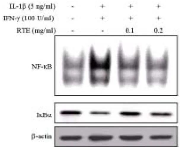
3. 천화분 추출물의 NF-κB 활성에 대한 효과
RINm5F 세포에 싸이토카인인 IL-1β와 IFN-γ를 병합하여 30분 동안 처리한 전 3시간 동안 천화분 추출물을 처리한 후 NF-κB와 이에 상응하는 κB oligomer와의 결합 정도, 세포질의 I κBα의 분해 정도를 조사하였다.
그 결과 싸이토카인을 처리한 군은 NF-κB 활성도가 증가 하였으며 IκBα의 분해 증가에 의한 것임을 확인할 수 있었다. 그 러나 천화분 추출물을 전처리한 군은 싸이토카인을 처리한 군에 비하여 NF-κB의 활성도가 감소하였으며, 이러한 효과는 싸이토 카인에 의한 IκBα의 분해를 억제하여 나타난 것으로 확인되었다 (Fig. 3).
Fig. 3. Effects of Radix Trichosanthis extract (RTE) on cytokine-induced NF-κB activation and IκBα degradation in cytosolic fractions of RINm5F cells. Cells were pretreated with various concentrations of RAE for 3 h, and then treated with IL-1β and IFN-γ for 30 min. NF-κB activities and IκBα in cytosolic fractions expressions were determined by EMSA and Western blotting as decribed in
4. 천화분 추출물의 GSIS에 대한 효과
싸이토카인인 IL-1β와 IFN-γ를 병합하여 24시간 동안 처리 한 전 3시간 동안 0.1, 0.2 ㎎/㎖의 천화분 추출물을 처리한 후 Krebs-Ringer bicarbonate buffer에 20 mM의 포도당을 30분 동 안 노출시켜 완충액내로 분비된 insulin (Glucose-stimulated insulin secretion, GSIS)의 양을 조사하였다.
Fig. 4. Effects of Radix Trichosanthis extract (RTE) on cytokine-induced inhibition of glucose-stimulated insulin secretion in pancreatic islets. Rat islets (10 islets/500 ㎕) were treated with IL-1β and IFN-γ in the presense or absence of RAE for 24 h. Following the incubation period, GSIS was assayed as described in independent experiments. **P<0.01 vs control, #p<0.05, ##p<0.01 vs cytokine-treated group.
그 결과 5.5 mM의 포도당으로 자극한 군의 insulin 분비량
은 유의한 변화가 나타나지 않았다. 20 mM의 포도당으로 자극 한 군에서 싸이토카인을 처리한 군은 insulin 분비량은 10.32 ng/10 islets으로 나타나 대조군(18.62 ng/10 slets)에 대하여 유 의한 감소를 나타냈다. 그러나 천화분 추출물 0.1, 0.2 mg/ml을 전처리 한 군의 insulin 분비량은 각각 14.56, 16.32 ng/10 islets 으로 싸이토카인에 의한 insulin 분비의 감소를 유의하게 억제하 였다(Fig. 4).
고찰 및 결론
본 연구에서는 제1형 당뇨병 in vitro 실험 모델을 이용하여 천화분 추출물의 항당뇨 효과를 실험적으로 규명하고자 하였다.
제1형 당뇨병의 췌장 β세포에 대한 독성 원인은 아직 규명되지 않았으나 지금까지 알려진 원인으로 췌장 β세포 주위로 대식세 포, T림프구 등과 같은 면역세포들이 침습하여 IL-1β, IFN-γ, TNF-α 등과 같은 싸이토카인이 분비되고 이는 췌장 β세포를 자 극하여 산화질소, prostaglandin E2(PGE2)와 같은 물질들이 생산 되어 췌장 β세포가 파괴되는 것으로 알려져 있다3-6). 이러한 싸 이토카인에 의한 췌장 β세포 독성에 대하여 계피13), 사인14), 동과
15), 황련16), 인진17) 등의 방어효과가 보고되었다. Fig. 1에서 나타 났듯이 천화분 추출물을 췌장 β세포인 RINm5F 세포에 3시간 동 안 전처리한 군은 싸이토카인을 처리한 군에 비하여 세포 독성 이 감소하였다. 이는 천화분 추출물은 싸이토카인에 의한 RINm5F 세포의 독성을 방어함을 의미한다. 싸이토카인에 의한 췌장 β세포 독성을 유발하는 물질로 알려진 것 중의 하나가 산 화질소 이다. 제1형 당뇨병 모델에서 산화질소의 중요성은 산화 질소를 생성시키는 유전자로 알려진 inducible nitric oxide synthase(iNOS)의 억제제인 Nw-nitro-L-arginice methylester (L-NAME)와 aminoguanidine이 싸이토카인에 의한 β세포 이상 을 억제하는 보고18-20)에 의해서도 입증되었다. 본 연구에서 천화 분 추출물은 싸이토카인에 의한 iNOS 단백질 단백질 발현의 억 제에 의한 것이었다. 이상의 결과는 천화분 추출물의 싸이토카인 에 의한 췌장 β세포 독성에 대한 방어효과는 산화질소 생성의 억제에 의한 것임을 시사한다.
NF(nuclear factor)-κB는 iNOS 유전자를 조절하는 전사인자 (transcription factor)로서3,4) 불활성화 상태(inactive form)에서는 IκB(inhibitory factor of NF-κB)와 같이 복합체를 이뤄 세포질내 에 존재하다가 외부에서 자극이 가해지면 IκB가 인산화 (phosphorylation)되어 분해(degradation)되고 NF-κB는 핵내로 이동하여 유전자 발현을 조절하게 된다21,22). 이러한 NF-κB 활성 화에 대한 천화분 추출물의 효과를 조사한 결과 싸이토카인에 의한 NF-κB 활성도의 증가는 천화분 추출물에 의해 유의하게 억 제되었다. 이는 싸이토카인에 의한 IκBα 분해의 억제로 인하여 나타났다.
천화분 추출물의 싸이토카인에 의한 췌장 β세포 독성에 대 한 방어 효과는 포도당에 의해 자극되어 insulin를 분비하는 췌 장 β세포의 기능에 있어서도 효과가 있는지 조사하였다. Fig. 4 에 의하면 고농도의 포도당(20 mM) 자극에 의한 insulin 분비는
싸이토카인에 의해 감소하였으나 천화분 추출물을 전처리한 군 은 싸이토카인에 의한 insulin 분비의 감소를 유의하게 억제하였 다. 이러한 결과는 천화분 추출물의 췌장 β세포 독성에 대한 방 어 효과가 β세포의 insulin을 분비하는 기능적인 면에도 나타남 을 시사한다 하겠다.
감사의 글
이 논문은 원광대학교 2007년도 교내연구비 지원에 의해 수 행되었습니다.
참고문헌
1. Bach, J.F. Insulin-dependent diabetes mellitus as a β-cell targeted disease of immunoregulation. Journal of Autoimmunity 8(4):39-63, 1995.
2. Foulis, A.K., Liddle, C.N., Farquharson, M.A., Richmond, J.A., Weir, R.S. The histopathology of the pancreas in type 1 (insulin-dependent) diabetes mellitus: a 25-year review of deaths in patients under 20 years of age in the United Kingdom. Diabetologia 29: 267-274, 1986.
3. Eizirik, D.L., Flodstrom, M., Karlsen, A.E., Welsh, N. The harmony of the spheres: inducible nitric oxide synthase and related genes in pancreatic β cells. Diabetologia 39:
875-890, 1996.
4. Mandrup-Poulsen, T. The role of interleukin-1β in the pathogenesis of IDDM. Diabetologia 39: 1005-1029, 1996.
5. Southern, C., Schulster, D., Green, I.C. Inhibition of insulin secretion by interleukin-1β and tumour necrosis factor-α via an L-arginine-dependent nitric oxide generating mechanism. FEBS Letters 276: 42-44, 1990.
6. Eizirik, D.L., Sandler, S., Welsh, N., Cetkovic-Cvrlje, M., Nieman, A., Geller, D.A., Pipeleers, D.G., Bendtzen, K., Hellerstrom, C. Cytokines suppress human islet function irrespective of their effects on nitric oxide generation.
Journal of Clinical Investigation 93: 1968-1974, 1994.
7. 신민교. 원색 임상본초학, 영림출판사, pp 241-242, 250-252, 283-284, 1983.
8. 노진구, 박정배, 이선동. 천화분이 실험적 당뇨 흰쥐의 췌장 내분비세포에 미치는 영향에 관한 면역세포화학적 연구. 방 제학회지 2(1):97-105, 1991.
9. Xie, Q.W., Cho, H.J., Calaycay, J., Mumford, R.A., Swiderek, K.M., Lee, T.D., Ding, A., Troso, T., Nathan, C. Cloning and characterization of inducible nitric oxide synthase from mouse macrophages. Science 256: 225-228, 1992.
10. Jeong, J.Y., Jue, D.M. Chloroquine inhibits processing of tumor necrosis factor in lipopolysaccharide-stimulated RAW 264.7 macrophages. Journal of Immunology 158:
4901-4907, 1997.
11. Kim, H.R., Rho, H.W., Park, B.H., Park, J.W., Kim, J.S., Kim, U.H., Chung, M.Y. Role of Ca2+ in alloxan-induced pancreatic β-cell damage. Biochimica et biophysica acta 1227: 87-91, 1994.
12. Bradford, M.M. A rapid and sensitive method for the quantitation of microgram quantities of protein utilizing the principle of protein-dye binding. Analasis Biochememistry 72: 248-254, 1976.
13. Kwon, K.B., Kim, E.K., Jeong, E.S., Lee, Y.H., Lee, Y.R., Park, J.W., Ryu, D.G., Park, B.H. Cortex cinnamomi extract prevents streptozotocin- and cytokine-induced β-cell damage by inhibiting NF-κB. World Journal of Gastroenterol 12: 4331-4337, 2006.
14. Kwon, K.B., Kim, J.H., Lee, Y.R., Lee, H.Y., Jeong, Y.J., Rho, H.W., Ryu, D.G., Park, J.W., Park, B.H. Amomum xanthoides extract prevents cytokine-induced cell death of RINm5F cells through the inhibition of nitric oxide formation. Life Sciences 73: 181-191, 2003.
15. Kwon, K.B., Ryu, D.G., Shin, M.K., Shin, B.C., Hwang, W.J., Lee, Y.R., Park, J.W., Park, B.H. Fructus Benincasae Recens extract prevents cytokine-induced nitric oxide formation and cytotoxicity of RINm5F cells. Immunopharmacology &
Immunotoxicology 25: 615-625, 2003b.
16. Kim, E.K., Kwon, K.B., Han, M.J., Song, M.Y., Lee, J.H., Lv, N., Ka, S.O., Yeom, S.R., Kwon, Y.D., Ryu, D.G., Kim, K.S., Park, J.W., Park, B.H. Coptidis rhizoma extract protects
against cytokine-induced viability reduction in pancreatic β -cells through suppression of NF-κB activation.
Experimental and Molecular Medicine 39: 149-159, 2007.
17. Kim, E.K., Kwon, K.B., Han, M.J., Song, M.Y., Lee, J.H., Lv, N., Choi, K.B., Ryu, D.G., Kim, K.S., Park, J.W., Park, B.H.
Inhibitory effect of Artemisia capillaris extract on cytokine-induced nitric oxide formation and cytotoxicity of RINm5F cells. International Journal of Molecular Medicine, 19(3):535-540, 2007.
18. Southern, C., Schulster, D., Green, I.C. Inhibition of insulin secretion by interleukin-1β and tumour necrosis factor-α via an L-arginine-dependent nitric oxide generating mechanism. FEBS Lett 276: 42-44, 1990.
19. Welsh, N., Eizirik, D.L., Bendtzen, K., Sandler, S.
Interleukin-1-induced nitric oxide production in isolated rat pancreatic islets requires gene transcription and may lead to inhibition of the Krebs cycle enzyme aconitase.
Endocrinology 129: 3167-3173, 1991.
20. Eizirik, D.L., Flodstrom, M., Karlsen, A.E., Welsh, N. The harmony of the spheres: inducible nitric oxide synthase and related genes in pancreatic β cells. Diabetologia 39:
875-890, 1996.
21. Baeuerle, P.A., Henkel, T. Function and activation of NF-κB in the immune system. Annual Review of Immunology 12:
141-179, 1994.
22. Baldwin, A.S. Jr. The NF-κB and IκB proteins: new discoveries and insights. Annual Review of Immunology 14: 649-683, 1996.