• 검색 결과가 없습니다.

A Case of Myxoid Nerve Sheath Myxoma of the Lower Lip

N/A
N/A
Protected

Academic year: 2021

Share "A Case of Myxoid Nerve Sheath Myxoma of the Lower Lip"

Copied!
3
0
0

로드 중.... (전체 텍스트 보기)

전체 글

(1)

- 63 -

서 론14

신경초 점액종은 말초 신경의 슈반세포(Schwann cell)에 서 기원한 매우 드문 양성 진피 종양이다.

1)

대개 증상이 없는 평균 1 cm 크기의 구진 또는 결절로 나타나며 안면이 나 상지에 호발하나 신체 어느 부위에서도 발생할 수 있으 며 병리조직학적으로 점액양(myxoid), 세포형(cellular) 그 리고 이 두가지 범주의 특징이 혼재되어 있는 혼합형 (mixed)으로 분류된다.

2)

지금까지 국내 문헌에서는 1990년 김 등이 두피에서 발 생한 신경초 점액종 1예

3)

를 처음 보고한 이후로 몇 예가 보고되었으나, 병변의 위치가 입술인 예는 없었다. 저자들 은 입술에 발생한 점액양 신경초 점액종 1예를 경험하고 문헌 고찰과 함께 보고하는 바이다.

증 례

34세 여자 환자가 내원 1개월 전부터 좌측 아랫입술에

Received : October 1, 2015 / Revised : October 20, 2015 Accepted : October 29, 2015

교신저자 : 최소희, 48972 부산광역시 중구 중구로 121 메리놀병원 이비인후과

전화 : (051) 461-2692·전송 : (051) 462-9419 E-mail : [email protected]

발생하여 서서히 크기가 증가하는 종물을 주소로 내원하 였다. 과거력 및 가족력 상 특이사항은 없었고 평소 기저질 환이나 다른 이비인후과적 이상 소견은 관찰되지 않았다.

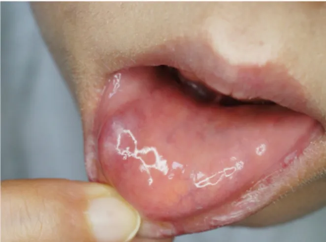
병변은 아랫입술 점막에 비교적 경계가 명확한 1.4 cm × 1.0 cm 크기로 돌출된 선홍색의 종물로 동통이나 소양증 등의 자각 증상은 동반하지 않았다(Fig. 1). 종괴는 국소마 취 하에 절제 생검하였으며 조직검사 상 진피층에 존재하 며 피막은 없으나 주위와 경계가 명확하게 관찰되었다. 종 물은 섬유성 중격에 의해 분리된 다수의 결절로 구성되어 있었고 각 결절은 풍부한 점액선 기질 내에 다양한 세포 밀도의 방추상 혹은 성상 세포들이 나선모양으로 존재하 였으며 염증세포의 침윤은 거의 없었다(Fig. 2).

면역조직화학 검사에서 S-100 단백 염색에 양성을 보여 점액양 신경초 점액종으로 진단하였다(Fig. 3).

병변은 외과적으로 완전 절제하였으며 현재까지 12개월 간 추적 관찰 중으로 임상적으로 재발한 소견은 관찰되지 않고 있다.

고 찰

신경초 점액종은 드물게 발생하는 양성 진피 종양으로 서 1969년 Harkin과 Reed가 처음으로 기술한 후,

2)

neuro- thekeoma,

3,4)

pacinian neurofibroma,

5)

perineurial myxoma,

6)

bizarre cutaneous neurofibroma

7)

등의 다른 명칭으로도 보 대한두경부종양학회지

제 31 권 제 2 호 2015

아랫입술에 발생한 점액양 신경초 점액종 1예

메리놀병원 이비인후과

박태정·김보영·최소희

= Abstract =

A Case of Myxoid Nerve Sheath Myxoma of the Lower Lip

Taejung Park, MD, Boyoung Kim, MD, PhD, Sohee Choi, MD, PhD Department of Otolaryngology, Maryknoll Medical Center, Busan, Korea

Nerve sheath myxoma is a benign tumor of the peripheral nerves that rarely occurs in the lip area. Among the few reported cases, no lesion has previously been reported on the lip in Korea. We report a case of nerve sheath myxoma occurring on the lip of a 34 year-old woman with a brief review of the literature.

KEY WORDS : Nerve sheath myxomaㆍLip.

(2)

- 64 -

고되고 있다. 이는 신경초 점액종의 발생이 말초 신경의 슈반세포인지 혹은 신경주위 세포 분화(perineural differ- entiation)에서 기원한 것인지에 대해서 면역조직학적 검사 나 전자현미경적 구조 모두 확실하게 밝히지 못하고 있고 보고가 드물기 때문이다.

8)

신경초 점액종은 병리조직학적으로 성숙한 점액형과 미 성숙한 세포형을 양단으로 하는 형태학적 스펙트럼을 가 지는데 세포 충실도, 점액 함유 정도 및 성장 양상에 따라 점액양, 세포형, 혼합형의 3가지 유형으로 구분한다.

9)

점액양 신경초 점액종은 주위 조직과 경계가 명확하며 방추형 세포와 점액으로 구성된 여러 개의 소엽으로 구성 된 종양으로 세포간질 내에 점액이 풍부한 반면, 세포형 신경초 점액종은 경계가 불분명하며 원형의 핵과 호산성 의 세포질을 갖는 상피양 세포로 이루어진 다수의 소엽이 관찰되며 세포의 기질 내에 점액 성분은 거의 없다.

2,9,10)

혼합형 신경초 점액종은 점액양 신경초 점액종과 유사한 조직 소견을 나타내지만 피낭의 형성이 뚜렷하지 않고 국 소적으로 세포가 밀집된 부분이 있는 점에서 다르며 세포 형 신경초 점액종과는 점액질이 풍부하고 방추형 세포가 우세한 것에서 차이를 보인다.

2,8)

Vimentin에 대한 면역조직화학 염색시 모든 아형에서 미만성으로 양성반응을 보이나 점액양 신경초 점액종은 S-100 단백, 교원섬유 4형과 epithelial membrane anti- gen(EMA)에 양성을 보이는 반면,

11)

세포형 신경초 점액종 인 경우 S-100 단백과 EMA에 음성을 보이고 평활근 특이 액틴(smooth muscle actin)에 대해서는 양성을 보인다.

12)

본 증례에서는 조직학적으로 방추형 혹은 성상의 세포 들로 구성되며 점액이 풍부하고 섬유 조직에 의해 주위와 경계가 명확한 미세결절들이 관찰되었고 면역조직화학 검 사에서 S-100 단백에 양성을 보여 점액양 신경초 점액종으 로 진단할 수 있었다.

신경초 점액종은 주로 젊은 연령층에 발생하고 여성에 서 남성에 비해 2배 정도 많은 발생을 보인다고 알려져 있고, 대부분 1 cm 미만의 증상이 없는 종양으로 안면과 상지에 호발하나 신체 어느 부위에서도 발생할 수 있다.

2)

하지만 원격 전이는 보고된 바 없다.

6)

구강의 신경초 점액 종은 주로 중장년층에 발생하고 피부의 신경초 점액종과

Fig. 1. Solitary and pinkish colored nodule on the lower lip.

A

B

Fig. 2. The tumors were composed of well defined encapsu- lated lobules (H&E stain, ×40 )(A). The lobules have prominent myxoid stroma seperated by thin fibrous septa in dermal layer (H&E stain, x200)(B).

Fig. 3. Tumor cells were strong positive for S-100 protein(x 400).

(3)

- 65 -

마찬가지로 여성에서 많은 발생을 보이며 정상 색상의 탄 력 있는 결절 형태로 대개 서서히 자라며 동통이 없다.

13,14)

신경초 점액종은 대부분 양성 경과를 취하며 치료는 외 과적으로 절제하는 것이고 불완전하게 절제할 경우에는 재발하므로 완전히 절제해야 한다.

14-16)

저자들은 좌측 아랫입술에 동통을 동반하지 않는 결절 로 임상적으로 점액낭종이나 점액종, 신경섬유종과 유사 하나 조직학적으로 전형적인 점액양 신경초 점액종 1예를 치험하였기에 문헌고찰과 함께 보고하는 바이다.

중심 단어:신경초 점액종ㆍ입술.

References

1) Harkin JC, Reed RJ. Tumor of the peripheral nervous system.

In: Atlas of tumor Pathology. 2nd Series. Washington DC:

Armed Forces Institute of Pathology; 1969. p.60-64.

2) Prieto VG, Reed RJ. Tumors of neural tissue. In: Elder DE, Murphy GF, Elenitsas R, et al, eds. Lever’s Histopathology of the skin, 11th ed. Philadelphia: Lippincott Williams & Wilkins;

2014. p.1369-1417.

3) Kim YC, Chun SI, Lee JB. Neurothekeoma. Ann Dermatol 1990;2:117-120.

4) Gallager RL, Helwig EB. Neurothekeoma--a benign cutaneous tumor of neural origin. Am J Clin Pathol. 1980 ;74(6):759-764.

5) MacLennan SE, Melín-Aldana H, Yakuboff KP. Pacinian neu- rofibroma of the hand: a case report and literature review. J Hand Surg Am. 1999;24(2):413-416.

6) Pulitzer DR, Reed RJ. Nerve-sheath myxoma (perineurial myx- oma). Am J Dermatopathol. 1985;7(5):409-421.

7) King DT, Barr RJ. Bizarre cutaneous neurofibromas. J Cutan Pathol. 1980;7(1):21-31.

8) Barnhill RL, Dickersin GR, Nickeleit V, Bhan AK, Muhlbauer JE, Phillips ME, et al. Studies on the cellular origin of neuro- thekeoma: clinical, light microscopic, immunohistochemical, and ultrastructural observations. J Am Acad Dermatol. 1991;25 (1 Pt 1):80-88.

9) Müller CS, Tilgen W, Kutzner H, Pföhler C. Recurring mixed-type neurothekeoma of the face. Dermatoendocrinol.

2009;1(4):220-222.

10) Argenyi ZB, LeBoit PE, Santa Cruz D, Swanson PE, Kutzner H.

Nerve sheath myxoma (neurothekeoma) of the skin: light micro- scopic and immunohistochemical reappraisal of the cellular variant. J Cutan Pathol. 1993;20(4):294-303.

11) Sheth S, Li X, Binder S, Dry SM. Differential gene expression profiles of neurothekeomas and nerve sheath myxomas by mi- croarray analysis. Mod Pathol. 2011;24:343

354.

12) Hornick JL, Fletcher CD. Cellular neurothekeoma: detailed characterization in a series of 133 cases. Am J Surg Pathol 2007;31:329-340.

13) Rozza-de-Menezes RE, Andrade RM, Israel MS, Gonçalves Cunha KS. Intraoral nerve sheath myxoma: case report and sys- tematic review of the literature. Head Neck. 2013;35(12):E397- E404.

14) Fetsch JF, Laskin WB, Miettinen M. Nerve sheath myxoma: a clinicopathologic and immunohistochemical analysis of 57 morphologically distinctive, S-100 protein- and GFAP-positive, myxoid peripheral nerve sheath tumors with a predilection for the extremities and a high local recurrence rate. Am J Surg Pathol. 2005;29(12):1615-1624.

15) Strumìa R, Lombardi AR, Cavazzini L. S-100 negative myxoid neurothekeoma. Am J Dermatopathol. 2001;23(1):82-83.

16) Prado JD, Andrade RG, Silva–Sousa YT, Andrade MF, Soares FA, Perez DE. Nerve sheath myxoma of the gingiva: report of a rare case and review of the literature. J Periodontol 2007;78:

1639

1643.

수치

Fig. 2. The tumors were composed of well defined encapsu- encapsu-lated lobules (H&E stain, ×40 )(A)

참조

관련 문서

Key words: cecal mass, dog, gastrointestinal mass, immunohistochemistry, malignant peripheral nerve sheath tumor.... Materials and

구순 결손부위의 성공적인 재건을 위해 적절한 종양학적 원칙의 준수, 예측할 수 있는 기능적인 결과, 환자의 심미적 인 기대에 대한 인정이 요구된다.

We present here a rare location for a soft tissue chondroma that originated from the dura mater in the lumbar spinal canal, and it mimicked a nerve sheath tumor.. We present here

In our case, US showed a spindle-shaped mass with a long distal tubular struc- ture, and we initially misdiagnosed this lesion as a pe- ripheral nerve sheath tumor due to

키는 등의 기계적 잇점을 부여하는 기능을 하며, 1) 건 조 직과 별도로 관절 내에 존재하는 부골을 소골(ossicle) 이라 구별한다. 족무지 지관절 부골은 지관절의 족저부

본 증례에서도 팽창된 정중 신경 내에 지방 조직의 침윤이 있는 섬유 신경속이 관 상면과 시상면에서는 특징적인 스파게티 양상으로 축 상 영상에서는 동축

대상포진은 수두 대상포진 바이러스(Varicella Zoster Virus, VZV)가 후근신경절(dorsal root ganglia), 삼차신경절(trigeminal ganglia) 또는 무릎신경절