Archives of Craniofacial Surgery
www.kcpca.or.kr ISSN 2287-1152
72 Copyright © 2012 The Korean Cleft Palate-Craniofacial Association
This is an Open Access article distributed under the terms of the Creative Commons Attribution Non-Commercial License (http://creativecommons.org/
licenses/by-nc/3.0/) which permits unrestricted non-commercial use, distribution, and reproduction in any medium, provided the original work is properly cited.
하구순부에 발생한 피부섬유종의 치험례
여관구·이지환·장충현
성균관대학교 의과대학 강북삼성병원 성형외과학교실
Purpose: Dermatofibroma is a common benign dermal tumor characterized by a proliferation of fibroblast-like spindle cells.
It is commonly localized on the skin of extremities and presents as a slow growing solitary nodule. To our knowledge, the occurrence of dermatofibroma in the oral cavity is rare. Herein, we report a rare case of dermatofibroma on the lower lip.
Methods: A 60-year-old woman presented with a slow growing mass that measured 1 × 0.8 cm in diameter on the lower lip. The mass was surgically excised with clear margins.
Results: Histologically, the mass was characterized by a nodular tumor composed of collagen bundles, fibroblasts, and histiocytes, which were findings consistent with dermatofibroma. The postoperative course was uneventful without any complications.
Conclusion: When evaluating nodular tumors of the oral area, dermatofibroma should be considered in the differential diagnosis.
Keywords: Dermatofibroma, Lower lip
Dermatofibroma of the Lower Lip: A Case Report
Kwan Koo Yeo, Ji Hwan Lee, Choong Hyun Chang
Department of Plastic and Reconstructive Surgery, Kangbuk Samsung Hospital, Sungkyunkwan University School of Medicine, Seoul, Korea
서 론
피부섬유종(dermatofibroma)은 적색 또는 적갈색을 띠 는 비교적 흔한 양성 종양으로, 진피 내에 섬유모세포, 조직 구, 혈관이 증식된 소견을 특징으로 한다.1 1894년 Unna는 단순 섬유종으로 기술하였고, 1973년 Mihatsch–Konz 등2이 처음 피부섬유종이라는 용어로 표현하였다. 피부섬유종은 대개 1 cm 미만 크기의 구진이나 결절의 형태로 나타난다.
호발부위로는 하지에 가장 많이 발생하고 상지, 배, 어깨 및
등에도 발생하는 것으로 알려져 있지만, 안면부의 구순부 에 발생하는 것은 매우 드물다.1 저자들은 성인 여성의 하 구순부에 발생한 결절성 피부 병변에 대하여 절제 및 조직 검사를 통하여 피부섬유종으로 진단하였고, 발생 부위가 매우 드문 경우라고 생각되어 문헌고찰과 함께 보고하고자 한다.
증 례
60세 여자 환자가 4–5개월 전부터 생긴 하구순부의 결절 성 피부 병변을 주소로 내원하였다. 과거력 및 가족력에 특 이사항은 없었으며, 외상의 병력도 없었다. 이학적 검사상 하구순부의 내측 점막 부위에 주위와 경계가 비교적 명확 하고 돌출된 1 × 0.8 cm 크기의 단단한 결절형태의 종괴가 관찰되었으며, 압통이나 열감, 다른 부위의 림프절 종대 및
Arch Craniofac Surg Vol.13 No.1, 72-75 http://dx.doi.org/10.7181/acfs.2012.13.1.72
Correspondence: Choong Hyun Chang
Department of Plastic and Reconstructive Surgery, Kangbuk Samsung Hospital, Sungkyunkwan University School of Medicine, 29 Saemunan-ro, Jongno-gu, Seoul 110-746, Korea
Tel: +82-2-2001-2178 / Fax: +82-2-2001-2177 / E-mail: [email protected] Received February 6, 2012 / Revised March 9, 2012 / March 16, 2012 Accepted March 19, 2012
Case Report
73
www.kcpca.or.kr
혈액 검사상의 이상 소견은 보이지 않았다(Fig. 1). 국소마 취 후 종양을 일부 정상 피부 및 주위 조직을 포함하여 완 전 절제하고 vicryl 5–0를 이용하여 일차봉합술을 시행하 였다. 절제한 종양은 조직 검사를 시행하였다.
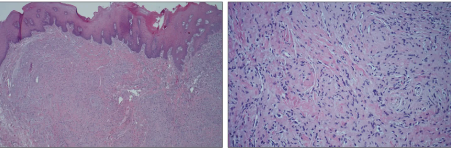
Hematoxylin and eosin 염색을 이용한 조직 검사 소견상 표피는 과각화증, 극세포증과 함께 능선의 연장 소견을 보 였다. 또한 진피는 불규칙한 모양의 방추형세포가 소용돌 이치듯이 증식하는 소견을 보였고 주변부에서는 교원섬유 를 에워싸는 듯한 피부섬유종의 특징적인 소견도 보였다.
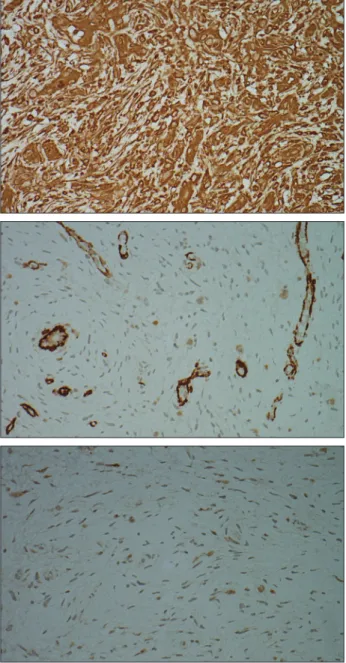
악성화를 의심할 만한 조직괴사나 핵의 다형성 및 세포이 형성은 관찰되지 않았다(Fig. 2). 면역조직화학염색상 종양 세포는 vimentin에 양성 소견을 보였고, smooth muscle ac- tin (SMA) 및 S–100 단백에는 음성 소견을 보였다(Fig. 3). 이
상의 조직 소견으로 피부섬유종으로 진단하였으며, 환자 는 종양의 완전 절제술 후 재발 및 합병증 없이 경과관찰 중 이다.
고 찰
피부섬유종은 임상적으로 비교적 흔하게 관찰되는 진피 의 섬유모세포와 조직구가 증식된 양성 종양으로, 1894년 Unna에 의하여 단순 섬유종(fibroma simplex)으로 불린 이 후, 조직구종(histiocytoma), 피부결절(nodulous cutaneous), 섬유성 조직구종(fibrous histiocytoma), 표피하 결절성 섬 유증(nodular subepidermal fibrosis) 등의 다양한 용어로 기 술되어 왔다. 1973년 Mihatsch–Konz 등2이 미세구조학적 연 구를 통하여 본 질환은 섬유모세포가 때때로 식세포성 작 용을 가져 생겨난다고 주장하였으며, 이때부터 피부섬유 종이라는 명칭으로 표현되었다. 피부섬유종의 기원에 대하 여서는 섬유모세포 또는 조직구에서 발생한 것이라는 주 장들이 있으나, 현재는 다분화능력을 가진 간엽세포에서 발생한 것으로 생각되고 있다.1 병변은 대개 1 cm 미만의 크 기를 가지며 대부분 단일병변으로 나타나고, 주로 젊은 여 성에서 호발하는 것으로 알려져있다. 1970년 Niemi1에 의하 면 하지에서 가장 호발하는 것으로 보고되었고, 그 외에 상 지, 배, 어깨, 등, 손, 목, 엉덩이 순으로 많이 발생하는 것으 로 관찰되었다. 본 증례와 같이 구순부에 발생한 경우는 Pubmed 검색 결과 지금까지 보고된 예가 7건에 불과할 정
Fig. 2. Microscopic examination. (Left) Histological findings showed hyperkeratosis, acanthosis, and elongation of rete ridges in the epidermis and storiform patterned spindle-shaped, cellular infiltration in the dermis (H&E, ×40). (Right) The biopsy specimen was found to be composed of a proliferation of fibroblast-like spindle cells and collagen bundles in a storiform arrangement (H&E, ×200).
Fig. 1. Preoperative appearance of the lesion. An approximately 1 × 0.8 cm sized, firm, non-tender, nodule in her left inner mucosal area on the lower lip with overlying mucosa.
Kwan Koo Yeo, et al. Dermatofibroma of the lower lip
Archives of Craniofacial Surgery Vol. 13, No. 1, 2012
www.kcpca.or.kr
74
도로 매우 드물다(TableⅠ).3–8 구순부에 발생한 피부섬유종 은 임상적으로 결절성 근막염, 외상성 섬유종, 지방종, 신경 섬유종, 신경초종 및 타액선 종양 등과의 감별이 필요하 다.6–8 위와 같은 질환들은 임상적으로는 정확히 감별이 어 렵고, 병리조직학적인 검사에 의해서만 정확한 진단이 가 능하다.
병리조직학적 분류에 따르면 피부섬유종은 소용돌이 모양을 이루는 섬유모세포와 교원섬유가 특징인 섬유형, 지질 또는 혈색소를 탐식한 조직구세포의 형태로 나타나 는 세포형, 이 두가지 유형이 동시에 나타나는 혼합형, 혈관 증식이 많은 혈관종형 등으로 분류되고 있다. 이 중 섬유형 이 가장 흔한 아형으로 보고되고 있고, 본 증례 또한 섬유형 의 양상을 보였다. 피부섬유종의 아형 중 세포형 피부섬유 종은 일반적인 섬유형 피부섬유종에 비하여 세포충실도 가 높고, 다형태(polymorphic form)가 드물며, 세포 사이에 교원섬유가 관찰되지 않는 등 저배율상에서 비교적 단형성 (monomorphic) 형태로 관찰이 되며, 임상적으로 다른 아형 에 비해 재발을 잘하기 때문에 융기성 피부섬유육종과 같 은 악성 종양과의 감별이 필수적이다. 피부섬유종의 특징 적인 병리조직 소견으로 표피의 다양한 변화를 들 수 있는 데, 특히 표피의 과형성과 이로 인한 기저세포암양 변화가 특이하다. 과반수 이상의 환자에서 극세포증, 과각화증, 기 저층의 과색소침착을 보이며, 극세포증은 표피능의 비후로 인해 다양한 변화를 보이기도 한다.1 이러한 표피 과형성의 원인은 확실치 않으나 진피 내 상당량 존재하는 기질과 미 성숙 교원질의 작용에 의해 기저세포가 증식되어 다양한 표피 소견을 보일 것으로 설명하고 있다.
면역조직화학적 검사에서는 vimentin, factor XIIIa에는 양성, CD–68, SMA에는 일부 양성 소견을 보이고, CD–34, S–100 protein, cytokeratin, epithelial membrane antigen에는
Fig. 3. Immunohistochemical examination. (Above) Strong immu-
nohistochemical expression of vimentin in neoplastic cells (×400).
(Center) Tumor cells were negative for smooth muscle actin (×400).
(Below) Tumor cells were negative for S-100 protein (× 400).
Table I. Summary of Published Data on Patients with Dermatofibroma of the Lip
Author Year Sex/Age (yr) Location Size (mm) Treatment method Recurrence
Hillis and Beasley
31975 M/52 Lower lip 5×10 Excision -
Gray et al.
41992 M/45 Lower lip 20-30 Excision -
Gray et al.
41992 F/45 Lower lip 20 Excision -
MacLeod and Jones
51992 F/22 Upper lip Not stated Excision -
Yamada et al.
62002 M/6 mo Upper lip 15 Excision -
Lee et al.
72010 F/ 41 Upper lip 10×10 Excision -
Migliario et al.
82010 F/ 61 Lower lip 12×9 Excision -
75
www.kcpca.or.kr
음성 소견을 보인다.7
피부섬유종은 수십년 지속되나 드물게는 자연 소실되기 도 한다. 치료는 다른 종양과 감별진단의 필요성이나 외모 개선을 위해 절제술과 생검이 추천된다. 악성화는 거의 없 으나, 불완전한 절제 시에는 국소재발을 할 수 있으며, 보통 절제술 후 약 1% 미만에서 재발하는 것으로 알려져 있다.
본 증례에서는 절제술 후 4년간의 추적관찰 기간 중 재발 없이 양호한 경과를 보였다.