Introduction
The newly published 2015 World Health Organization (WHO) classification of lung tumors reclassified squamous cell carcinomas (SCC) into keratinizing, nonkeratinizing, and basaloid subtypes
1, similar to the 2005 Head and Neck WHO Classification of nasopharyngeal carcinomas. Typically, keratinization implies lung SCC, although in the absence of unequivocal keratinization, immunohistochemistry is usually required to distinguish nonkeratinizing SCC from adenocarci- noma. Similar to the head and neck cancer classification, the new lung SCC classification was upgraded to address these pathological issues
2. However, the prognostic or other clini- cal significance of this new lung SCC subtype classification is unclear, although recent studies of head and neck cancer have
Keratinization of Lung Squamous Cell
Carcinoma Is Associated with Poor Clinical Outcome
Hye Jung Park, M.D., Ph.D.
1, Yoon-Jin Cha, M.D., Ph.D.
2, Seong Han Kim, M.D.
1, Arum Kim, M.S.
1, Eun Young Kim, M.D., Ph.D.
1and Yoon Soo Chang, M.D., Ph.D.
1Departments of
1Internal Medicine and
2Pathology, Yonsei University College of Medicine, Seoul, Korea
Background: Although the World Health Organization (WHO) classification of lung squamous cell carcinoma (SCC) was revised in 2015, its clinical implications for lung SCC subsets remain unclear. We investigated whether the morphologic characteristics of lung SCC, including keratinization, were associated with clinical parameters and clinical outcome of patients.
Methods: A total of 81 patients who underwent curative surgical resection of diagnosed lung SCC, were enrolled in this study. Attributes such as keratinization, tumor budding, single cell invasion, and nuclear size within the tumor, as well as immunohistochemistry of Bcl-xL and pS6 expressions, were evaluated.
Results: The keratinizing and nonkeratinizing subtypes did not differ with respect to age, sex, TNM stage, and morphologic parameters such as nuclear diameter, tumor budding, and single cell invasion at the tumor edge. Most patients with the keratinizing subtype (98.0%) had a history of smoking, whereas the nonkeratinizing group had a relatively higher proportion of never-smokers relative to the keratinizing group (24.0% vs. 2.0%; p=0.008, chi-square test).
Expression of pS6 (a surrogate marker of mammalian target of rapamycin complex 1 [mTORC1] signaling that regulates keratinocyte differentiation), and Bcl-xL (a key anti-apoptotic molecule that may inhibit keratinization), did not correlate significantly with the presence of keratinization. Patients with the keratinizing subtype had a significantly shorter overall survival (85.2 months vs. 135.7 months, p=0.010, log-rank test), and a multivariate analysis showed that keratinization was an independent, poor prognostic factor (hazard ratio, 2.389; 95% confidence interval, 1.090–5.233; p=0.030).
Conclusion: In lung SCC, keratinization is associated with a poor prognosis, and might be associated with smoking.
Keywords: Lung; Carcinoma, Squamous Cell; Bcl-X Protein
Address for correspondence: Eun Young Kim, M.D., Ph.D.
Department of Internal Medicine, Yonsei University College of Medicine, 50-1 Yonsei-ro, Seodaemun-gu, Seoul 03722, Korea
Phone: 82-2-2228-2267, Fax: 82-2-393-6884 E-mail: [email protected]
Received: Sep. 7, 2016 Revised: Jan. 10, 2017 Accepted: Jan. 16, 2017
cc It is identical to the Creative Commons Attribution Non-Commercial License (http://creativecommons.org/licenses/by-nc/4.0/).
Copyright © 2017
The Korean Academy of Tuberculosis and Respiratory Diseases.
All rights reserved.
revealed that compared to the non-keratinizing subtype, the keratinizing subtype is associated with a poorer prognosis
3,4. In contrast, studies of the relationship between the keratiniz- ing subtype and prognosis in lung SCC are rare, and one such study reported that the presence of keratinization was not a significant prognostic factor
5.
According to previous literature, keratinization is accom- panied by apoptosis and is ultimately associated with tumor progression in patients with esophageal SCC
6. The expression of B-cell lymphoma (Bcl)-xL, an oncoprotein involved in lung SCC tumorigenesis, is known to correlate with apoptosis
7-9; furthermore, deactivation of the tumorigenic mammalian target of rapamycin (mTOR) signaling pathway, which plays a key role in regulating cellular proliferation, survival, and angio- genesis, also affects apoptosis in lung cancers
10-13. These key apoptotic factors might correlate with keratinization and thus might affect prognosis. However, potential direct correlations of keratinization with the mTOR pathway and Bcl-xL expres- sion have not been studied in lung SCC.
In the present study, we aimed to characterize the keratin- izing and nonkeratinizing subtypes of lung SCC and confirm the effects of keratinization on overall survival (OS). In addi- tion, we aimed to investigate correlations of keratinization with mTOR pathway activation and Bcl-xL expression.
Materials and Methods
1. Patients
Eighty-one patients who underwent surgical treatment of lung SCC between 1993 and 2016 were randomly selected from the Severance Hospital (Seoul, Korea) lung cancer data- base. To obtain clinical data, we retrospectively reviewed the patients’ electronic medical records. Tumor stage was re-eval- uated according to the seventh edition of the American Joint Committee on Cancer TNM Staging Manual
14. This study was approved by the Institutional Review Board (IRB) of Sever- ance Hospital (No. 3-2016-0019).
2. Histologic evaluation
All tissue slides were subjected to hematoxylin and eo- sin staining and evaluated for the presence of the following recently identified poor prognostic factors: tumor budding, single cell invasion, and large nuclei
5. Initially, the entire tumor set was scanned at ×100 magnification and subjected to a detailed review. First, tumor budding, or the presence of small tumor nests comprising fewer than five tumor cells, were counted in 10 high-power fields (HPFs) at ×200 magnification.
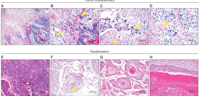
We defined a high grade of tumor budding as more than eight tumor budding events per 10 HPFs (Figure 1A, B). Single cell
Figure 1. Histologic parameters and patterns of keratinization applied in this study. Tumor budding is observed along tumor edge (A); this is defined as the presence of structures comprising fewer than five tumor cells (arrowheads) at higher magnification (B). Single cell invasion (C) and large nuclei (cancer cell nucleus >4 times than that of a small lymphocyte) (D), nonkeratinization (entire tumor area, <5% keratinization) (E), cytoplasmic keratinization (F), keratin pearl (G), and layered keratinization (H) (H&E stain; A, ×40; B–D, ×400; E–H, ×200).
Tumor characteristics
Keratinization
A B C D
E F G H
50 m 50 m
40 m 200 m
100 m 100 m
100 m 100 m
invasion was also evaluated at ×200 magnification (Figure 1C), and nuclear features were assessed at ×400 magnification. We calculated the average nuclear diameter of at least 100 tumor cells in at least three HPFs per sample. A large nucleus was defined as a diameter greater than that of four small lympho- cytes (Figure 1D).
The keratinization grade was determined, and tumors were classified accordingly as the keratinizing subtype, defined as a keratinizing pattern comprising ≥5% of the entire tumor, or the nonkeratinizing subtype, defined as a keratinizing pattern comprising <5% of the tumor (Figure 1E). Subsequently, the keratinization grades were refined to nonkeratinization (<5%) and low (5%–20%), moderate (20%–50%), or severe (>50% of the entire tumor) keratinization, similar to the classification used for head and neck cancers
15. The keratinization patterns included cytoplasmic keratinization (Figure 1F), keratin pearl (Figure 1G), and layered keratinization (Figure 1H). All histo- logic parameters were evaluated independently by a patholo- gist (Y.J.C.).
3. Immunohistochemistry
To evaluate mTOR complex 1 (mTORC1) and Bcl-xL ex- pression, immunohistochemistry (IHC) staining for pS6 and Bcl-xL proteins was performed using an EnVision+ system (Dako Corp., Carpinteria, CA, USA) according to the manu- facturer’s instructions. Briefly, sections were deparaffinized, rehydrated, and subjected to antigen retrieval via microwave heating for 10 minutes. Sections were then immersed in a H
2O
2–phosphate-buffered saline solution prior to overnight incubation with primary anti-pS6 (1:400, Cell Signaling Tech- nology, Danvers, MA, USA) or Bcl-xL antibodies (1:600, Cell Signaling Technology). Subsequently, the sections were in- cubated with a peroxidase-labeled polymer for 1 hour at 4°C.
IHC staining was scored independently at ×200 magnification by Y.J.C. and S.H.K., who were blinded to the clinicopathologi- cal data. A semiquantitative evaluation of pS6 and Bcl-xL was performed according to the method described in a previous study
16. The staining intensity was classified as 0 (negative), 1 (trace), 2 (moderate), or 3 (strong), and the frequency of positive cells was classified as 0 (<10%), 1 (10%–50%), 2 (50%–
80%), or 3 (>80%). The expression score was determined as the product of the staining intensity and frequency of positive cells.
4. Statistical analysis
Associations between categorical variables were analyzed using the chi-square test; this test was also used to assess the linear correlation trend of the keratinization grade with IHC scoring. Disease-free survival (DFS) and OS were estimated using the Kaplan-Meier method, and associations between factors and survival outcomes (OS) were analyzed using the
log-rank test. The Cox proportional hazards model was used for multivariate analyses. SPSS version 18.0 (SPSS Inc., Chi- cago, IL, USA) was used for all statistical analyses, and signifi- cance was defined as a p-value of <0.05.
Results
1. Smoking correlates with the keratinization of lung SCC
Among the 81 patients enrolled in this study, 56 (69.1%) and 25 (30.9%) were classified as having the keratinizing and nonkeratinizing subtypes of lung SCC, respectively, as con- firmed by a pathologic review of tumor slides. The average age of the subjects was 62.8±8.7 years (mean±standard deviation);
77 (95.1%) were male, and the majority had stage T2 or T3 dis- ease (84.0%). Among patients with the keratinizing subtype, 21 (37.5%), 19 (33.9%), and 16 patients (28.6%) were classified as having low, moderate, and severe keratinization, respec- tively. The keratinizing and nonkeratinizing groups did not dif- fer significantly with respect to the distributions of age, sex, T-, N-, pStage, type of surgery, and adjuvant chemotherapy. How- ever, most patients with the keratinizing subtype (98.0%) had a history of smoking, whereas never smokers (24.0%) were significantly more prevalent in the nonkeratinizing subtype group (Pearson’s R=0.262, p=0.008, chi-square test). In a sub- group analysis of smokers, keratinization was more frequently observed among heavy smokers (i.e., ≥30 pack-year history of smoking) (p=0.012, chi-square test). This finding suggests that keratinization of lung SCC might be related to smoking (Table 1). Other morphologic parameters, such as nuclear diameter, tumor budding, and single cell invasion at the tumor edge, were not found to correlate with the keratinization of lung SCC.
2. mTORC1 and Bcl-xL expression did not correlate with lung SCC keratinization
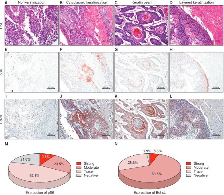
Because mTOR signaling is known to regulate keratino- cyte differentiation, we reviewed the relationship between keratinization and mTORC1 in 51 available lung SCC tissues, using pS6 expression as a surrogate marker. Among these 51 cases, five (9.8%), 12 (23.5%), and 23 (45.1%) exhibited strong, moderate, and trace expression, respectively. An additional 11 cases (21.6%) were negative for pS6. Viable tumors expressed pS6 beneath the shedding area, where keratinization initiates, regardless of keratinization subtype (Figure 2). However, the correlation between keratinization grade and pS6 expression failed to reach statistical significance (p=0.153, chi-square test).
In tumors, keratinization is generated by the epithelial layer
and is subsequently shed from the tumor margin by apopto-
Table 1. Demographics and pathologic characteristics of patients with and without keratinization
Variable Nonkeratinization (n=25) Keratinization (n=56) Total (n=81) p-value
Age, yr 0.807
≤65 16 (64.0) 33 (58.9) 49 (60.5)
>65 9 (36.0) 23 (41.1) 32 (39.5)
Sex 0.583
Male 23 (92.0) 54 (96.4) 77 (95.1)
Female 2 (8.0) 2 (3.6) 4 (4.9)
Smoking status 0.008
Never smoker 6 (24.0) 1 (2.0) 7 (8.6)
Former smoker 7 (28.0) 18 (35.3) 25 (30.9)
Current smoker 12 (48.0) 32 (62.7) 44 (54.3)
Unknown - - 5 (6.2)
Smoking pack-years 0.012
<30 12 (48.0) 9 (17.6) 21 (27.6)
≥30 13 (52.0) 42 (82.4) 55 (72.4)
T classification 0.605
T1 4 (16.0) 4 (7.1) 8 (9.9)
T2 15 (60.0) 39 (69.6) 54 (66.7)
T3 4 (16.0) 10 (17.9) 14 (17.3)
T4 2 (8.0) 3 (5.4) 5 (6.2)
N classification 0.811
N0 16 (64.0) 35 (62.5) 51 (63.0)
N1 5 (20.0) 9 (16.1) 14 (17.3)
N2 4 (16.0) 12 (21.4) 16 (19.8)
pStage 0.938
pStage I 9 (36.0) 22 (39.3) 31 (38.3)
pStage II 9 (36.0) 18 (32.1) 27 (33.3)
pStage III 7 (28.0) 16 (28.6) 23 (28.4)
Type of surgery 0.806
Lobectomy 16 (64.0) 32 (58.2) 48 (60.0)
Pneumonectomy 9 (36.0) 23 (41.8) 32 (40.0)
Adjuvant chemotherapy >0.999
No 15 (60.0) 33 (58.9) 48 (59.3)
Yes 10 (40.0) 23 (41.1) 33 (40.7)
Nuclear diameter 0.111
Small 6 (31.6) 31 (55.4) 37 (49.3)
Large 13 (68.4) 25 (44.6) 38 (50.7)
Tumor budding 0.493
Low (<8/10 HPFs) 18 (90.0) 44 (80.0) 62 (82.7)
High (≥8/10 HPFs) 2 (10.0) 11 (20.0) 13 (17.3)
Single cell invasion >0.999
Absent 14 (73.7) 40 (72.7) 54 (73.0)
Present 5 (26.3) 15 (27.3) 20 (27.0)
Values are presented as number (%).
HPFs: high-power fields.
sis. Under a hypothesis that Bcl-xL, a key anti-apoptotic mole- cule, might inhibit keratinization, we investigated the relation- ship between keratinization and apoptosis in the keratinized area. Although we observed a negative correlation between Bcl-xL and tumor cell keratinization, this relationship did not reach statistical significance (Pearson’s R=–0.264, p=0.062, chi- square test) (Figure 2).
3. Keratinization was associated with a poor clinical outcome in lung SCC
We next analyzed the clinical outcomes of lung SCC ac- cording to keratinization. Although the keratinizing subtype was associated with a reduced DFS, this difference did not reach statistical significance (119.6 months vs. 122.7 months, p=0.459, log-rank test). However, the keratinizing subtype was associated with significantly shorter OS relative to the non- keratinizing subtype (85.2 months vs. 135.7 months, p=0.010, log-rank test) (Figure 3).
A B C D
E F G H
Nonkeratinization Cytoplasmic keratinization Keratin pearl Layered keratinization
H&EpS6Bcl-xL
100 m
I J K L
100 m 100 m
100 m
100 m 100 m
100 m 100 m
100 m 100 m
100 m 100 m
Strong Moderate Trace Negative Strong
Moderate Trace Negative
M N
Expression of pS6 21.6%
45.1%
23.5%
9.8%
Expression of Bcl-xL 28.8%
63.5%
1.9% 5.8%
Figure 2. Relationship of keratinization (A–D, H&E stain, ×200) with the expression of pS6 ribosomal protein (Ser235/236; E–H, ×200) and
Bcl-xL (I–L, ×200). Images represent nonkeratinization (A, E, I), cytoplasmic keratinization (B, F, J), keratin pearl (C, G, K), and layered kerati-
nization (D, H, L). Distribution of pS6 (M) and Bcl-xL (N) expression in lung squamous cell carcinoma.
In a univariate analysis, old age (p=0.039) and a history of smoking (p=0.036) were associated with a significantly short- er OS. Among histologic parameters, a high tumor budding
grade (p=0.037), presence of single cell invasion (p=0.006), and keratinizing subtype (p=0.013) were associated with a significantly poorer OS. To confirm keratinization as an inde-
Probabilityofsurvival
A B
0 1.0
0.8
0.6
0.4
0.2
250 Months since surgery
0.0
Nonkeratinization Keratinization Disease-free survival
p=0.459
200 150
100 50
Probabilityofsurvival
0 1.0
0.8
0.6
0.4
0.2
250 Months since surgery
0.0
Nonkeratinization Keratinization Overall survival
p=0.010
200 150
100 50