Ⅰ. 서 론
치아단백질은 여러 가지 유전자에서 발현되는 다양한 종류의 치아단백질을 함유하고 있다. 하지만 정확한 구성요소에 대한 연구는 아직 미약한 상태이고 많은 선현들이 생화학적 정제를 시도하였지만 현재로서는 몇 가지 해결해야 할 것이 남아있는 상태이다1-6). 첫째로 치아 내에 존재하는 단백질의 양이 소량이 다는 점과, 둘째로 정제한 후 검정하는 과정이 시간이 많이 소요 된다는 점이다. 이런 장애요소를 극복하기 위해 단백질 추출 효
사람치아 단백질을 분리 흡착한 PVDF막의 생체반응에 관한 연구
강나라1,2∙홍종락1,3∙정필훈1
서울대학교 치과대학 구강악안면외과학교실1, 이화여자대학교 의과대학 구강악안면외과2 삼성서울병원 구강악안면외과3
Abstract (J. Kor. Oral Maxillofac. Surg. 2004;30:186-192)
강 나 라
158-710 서울시 양천구 목6동 911-1번지 이화여자대학교 의과대학 구강악안면외과 Nara Kang
Dept. of OMFS, Ewha Womans University, College of Medicine 911-1 Mok 6-dong, Yangcheon-gu, Seoul 158-710, South Korea Tel : 82-17-336-8478 Fax : 82-2-2650-2687
E-mail : [email protected]
BIOASSAY OF HUMNA TOOTH PROTEIN BLOTTED POLYVINYLIDENE DIFLUORIDE(PVDF)MEMBRANE
Nara Kang
1,2, Jong-Rak Hong
1,3, Pill-Hoon Choung
11
Department of Oral and Maxillofacial Surgery, College of Dentistry, Seoul National University
2
Department of Oral and Maxillofacial Surgery, College of Medicine, Ewha Womans University
3
Department of Oral and Maxillofacial Surgery, Samsung Medical Center
Purpose: Human tooth proteins are highly heterogeneous, comprising diverse proteins derived from a number of genes. The attempts to identify protein for activity of tooth matrix proteins have been defied by several factors. First, the amount of proteins within teeth is very small relative to many extracellular matrix proteins of other tissues. Second, the bioassay system is tedious and needed for long time. Therefore we tried to find easy techniques, which increase the product rate, and an assay of small proteins, with which amino acid sequence is possible without additional procedures.
Materials and Methods: Total protein were extracted from 300 g enamel removed teeth and 600 g teeth with 4 mol/L guanidine HCl and purified by gel chromatography. Aliquot of proteins was implanted into muscle pouches in Sprague-Dawley rats for bioassay. By SDS-PAGE and membrane blotting, molecular weight of each protein was estimated and a partial amino acid sequence was obtained.
Each fraction blotted on the membrane was cut out and inserted in rat ectopic model.
Results: In dissociative method, total tooth proteins were obtained 1mg/ml from enamel removed teeth and 3.5 mg/ml from teeth. In SDS-PAGE, four clear bands at the sites corresponding to 66, 40, 20 and 18 kD. Especially The 66 kD band was clearly exhibited.
Amino acid sequencing from tooth could be possible using PVDF membrane blotting technique. In amino acid sequencing, 66 kD pro- tein was identified as albumin.
Conclusion: Compared with conventional method for extraction of teeth protein and bioassay of proteins, the methods in this study were easy, time-saving and more productive technique. The matured tooth proteins omitting additional procedure of mechanical removal of enamel were simply analyzed using blotted PVDF membrane. This method seems to make a contribution as a technique for bioassay and amino acid sequencing of protein.
Key words : Human Teeth, Tooth Protein, Albumin, Bioassay, PVDF membrane, Blotting
※ This study was supported by a grant of the Korea Health 21 R&D project, Ministry of Health & Welfare, Republic of Korea (00-PJ1-PG1- CH11-0004)
율을 증대시키고, 동시에 PVDF막을 이용하여 추가적인 술식없 이 아미노산 서열을 확인할 수 있는 방법과 소량의 단백질을 이 용한 생체반응 시편제작을 간소화하는 방법을 찾고자 시도하 였다.
Ⅱ. 재료 및 방법 1. Dissociative extraction
서울대학교 치과대학 구강악안면외과에서 발거한 사람의 치 아를 2가지 처리방법에 따라 분류하였다. 시편 1: 법랑질을 기계 적으로 제거한 후 남아있는 상아질을 증류에 세척하였다(300g).
시편 2: 법랑질을 제거하지 않은 치아를 증류수로 세척하였다 (600g). 액화질소하에서1 mm3크기까지 분쇄한후 증류수로 수세 한후 이를 메탄올과 클로로포름(1:1)을 사용하여 12시간 동안 탈 지하고, 0.5N HCl로 4�C에서 72 시간 동안 탈회하였다. 탈회된 시 편을 다시 6시간 동안 메탄올과 클로로포름(1:1)을 사용하여 탈 지하고, 수세한 후 총량의 10배의 4 M guanidine HCl(pH 5.2, 4�C) 을 이용하여 96시간동안 추출하고, 추출용액을 4�C에서10,000 rpm으로 1시간 동안 원심분리한 후 상층액을 흡광분석기 (Spectrophotometer)를 이용하여 단백질의 농도를 확인하고 농축 기(Diafol membrane YM-10: 10,000 molecular weight cut-off, Amicon, Bedford, MA)를 이용하여 1:5로 농축시킨다7).
2. Fractionation by gel chromatography
농축된 추출물을 4 mmol/L guanidine-HCl (pH 5.2,sample 1: 0.1 mg/ml, sample 2: 1 mg/ml)에 녹여 Sephadex G-75 gel chromatogra- phy(2.5×100 cm)를 이용하여 flow rate를30 ml/L로 조정하여 4.0 ml분획하고, 280 nm에서UV Spectrophotometer(Ultrospec2000 UV/Visible Spectrophotometer, Amersham Pharmacia Biotech, Piscataway, NJ, USA)를 이용하여 분석하였다.
3. Bioassay of total protein
50-200 μm의 다공성 표면을 가진 성형 가공된 키토산을 단백 질전달 물질로 사용하였다8). 체중 200 g내외의 수컷흰쥐 (Sprague-Dawley rat)를 럼푼(20 mg/ml, Rompun�, Baeyer Korea Co., Seoul, Korea)과 케타민(100 mg/ml, Ketalar�, Yuhan Co., Seoul, Korea)을 1:4로 혼합한 주사액을 0.1 ml/100g용량으로 복 강 내 주입하여 전신마취를 유도한 후 fraction I 단백질(200 μg) 을 24시간 흡착한 지름 8.0 mm, 높이 2.0 mm의 키토산 원판과 fraction II 단백질(200 μg)을 24시간 흡착한 지름 8.0 mm, 높이 2.0 mm의 키토산 원판을 허벅지 근육에 매식하였다9). 수술 후 2, 4, 6, 8주 되는 시기에 방사선사진(20 cm거리, 70 kVp, 10mA, Yoshida, Osaka, Japan)상기의 전신마취주사액을 과량 주사하여 각각 2마리씩 희생한 후 방사선사진을 촬영하고 조직 편을 채
취하여 10% 중성formalin에 24시간 고정한 후 5% nitric acid에 넣 어 탈회과정을 거치고 paraffin 포매를 하여 4 μm 두께로 절단한 다음 hematoxilin-eosin(HE) 염색법으로 염색하고 광학현미경하 에서 관찰하였다10).
4. SDS-PAGE (Sodium dodecyl sulfate polyacrylamide gel electrophoresis)
주요 분획을 Laemmli system을 따른 Hoefer SE 280 Tall Mighty Small Electrophoresis unit (Hoefer Scientific Instruments, San Francisco, CA, USA)을 사용하여 12% acryl amide gradient gel SDS- PAGE(Sodium dodecyl sulfate-polyacrylamide gel electrophoresis)법 으로 전기영동 하였다(20 mmol/l Tris-glycine, pH 8.5, per 0.1% SDS at 70 V at 4�C for 3 hours). 전기영동 후 Coomassie Brilliant Blue R 250 (30% methanol/10% acetic acid)을 사용하여 2 시간동안 염색을 한 후5% methanol / 7% acetic acid을 사용하여 탈색하였다11,12).
5. Blotting method
전기영동으로 분리된 단백질을 PVDF(polyvinylidene difluoride, 0.45 μm, Bio-Rad Laboratories, Hercules, CA, USA)막에 semi dry blotting unit (mini VE, Hoefer Scientific Instruments, San Francisco, CA, USA)을 이용하여 흡착시켰다(0.8 mA/cm2). PVDF막을 증류수 에 수세한후Coomassie BR을 사용하여 염색한 후 각각의 단백질 의 위치를 확인하였다. 필요한 부위를 잘라서 50% ethanol로 소 독을 한 후 20�C 에서 보관하였다13,14).
6. Bioassay of different proteins
PVDF막에 분리 흡착된 각 단백질의 조직반응을 관찰하기 위 해, 주요 단백질띠 부분을 잘라내어 흰쥐근육 내에 삽입하고 1, 2, 3, 4 주후 그 조직반응을 방사선학적 방법 및 조직학적 방법으 로 관찰하였다.
7. Amino acid sequencing
흡착된 단백질은 PVDF막에서 1 mm크기로 잘라서 아미노산 분석기(173A cLC MicroBlotter, Biosystems, Foster City, CA, USA)에 서 아미노산 순서를 확인하였다.
Ⅲ. 결 과 1. Dissociative extraction of tooth protein
시편 1(법랑질을 기계적으로 제거한 후 남아있는 상아질)에서 추출된 단백질량은1.0 mg/ml이었고, 시편 2(법랑질을 제거하지 않은 치아)에서는 3.5 mg/ml이었다(Table 1).
2. Gel chromatography
Sephadex G-75를 이용하여 fraction I (29-30) and fraction II (38- 39)을 얻었고, rat ectopic assay을 통해서 fraction I을 bioassay및 amino acid sequencing을 하였다(Fig. 1).
Table 1. The amount of total protein extracted from teeth using two different methods
Initial teeth GuHCl extracts Total proteins
(g) (L) (mg/ml)
Enamel - removed
teeth 300 2 1.0
Total Teeth 600 4 3.5
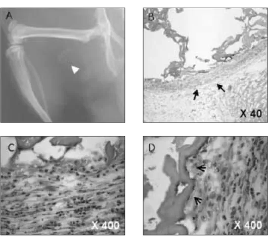
Fig. 2. X-ray radiograph are taken 6 weeks after implantation of block chitosan soaked with fraction I protein extracted from teeth. Radiopaque material (arrow head) was observed in experimental group at 6 weeks (A) (20 cm distance from the cone, 70kVp, 10mA), the chitosan block was surrounded by fibrous tissues (arrows) (B) (decalcified section: H & E stain, original magnification x 40), high magnification of B (C) (decalcified section: H & E stain, original magnification x 400), thin osteoblastic rim (open arrows) are observed toward chitosan block area (D) (decalcified section: H &
E stain, original magnification x 400)
Fig. 1. Gel chromatography of teeth protein in the excluded peak of Sephadex G-75.
Two peaks on the curve are observed. Fraction I
(29-30) and Fraction II (38-39)
3. Bioassay of total proteins
Fraction I을 매식 후 2, 4, 6, 8주 후에 채취한 표본에서 Calcification inducing activity를 보였다. 성형 가공된 키토산만을 매식한 군에서는 골 형성의 소견을 보이지 않았고(Fig. 2), 단백질 을 흡착한 군에서는 6 주 방사선 사진에서 방사선 불투과성을 보 였다 (Fig. 3).
4. SDS-PAGE (Sodium dodecyl sulfate-polyacrylamide gel electrophoresis)
SDS-PAGE에서 분자량 14-66kD사이에 명확한 단백질 띠로 분 리되었고(Fig. 4), 아미노산 분석결과 66 kD의 분자량을 갖는 단 백질이 가장 뚜렷하게 나타났다(Fig. 5). 이 단백질의 아미노산 분 석과 bioassay를 실시하였다.
Fig. 4. SDS PAGE of fraction I protein extracted from human teeth. Gel running exhibited four clear bands at the sites corresponding to 66, 40, 20 and 18 kD. Proteins extracted from total teeth (1, 2), Proteins extracted from enamel mechanically removed teeth (3, 4), size marker (SM).
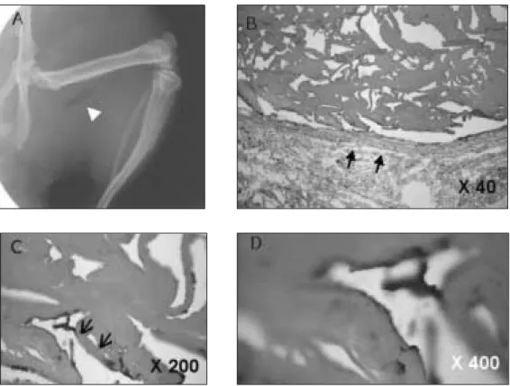
Fig. 3. X-ray radiograph shows radiolucent appearance (arrow head) at 6 weeks in the control
group (A) (20 cm distance from the cone, 70 kVp, 10 mA), the chitosan block was
surrounded by fibrous tissues (arrows) (B) (decalcified section: H & E stain, original
magnification x 40) no lining cells (open arrows) are observed around the chitosan block
(C) (decalcified section: H & E stain, original magnification x 200), high magnification of C
(D) (decalcified section: H & E stain, original magnification x 400)
5. Histological features
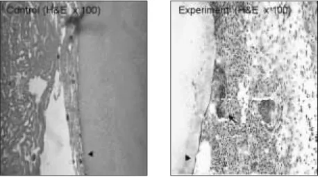
PVDF막 자체는 매식시 이물반응을 보이지 않았다. 그러나, 분 리 흡착시킨 치아단백질을 근육조직 내에 매식했을 때 알부민을 흡착시킨 PVDF막 주변에는 다핵거대세포가 출현하였으나, 사람 치아에서 추출한 단백질과 알부민을 혼합하여 흡착시킬 때에는 매식체 주변에서 석회화양상을 관찰할 수 있었다
실험군1주 소견에서 PVDF막 주변에 fibroblast와mesenchymal cell 이 모여있는 소견을 보였고, 2주 소견에서는 알부민을 흡착 시킨 PVDF막 주변에는 다핵거대세포가 출현하였다. 3, 4주에서 PVDF막 주위로 섬유성막이 형성되었다.
대조군으로 PVDF막만 매식한 경우에는 염증반응은 보이지 않 았고, 2, 3, 4주가 경과되어도 염증반응은 보이지않고 섬유화만 관찰되었다(Fig. 6).
6. Amino acid sequencing
Fraction I 을 PVDF막에 흡착시킨 것 중 가장 뚜렸한 양상을 보 인 band를 아미노산 서열을 분석하여 처음 12개의 순서를 얻어 전체 서열을 찾아보았다. 66 kD 단백질은 human albumin으로 판 명되었다(Table 2).
Fig. 5. 66 kD tooth protein (1-9) is blotted on the PVDF membrane. Size marker (SM)
Fig. 6. 66kD protein is blotted on PVDF membrane (arrow head) and inserted in muscles pouch of Sprague Dawley rat. The control group (PVDF strip is only inserted) show no inflammatory appearance at 2 weeks postoperatively. (Control) (Decalcified section: H & E stain, original magnification x 100), Fibroblast and mesenchymal cells had accumulated at the surface of PVDF membrane. They are surrounded by large multinuclear giant cells (arrow). (Experiment) (Decalcified section: H & E stain, original magnification x 100).
Table 2. The amino acid sequence of 66kD protein from fraction I
66kD 1 5 10
protein Ala-His-Lys-Ser-Glu-Val-Ala-His-Arg-Phe-Lys-Asp
Ⅳ. 고 찰
치아단백질을 추출하는 통상적인 방법에서는 법랑질을 기계 적으로 제거하고 상아질만을 사용하였지만 상아질에서 추출한 양은 1 mg/ml로 임상적으로 이용하기에는 소량이었다. 따라서 법랑질을 제거하는 과정을 생략하고 치아전체로 단백질을 추출 한 경우 3.5 mg/ml의 효율을 나타냈다. 이 두 가지 방법에서 추출 한 단백질을 SDS-gel을 이용하여 비교한 결과 유사한 band를 보 였다. 하지만 정확한 구성 요소간의 비교는 추가적인 HPLC(high performance liquid chromatography)을 통하여 알 수 있을 것이다.
치아단백질을 추출하기위한 법랑질을 제거한 술식을 생략해서 간단하고, 시간이 절약되면서도 추출효율도 더 높은 방법이라고 사료된다.
단백질 정제 시 순도를 높여감에 따라 단백질의 양과 기능이 저하되어가기 때문에 정확한 단백질의 특성을 알 수 없게 된다.
이런 단점을 보완하기 위해 소량의 단백질을 이용하여 자체의 기능을 저하시키지 않고 생체반응을 연구할 수 있는 방법을 모 색하였다. 기존의 방법에서는 생체반응을 조사하기위해서 매식 제를 주위로 분산되는 것을 막기위해 젤라틴 캡슐에 포장하여 매식하였다. 이 방법에는 약 3�5 mg의 단백질이 필요하기 때문 에 소량의 단백질인 경우 사용하기가 힘들다15). 그리고 일반적으 로 전달체로 사용하는 것 collagen gel, hydroxyapatite, plaster, titanium, fibrin 등이 사용되었다16-20). 이 전달체의 기능은 매식체 를 매식한 부위에서 지속적으로 유지하게 하고 같이 매식된 단 백질의 기능이 발현될 수 있어야 하는 것이다21,22). 이런 기능을 하기위해 막이 주로 사요 된다. 특히 nitrocellulose 막은 단백질과 핵산의 기능을 알기 위해 사용할 수 있고, 강력한 결합능력을 가 지고 있다. SDS-PAGE에 의해서 분리된 단백질을 nitrocellulose막 에 흡착시켜서 막 자체를 매식하여 생체반응을 조사할 수 있었 다. 이는 막이 단백질을 고정된 농도로 유지시켜서 단백질이 주 위 세포에 기능을 할 수 있는 환경이 형성되었기 때문이다23). 그 러나 이 방법도 아미노산 서열을 알기 위해서는 추출을 하는 단 계를 거쳐야 하는 불편을 가지고 있었다. 과거에는 흡착된 단백 질의 단백질간의 상호작용을 이용하여 특정한 분자만을 발견할 수 있었지만, PVDF막이 사용되면서 단백질 구조에 대한 정보를 알 수 있게 되었다. 특히 아미노산 서열과 전체 아미노산의 구성 에 대해서도 많은 정보를 얻을 수 있게 되었다24,25). PVDF 막은 다 른 막보다 초기 결합능력이 2배 이상 능가하고, nitrocellulose의 용량이 80�100 μg/cm2인 반면에 PVDF막은 170�200 μg/cm2이 었다. 또한 기계적 강도와 아미노산 서열에 확인을 위해 사용되 는 용제에 대한 화학적 저항력 크기와 결합능력이 크기 때문에 소 량 존재하는 단백질을 조사하는데 유용하게 사용할 수 있다26,27).
아미노산 서열을 확인하기 위해서는 PVDF막을 1 mm크기로 잘라서 분석할 수 있다. 일반적으로 Polybrene-coated PVDF 막에 흡착되는 양은 beta-lactoglobin (10-50 pmol)은 50�60%, bovine serum albumin 와 soybean trypsin inhibitor은 20�30% (50 pmol)이 다24). 이 막은 아미노산 서열 분석에도 높은 효율성을 보인다. 대 조군으로 사용한 PVDF막을 백서의 근육 속에 매식한 후 조직소
견상 염증반응을 보이지 않았지만, 치아단백질을 흡착한 막에서 는 거대세포들이 단백질의 작용으로 인하여 반응하였다. 단백질 의양은 주위조직에 영향을 미치기에 충분한 것으로 사료되고, PVDF막을 이용한 생체반응 검사는 유용한 것으로 사료된다.
Ⅴ. 결 론
PVDF막이 전기 영동으로 분리한 단백질을 쉽게 흡착할 수 있 고, 생체내 매식시 이물반응을 보이지 않기 때문에, 분리한 단백 질을 그대로 함유한 채로 약물 전달체로서 사용될 수 있음을 보 여주는 결과로서, 분리된 단백질의 생체검증에 PVDF막을 간편 하게 이용할 수 있다.
참고문헌