성균관대학교 의과대학 삼성서울병원 영상의학과 이주현, 이경수
Lobar Atelectasis: Radiographic-CT Correlation
Ju Hyun Lee, M.D., Kyung Soo Lee, M.D.
Department of Radiology, Samsung Medical Center, Sungkyunkwan University School of Medicine, Seoul, Korea
Address for correspondence : Kyung Soo Lee, MD Department of Radiology, Samsung Medical Center, Sungkyunkwan University School of Medicine 50, Ilwon-Dong, Kangnam-Ku Seoul 135-710, Korea Phone : 822-3410-2518 Fax : 822-3410-2559 E-mail : [email protected]
A B
A B
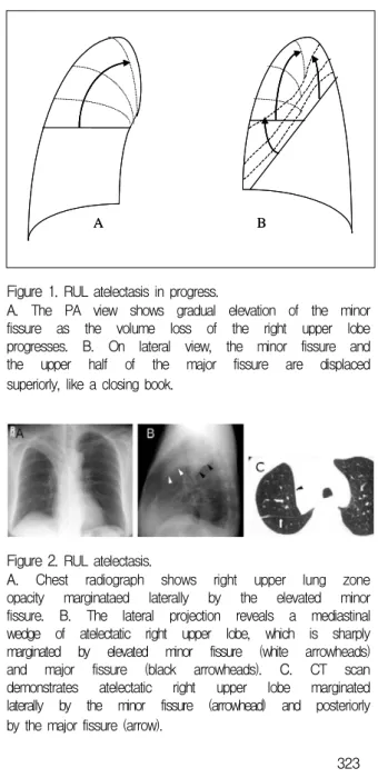
Figure 1. RUL atelectasis in progress.
A. The PA view shows gradual elevation of the minor fissure as the volume loss of the right upper lobe progresses. B. On lateral view, the minor fissure and the upper half of the major fissure are displaced superiorly, like a closing book.
Figure 2. RUL atelectasis.
A. Chest radiograph shows right upper lung zone opacity marginataed laterally by the elevated minor fissure. B. The lateral projection reveals a mediastinal wedge of atelectatic right upper lobe, which is sharply marginated by elevated minor fissure (white arrowheads) and major fissure (black arrowheads). C. CT scan demonstrates atelectatic right upper lobe marginated laterally by the minor fissure (arrowhead) and posteriorly by the major fissure (arrow).
Introduction
The characteristic radiographic and CT findings of lobar atelectasis are well known. However, lobar atelectasis is a dynamic process, and atypical pres
entations may occur due to a number of different causes. The purpose of this review article is to illustrate the spectrum of radiographic findings of lobar atelectasis and to correlate the radiographic findings with the CT findings.
Right Upper Lobar Atelectasis
Right upper lobe (RUL) atelectasis results in over
inflation of the right middle lobe and shift of the minor fissure superiorly and medially. It also results in compensatory overinflation of the right lower lobe (RLL) with shift of the major fissure anteriorly, superiorly and medially. The Golden’s S sign denotes a centrally located mass with associated lobar atelectasis. The mass should be large enough to be borderforming with the adjacent hyperexpanded lung. With complete atelectasis, the RUL is either pancaked medially, simulating mediastinal widening or a mediastinal mass, or superiorly simulating an apical pleural cap.
On the lateral chest radiograph, an ill-defined opacity
anterior to the trachea and obliteration of the anterior margin of the ascending aorta may sometimes be the only findings
1-3(Fig. 1).
The minor fissure changes its position more dra
matically than does the major fissure. With elevation
A B
A B
Figure 3. LUL atelectasis in progress.
A. On PA view, the collapsing left upper lobe manifests as a poorly defined, large perihilar opacity fading away as its periphery. B. The lateral view shows gradual anterosuperior displacement of the major fissure.
Figure 4. LUL atelectasis.
A. Chest radiograph shows ill-defined opacity in left hilar area. B. Lateral radiograph shows anterior displacement of the left major fissure (arrowheads).
C, D. CT scans show “V”-shaped posterior margin of atelectatic left upper lobe.
of the minor fissure, the middle lobe shifts up laterally alongside the atelectatic upper lobe. On CT, the middle and upper lobes can be seen side-by-side anterior to the major fissure with the superior segment of the lower lobe posterior to the fissure.
The major fissure maintains its previous contour, whether straight, concave, or convex
4-7(Fig. 2).
Left Upper Lobar Atelectasis
With LUL atelectasis, the direction of movement is anterosuperior rather than directly superior as in RUL atelectasis. The left pulmonary artery, which courses over the left main bronchus, restrains the bronchus and limits the superior migration of the atelectatic lobe
8. For this reason, the superior seg
ment of the LLL expands upward toward the apex of the left hemithorax. Therefore atelectasis of the left upper lobe is associated with increased opacity in the suprahilar region on the PA radiograph. As atelectasis progresses, it leads to increased opacity with poorly defined margins in the perihilar region.
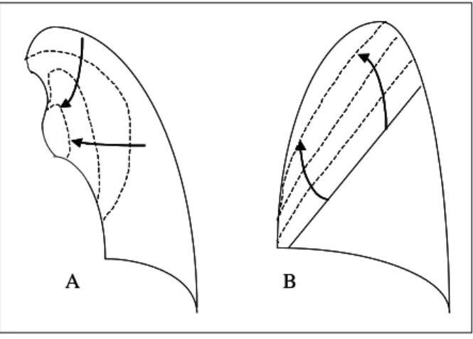
On the lateral radiograph, the lateral portion of the major fissure is displaced forward and is placed tangentially resulting in a sharp interface (Fig. 3).
On CT scans, the atelectatic LUL forms a homo
geneous opacity based on the anterior chest wall and the mediastinum. The posterior margin has a V-shaped contour from the lung apex to the hilum, where the apex of the V merges with the hilar vessels and bronchi. It is these hilar structures, which are relatively fixed in position, that tether the major fissure into the V-shape. The superior segment of the LLL is pulled forward along both the medial and lateral limbs of the V. The part of the superior segment that follows the medial limb forms a tongue of lung between the mediastinum and the atelectatic LUL. This tongue is visible on PA radiographs and has been called the Luftsichel (air-crescent) or periaortic lucency
7(Fig. 4). Less commonly, the major fissure may have a straight border rather than a V-shaped contour.
Occasionally the atelectatic lobe may have sharp margins on the PA radiograph simulating a hilar mass. With marked LUL atelectasis, the contour of the major fissure interface may appear continuous with that of the normal epipericardial fat on the lateral radiograph.
Right Middle Lobar Atelectasis
As the RML loses volume, the minor and major
fissures move toward each other in an inferomedial
A B
A B
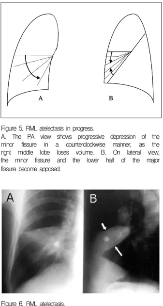
Figure 5. RML atelectasis in progress.
A. The PA view shows progressive depression of the minor fissure in a counterclockwise manner, as the right middle lobe loses volume. B. On lateral view, the minor fissure and the lower half of the major fissure become apposed.
Figure 6. RML atelectasis.
A. PA chest radiograph shows trianglar opacity in right lower lung zone with obliteration of right lower cardiac border. B. Lateral radiograph shows triangular opacity in lower anterior aspect of thorax with tis apex toward hilum. It is marginated superiorly by the minor fissure (small arrow) and inferiorly by the major fissure (large arrow).
and superomedial direction, respectively. The RML thus assumes an oblique orientation and on the PA radiograph results in a poorly defined increased opacity that obscures the right heart border. In general, the greater the atelectasis and the greater the reorientation of the RML, the more difficult it is to recognize the atelectasis on PA radiograph. On the lateral view, RML atelectasis is seen as a triangular
opacity marginated superiorly by the minor fissure and inferiorly by the major fissure. The apex of the triangle is in the hilar area, and the base is located peripherally (Fig. 5 and 6).
On CT scans, the RML is triangular or trapezoidal.
Its posterior border, demarcated by the major fissure, is usually well defined because the major fissure crosses the scan plane almost perpendicularly.
On the other hand, the interface between RML and RUL is often less distinct because of the dome- shaped contour of the minor fissure.
Lower Lobar Atelectasis
As the lower lobes become atelectatic, the lateral portion of the major fissure moves posteriorly to
ward the costophrenic angle and may be well del
ineated on the lateral radiograph. The medial portion of the major fissure relates to the mediastinal wedge of pulmonary attachment. The wedge is frequently difficult to detect on the lateral radiograph except for a slight area of increased opacity extending from the posterior costophrenic angle and may be well delineated on the lateral radiograph. The medial portion of the major fissure relates to the mediastinal wedge of pulmonary attachment. The wedge is fre
quently difficult to detect on the lateral radiograph except for a slight area of increased opacity extending from the posterior costophrenic angle toward the hilum. On PA radiographs, the lateral margin of the lobe may be ill- or well-defined, depending on whether or not the adjacent hyperexpanded lung has placed the fissural edge of the lower lobe tangential to the x-ray beam.
If marked atelectasis of the RLL has occurred, the triangular-shaped opacity may be difficult to detect through the mediastinum because of its small size.
In LLL atelectasis, the involved lobe may appear as
a left paraspinal mass instead of the more charac
A B
A B
Figure 7. RLL atelectasis in progress.
A. On PA view, the superolateral portion of the major fissure is displaced inferomedially. B. On lateral view, the lower and the upper half of the major fissure move like the halves of a closing book.
A B
A B
Figure 8. LLL atelectasis in progress.
A. On the PA view, the superolateral portion of major fissure is displaced inferomedially, manifesting as a thin convex, curvilinear line. B. On lateral view, the lower and the upper half of the major fissure are displaced to meet each other.
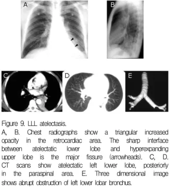
Figure 9. LLL atelectasis.
A, B. Chest radiographs show a triangular increased opacity in the retrocardiac area. The sharp interface between atelectatic lower lobe and hyperexpanding upper lobe is the major fissure (arrowheads). C, D.
CT scans show atelectatic left lower lobe, posteriorly in the paraspinal area. E. Three dimensional image shows abrupt obstruction of left lower lobar bronchus.
teristic triangular shape with the apex at the hilum and the base at the left hemidiaphragm. The appear
ance of lower lobar atelectasis as a paraspinal mass is believed to result from incomplete attachment of the inferior pulmonary ligament to the hemidiaphragm
9(Figs. 7,8 and 9).
On CT scans, the lower lobes lose volume in a posteromedial direction, pulling down the major fi
ssure. The lateral portion of this fissure demonstrates a greater degree of mobility, because the medial portion
is fixed to the mediastinum by the hilar structures and the inferior pulmonary ligament.
Combined Lobar Atelectasis
1. Combined Atelectasis of the Right Middle and Lower Lobes
Because the bronchus intermedius is the common pathway to the right middle and lower lobes, a single localized lesion involving the bronchus inter
medius gives rise to combined atelectasis of these lobes. The bronchus can be obstructed with a tumor, a foreign body, a mucous plug, or an inflammatory stricture
10.
On the PA radiograph, the atelectatic RLL obscures the right hemidiaphragm, whereas the atelectatic right middle lobe obscures the right cardiac border.
Depression of both the major and minor fissures is present, the depression being most marked laterally.
The fissures cross over each other; the major
fissure assumes more vertical orientation than the
minor fissure. However, both the right minor and
Figure 11. Combined atelectasis of RM and RLL due to mucus impaction in the bronchus intermedius.
A. Chest radiograph shows opacity in right lower lung zone obscuring right atrium and right hemidiaphragm.
Inferior displacement of major (arrows) and minor (arrowheads) fissures is present. B. Lateral radiograph shows opacification throughout right lower lobe obscuring right hemidiaphragm. Upper border of the opacity is bordered anteriorly by minor fissure (arrow
head) and posteriorly by major fissure (arrow).
A B
A B
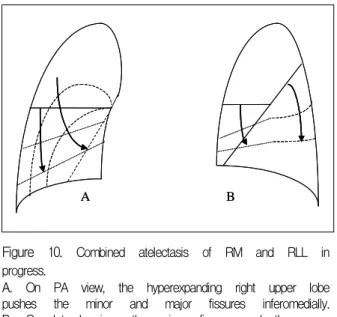
Figure 10. Combined atelectasis of RM and RLL in progress.
A. On PA view, the hyperexpanding right upper lobe pushes the minor and major fissures inferomedially.
B. On lateral view, the minor fissure and the upper half of the major fissure are displaced downward.
major fissures are usually obscured within medias
tinal shadow. Therefore, visualization of the right minor fissure should suggest the diagnosis of isolated atelectasis of the right lower lobe. Occasionally, the fissures may be seen as crossing double interface in close approximation on PA radiograph. On the lateral view, increased opacity is present throughout the lower part of the chest (Fig. 10 and 11).
On CT scans, the atelectatic RML and RLL occupy
the lower hemithorax and abut the right cardiac border medially and the right hemidiaphragm in
feriorly. The right major and minor fissures border the posterior and anterior margins of the atelectatic lobes, respectively. Complete combined RML and RLL atelectasis can be difficult to detect on PA and lateral radiographs. The diagnosis should be suspected in patients with a small right hilum and an appar
ently oligemic right lung which represents the hy
perexpanded RUL.
2. Combined Atelectasis of the Right Upper and Middle Lobes
For combined atelectasis of the RUL and RML to occur, the bronchi of both lobes must be narrowed or occluded by a single or two separate lesions while the bronchus intermedius remains patent, thus allowing the RLL to remain expanded. Combined atelectasis of the RUL and RML occurs most frequently in patients with bronchogenic carcinoma, in which the primary tumor can obstruct one bronchus and cause the other bronchus to be obstructed by direct extension through the lung parenchyma or peribronchial sheath or by lymphadenopathy
10.
On the PA radiograph, the atelectatic RUL and RML form an opacity that obscures the outline of the mediastinum and fades laterally. Combined atelectasis of the RUL and RML can lead to cephalad and lateral displacement and rotation of the hilar vessels. The silhouettes of the ascending aorta and the right atrium are usually obscured. On the lateral view, the major fissure can be seen displaced anteriorly.
The relative proximity of the major fissure to the anterior chest wall is dependent on the degree of atelectasis of the RUL and RML. The radiographic findings of combined atelectasis of the RUL and RML are similar to those of LUL atelectasis
10(Fig. 12).
On CT scan, the atelectatic RUL and RML cause
A B
A B
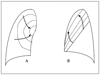
Figure 12. Combined RU and RML atelectasis in pro
gress.
A. On the PA view, the collapsing right upper and right middle lobe manifest as a poorly defined, large perihilar opacity fading away at its periphery. B. The lateral view shows gradual anterosuperior displacement of the major fissure.
a wedge-shaped area of soft-tissue attenuation abutting the chest wall anteriorly and the ascending aorta and right cardiac border medially. This wedge- shaped opacification extends inferiorly to the level of the right atrium. The major fissure is displaced anteriorly, and the hyperexpanded lower lobe fills most of the right hemithorax.
3. Combined Atelectasis of the Right Upper and Lower Lobes
Combined atelectasis of the RUL and RLL is rare.
It may be due to mucous plugs occurring simultan
eously in the bronchi of the RUL and RLL. The radiographic findings of combined atelectasis of RUL and RLL are similar to those of isolated atelectasis of either lobe. Upper lobe atelectasis leads to elevation of the minor fissure, whereas lower lobe atelectasis leads to downward and medial shift of the major fissure. On CT scans, the minor fissure is higher than normal because of the atelectasis of the RUL and more posterior than normal because of the at
electasis of the RLL. The middle lobe is overinflated
10.
Peripheral Lobar Atelectasis
Franken and Klatte
11described the radiographic findings of what they called "atypical (peripheral) right upper lobe atelectasis", mimicking apical pleural effusion. In this type of atelectasis of the RUL, the atelectatic lobe continues to lie adjacent to the lateral chest wall. The dense portion of the atelectatic lobe is sharply marginated medially. On CT in this form of atelectasis, the RML expands upward in front of the atelectatic RUL with the minor fissure adopting an almost coronal orientation.
The superior segment of the RLL herniates upward posterior and medial to the atelectatic RUL with the major fissure being repositioned to a more parasagittal orientation superiorly, presenting itself as a radiographic interface on the PA projection
11-13. The herniated superior segment of RLL forms the so-called Luftsichel (air crescent) medial to the atelectatic lobe. Recently two cases of peripheral atelectasis of left upper lobe, caused by bronchogenic carcinoma, have also been reported
13.
Migrating Lobar Atelectasis
A very heavy lobe, filled with fluid, chronic pne
umonia, or a tumor, may migrate in the hemithorax
with change in body position adopting a dependent
position. Heavy lobes and pedunculated benign fibrous
tumors of the pleura are the two likely causes of a
large migrating chest density
10,14. Migrating atelectasis
usually involves a single lobe (usually RUL),
however it has also been described with combined
RUL and RML atelectasis
15. Migratory lobar atelectasis
should be distinguished from lung torsion. Lobar
migration is mainly a shifting process within the
hemithorax, while lobar torsion is a rotatory or
twisting process around its pedicle (bronchovascular
bundle). Lobar migration has been regarded to
indicate a lobar torsion
16. Some degree of torsion may be associated with lobar migration. However, on CT, twisted or obliterated bronchovascular bundles due to torsion are not usually seen in patients with lobar migration. Patients with lobar migration usually have no symptoms as in those with spontaneous lobar torsion, but differently from those with postoperative or posttraumatic torsion
15.
Rounded Atelectasis
Rounded atelectasis is a form of peripheral pulmonary volume loss. Rounded atelectasis is hypothesized to be due to contraction of a focus of visceral pleural fibrosis that results in buckling of the pleura and atelectasis of underlying lung parenchyma
17. It usually results in volume loss of part of a lobe unrelated to the segmental anatomy. Rounded atelectasis usually presents as a mass that may simulate a pulmonary neoplasm on chest radiograph. The CT criteria for the diagnosis of rounded atelectasis include (1) a rounded or oval mass abutting a pleural surface, (2) vessels and bronchi curving into the mass, and (3) associated pleural thickening with or without calci
fication
18. Although rounded atelectasis is usually confined to a small portion of lung, occasionally it may involve the entire lobe and simulate a large mass.
References
1. Lee KS, Kim TS. Chapter 56. Atelectasis. In: Taveras JM, Ferrucci JT, editors. Radiology, diagnosis-im
aging-intervention. Vol. 1. Philadelphia: Lippincott- Raven Publishers; 1998. p. 1-32.
2. Mintzer RA, Sakowicz BA, Blonder JA. Lobar collapse:
usual and unusual forms. Chest 1988;94:615-20.
3. Lubert M, Krause GR. Further observations on lobar collapse. Radiol Clin North Am 1963;1:331-46.
4. Naidich DP, McCauley DI, Khouri NF, Leitman BS, Hulnick DH, Siegelman SS. Computed tomography of lobar collapse: 1. endobronchial obstruction. J Com
put Assist Tomogr 1983;7:745-57.
5. Naidich DP, McCauley DI, Khouri NF, Leitman BS, Hulnick DH, Siegelman SS. Computed tomography of lobar collapse: 2. collapse in the absence of endo
bronchial obstruction. J Comput Assist Tomogr 1983;7:758-67.
6. Naidich DP, Ettinger N, Leitman BS, McCauley DI.
CT of lobar collapse. Semin Roentgenol 1984;19:222 -35.
7. Raasch BN, Heitzman ER, Carsty EW, Lane EJ, Verlow ME, Niter G. A computed tomographic study of bronchopulmonary collapse. Radiographics 1985;
4:195-232.
8. Khoury MB, Godwin JD, Halvorsen RA Jr, Putman CE. CT of obstructive lobar collapse. Invest Radiol 1985;20:708-16.
9. Glay J, Palayew MJ. Unusual pattern of left lower lobe atelectasis. Radiology 1981:141:331-3.
10. Lee KS, Logan PM, Primack SL, Müller NL. Com
bined lobar atelectasis of the right lung: imaging findings. Am J Roentgenol 1994;163:43-7.
11. Franken EA Jr, Klatte EC. Atypical (peripheral) upper lobe collapse. Ann Radiol 1977;20:87-93.
12. Adler J, Cameron DC. CT correlation in peripheral right upper lobar collapse. J Comput Assist Tomogr 1988;12:510-1.
13. Don C, Desmarais R. Peripheral upper lobe collapse in adults. Radiology 1989;170:657-9.
14. Heitzman ER. The lung: radiologic-pathologic cor
relation. 2nd ed. St Louis: Mosby; 1984. p. 457-501.
15. Kim TS, Lee KS, Hwang JH, Choo IW, Lim JH.
Migrating lobar atelectasis of the right lung: radiologic findings in six patients. Korean J Radiol 2000;1:
33-7.
16. Felson B. Lung torsion: radiographic findings in nine cases. Radiology 1987:162:631-8.
17. Menzies R, Fraser R. Rounded atelectasis: pathologic and pathogenetic features. Am J Surg Pathol 1987;
11:674-81.