Note
http://dx.doi.org/10.4217/OPR.2016.38.1.081
절식이 나일 틸라피아 Oreochromis niloticus의 Kiss2, GnRH I mRNA 발현 및 성 스테로이드 호르몬 농도에 미치는 영향
박진우
1· 권준영
2· 진예화
2· 오승용
3*1한국해양과학기술원 생태기반연구센터 (15627) 경기도 안산시 상록구 해안로 787
2선문대학교 건강보건대학 수산생명의학과 (31466) 충청남도 아산시 탕정면 선문로 221
3한국해양과학기술원 통영해상과학기지 (15627) 경기도 안산시 상록구 해안로 787
Effects of Fasting on Brain Expression of Kiss2 and GnRH I and Plasma Levels of Sex Steroid Hormones, in Nile Tilapia Oreochromis niloticus
Jin Woo Park
1, Joon Yeong Kwon
2, Ye Hwa Jin
2, and Sung-Yong Oh
3*1Marine Ecosystem and Biological Research Center, KIOST Ansan 15627, Korea
2Department of Aquatic Life Medical Sciences, College of Health Science, Sunmoon University Asan 31466, Korea
3Tongyeong Marine Science Station, KIOST Ansan 15627, Korea
Abstract : In many fish species, including Nile tilapia (Oreochromis niloticus), gonadal development occurs at the expense of stored energy and nutrients. Therefore, reproductive systems are inhibited by limited food supply. It has been well established that reproductive function is highly sensitive to both metabolic status and energy balance. Nothing is known about the possible mediated connection between energy balance and reproduction. Kisspeptin, a neuropeptide product of the Kiss gene has emerged as an essential gatekeeper of reproduction and may be possibly be linked to energy balance and reproduction in non-mammalians. Thus, in this study, the effect of fasting (10 days) on the expression of kisspeptin and the gonadotropin-releasing hormone (GnRH) gene were assessed in Nile tilapia (male and female) using qRT- PCR. In addition, plasma levels of estradiol-17β (E
2) and 11-ketotestosterone (11-KT) in adult tilapia were measured by ELISA. In male tilapia, fasting reduced Kiss2 and GnRH I mRNA expression in the brain and 11-KT level in comparison with the fed tilapia (p < 0.05). In females, however, there were no significant differences in GnRH I mRNA expression and E
2between fish subjected to fasting and those fed (p > 0.05).
These data indicate the impact of nutritional states on kisspeptin as a potential regulatory mechanism for the control of reproduction in male Nile tilapia.
Key words : Oreochromis niloticus, Kiss2, GnRH I, sex steroid hormone, fasting
*Corresponding author. E-mail : [email protected]
1. 서 론
어류를 포함한 척추동물의 번식은 환경변화(온도, 광주 기 등)에 반응한 각 어체 뇌의 시상하부가 Gonadotropin- Releasing Hormone(GnRH) 를 분비함으로써 조절된다. 시 상하부(hypothalamus)에서 분비된 GnRH는 뇌하수체 전 엽(anterior pituitary)에 위치한 GnRH 수용체를 활성화시 키고, 두 종류의 서로 다른 gonadotropin(GTH)인 Follicle Stimulating Hormone(FSH)과 Luteinizing Hormone(LH) 의 분비를 조절함으로써 생식소 발달 및 성숙에 관여한 다. 이러한 성 성숙 과정은 체성장(somatic growth)에 이 용되던 에너지가 생식소 발달 및 번식 활동을 위해 소비 되는 생리학적 과정으로(Taranger et al. 2010) 상당한 에 너지가 요구된다. 따라서 풍부한 먹이 공급은 성 성숙을 유도할 수 있는 반면, 먹이 공급이 원활하지 않은 경우 성 성숙을 방해할 수 있다(Schneider 2004).
에너지 항상성(homeostasis)과 번식과의 관계는 다양한 척추동물에서 입증되었다(Mircea et al. 2007). 암컷 쥐 (rats)는 단기간 절식에 의해 LH의 맥동적(pulsatile) 분비 가 억제되었고(Cagampang et al. 1991; Kohsaka et al.
2001), 미성숙한 암컷 쥐에서 장기간 절식은 성 성숙개시 를 방해하였다(Castellano et al. 2005). 어류의 경우 절식 은 제브라피쉬(zebra fish, Danio rerio)의 GnRH II mRNA 발현을 감소시켰다(Nishiguchi et al. 2012). 이렇듯 먹이 는 번식 활성에 관여하는 주요한 외부요인으로 작용하며, 번식 활성과 먹이와의 상호영향은 신경내분비시스템에 의 해 조절되어질 가능성이 높다(Hoskins et al. 2008). 그러나 먹이 섭취와 GnRH 분비 사이에 존재하는 신호의 전달경 로는 확실하지 않다.
최근 번식 신경내분비 연구에서 kisspeptin이 주목을 받 고 있다. Kisspeptin은 Kiss 유전자의 세포내 translation에 의해 만들어지는 peptide이다. 이들은 처음에는 전이가 일 어나지 않는 암세포 집단에서 발견되었다(Lee et al.
1996). 이후 kisspeptin은 다수의 어류와 포유류에서 그 구 조가 밝혀졌으며 기능에 대한 다양한 연구가 진행되었다.
그 결과 경골어류를 포함한 다양한 척추동물의 GnRH, GTH 유전자(FSHβ and LHβ) 발현상승 및 GTH의 방출을 자극하는 강력한 조절자임이 밝혀졌다(Biran et al. 2008;
Filby et al. 2008; Felip et al. 2009; Kitahashi et al. 2009;
Lee et al. 2009; Li et al. 2009).
포유류의 연구에서는 추가적으로 kisspeptin이 영양과 대사 등 중요한 생물학적 기능들과 관련되어 있음이 확인 되면서(Messager et al. 2005; Patterson et al. 2006; Dhillo et al. 2007; Roa and Tena-Sempere 2007), 먹이 활동과 번식 활성의 연결고리로써 작용할 가능성이 있다고 보고 했다(Popa et al. 2008). 실제로 포유류에 대한 몇몇 연구
는 극심한 절식이 시상하부의 Kiss1 유전자의 발현을 감 소시켰다(Castellano et al. 2005; Iwasa et al. 2010). 이와 같은 연구 결과들은 kisspeptin이 먹이 활동에 의해 조절 되며 이를 통해 번식활성에 영향을 미칠 수 있음을 나타 낸다. 그러나 지금까지 연구결과들은 절식이 번식활성에 미치는 영향이나 또는 절식이 kisspeptin 유전자 발현에 미치는 영향들을 따로 따로 연구한 것들이어서, 절식→
kisspeptin → GnRH → sex steroid로 이어지는 연결 과정 을 한 번에 설명하지 못하였다. 또한 어류의 경우에는 번 식 활동에 주요한 역할을 하는 kisspeptin과 먹이 활동 의 연관성은 명확히 밝혀지지 않아 연구의 필요성이 제 기된다.
따라서 본 연구에서는 하등 척추동물의 연구를 위한 실 험어종으로 널리 사용되어지는 나일 틸라피아(Nile tilapia, Oreochromis niloticus)를 이용하여 절식에 따른 시상하부 Kiss2와 GnRH I 유전자 발현 변화 그리고 혈중 성 스테 로이드 호르몬의 변화를 함께 조사하였다.
2. 재료 및 방법
실험어
실험어는 수온 27 ± 1
oC, 광주기 14L:10D 조건의 순환 여과시스템에서 사육된 나일 틸라피아 성어(암: 전장 14.1 ± 0.9 cm, 체중 56.7 ± 7.4 g, 수: 전장 14.4 ± 1.4 cm, 체중 55.4 ± 7.6 g)를 사용하였다. 암·수 나일 틸라피아 모 두 성적 성숙이 이루어진 개체들(암컷=20, 수컷=20)을 사용하였다. 먹이는 1일 2회 상업용 부상사료(단백질 함 량 38%, Woosung, Korea)를 실험 전까지 만복으로 공 급하였다.
절식실험
암컷과 수컷을 각각 두 그룹으로 나누어 한 그룹은 먹 이 공급 후 10일 동안 절식시켰다. 10일 동안 절식한 암컷 (6마리)과 수컷(5마리)에서 뇌하수체를 포함한 뇌 조직을 적출하였다. 다른 한 그룹은 1일 2회 상업용 배합사료를 만복으로 10일 동안 공급하고, 먹이 공급 10일째 암컷과 수컷 각각 6마리씩 뇌하수체를 포함한 뇌 조직을 적출하 여 절식한 개체와 비교하였다.
모든 실험어의 뇌 조직은 실험어를 benzocaine(50 ppm)
에 마취하여 적출하였으며, total RNA를 추출하기 위해
추출 전까지 −70
oC에서 보관하였다. 또한 암컷의 혈장 내
estradiol-17β(E
2) 와 수컷의 혈장 내 11-ketotestosterone(11-
KT)를 조사하기 위해 뇌 조직 적출 전에 heparin sodium
처리 주사기(1 mL)를 사용하여 미부혈관에서 혈액을 채
취하였다. 채취한 혈액은 혈장을 얻기 위해 원심분리(4
oC
에서 1000 × g로 30분간) 하였다. 얻어진 혈장은 분석 전
까지 −70
oC 에서 보관하였다. 실험기간 동안 실험어는 순 환여과시스템에서 수온 27 ± 1
oC, 광주기 14L:10D 조건 아래 사육하였다.
RNA 추출 및 cDNA 합성
Total RNA 는 TRI reagent
®(Molecular Research Center Inc., USA) 를 사용하여 추출하였다. 적출한 뇌를 50 mL conical tube 에 넣고, 1 mL의 TRI reagent를 넣어 homogenizer(T 10, IKA, China)를 이용하여 얼음 위에서 조직을 분쇄하였다. 조직의 분쇄 후 실온에서 5분간 방치 하였다. 그 다음, chloroform(Sigma. Inc., USA) 200 µL을 첨가하여 강하게 vortexing한 후, 실온에서 5분간 반응시 켰다. 이 후 4
oC에서 12,000 × g로 15분간 원심분리하고, RNA가 포함된 aqueous phase를 잘 걷어내어 새 튜브에 옮겼다. RNA가 들어있는 새 튜브에 300 µL의 isopropanol (Sigma. Inc., USA) 을 넣고, inverting 후 실온에서 10분간 반응시켰다. 그 다음 4
oC에서 12,000 × g로 8분간 원심분 리하여 RNA pellet을 얻어내고 상층액을 제거 후 wash과 정을 거쳐 total RNA를 얻었다. 추출한 total RNA는 RQ1 RNase-free DNase(Promega, USA) 로 처리하여 genomic DNA에 의한 오염가능성을 최소화하였다. 이 total RNA (1 µg)와 M-MLV(Moloney Murine Leukemia Virus) reverse transcriptase(Promega, USA), oligo (dT)
15primer(Promega, USA) 를 이용하여 cDNA를 합성하였다.
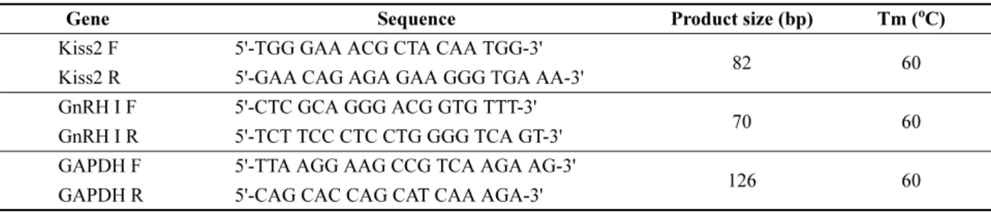
절식 후 GnRH I 및 Kiss2 mRNA 발현량 조사 뇌 조직에서 GnRH I과 Kiss2 유전자 발현량을 알아보 기 위해 quantitative real-time PCR(qRT-PCR)을 실시하였 다. 실험에 사용된 Kiss2, GnRH I, GAPDH (Glyceraldehyde- 3-Phosphate Dehydrogenase) 유전자 primer는 Beacon Designer software (Bio-Rad, Hercules, CA, USA) 를 이용 해 제작하였다(Table 1).
qRT-PCR 반응은 Topreal
TMqPCR 2X PreMIX SYBR Green(Enzynomics, Korea)을 이용하여 분석하였다. 반응 액은 5 µL의 cDNA(1:50 dilution)를 주형으로 사용하고,
7.5 µL Topreal
TMqPCR 2X PreMIX SYBR Green, 250 nM primer sets 그리고 N.F.W(Nuclease-free water)를 혼합하 여 총 15 µL의 volume으로 실시하였다. qRT-PCR의 수행 은 CFX96 Touch
TMReal-Time PCR Detection System (Bio-Rad, USA) 을 이용하여 95
oC 에서 15분간 initial denaturation하였으며, 95
oC 에서 15초 denaturation, 60
oC 에서 15초 annealing, 72
oC에서 30초 elongation하여 45 cycles 을 반응시켜 주었다. 반응이 끝난 후에는 melting curve를 분석하였다.
GAPDH를 대조유전자로 사용하였으며, 2
-ΔΔCt방법 (Livak and Schmittgen 2001) 을 이용하여 상대 정량하였 다. 모든 샘플은 2회 이상 반복 측정하였다.
절식 후 혈중 E
2및 11-KT 농도 조사
절식에 따른 성어 암컷(6마리) 혈장 내 E
2와 수컷(5−6 마리) 혈장 내 11-KT 변화를 조사하기 위해 Enzyme- Linked Immunosorbent Assay(ELISA)를 수행하였다. E
2는 어류 E
2ELISA kit(CUSABIO, China) 를 사용하여 경 쟁적 ELISA법(competitive inhibition technique)으로 측정 하였고, 11-KT는 어류 11-KT ELISA kit(MyBioSource, USA)를 사용하여 이중항체 샌드위치 ELISA법(double antibody sandwich technique) 으로 측정하였다. E
2와 11- KT 실험은 각 제조회사에서 제공한 실험방법에 따라 진 행하였으며, E
2와 11-KT 모두 EMax Endpoint ELISA Microplate Reader(Molecular Devices, USA) 를 이용하여 파장 450 nm에서 흡광도를 측정하였다. 이렇게 측정된 흡 광도 값과 SOFTmax Pro 4.0 software(Molecular Devices)에 서 만들어진 logistic(log-log) curve-fit standard curve를 사용하여 E
2와 11-KT의 농도를 계산하였다.
통계처리
각 실험결과로부터 얻어진 자료 값(mean ± S.E.M.) 사 이의 유의차 유무는 SPSS-통계패키지(version 18.0)을 이 용하여 절식그룹과 먹이 섭취 그룹 간에 independent t- test(p < 0.05) 로 검정하였다.
Table 1. List of Primers used for quantitative real-time PCR analysis
Gene Sequence Product size (bp) Tm (
oC)
Kiss2 F 5'-TGG GAA ACG CTA CAA TGG-3'
82 60 Kiss2 R 5'-GAA CAG AGA GAA GGG TGA AA-3'
GnRH I F 5'-CTC GCA GGG ACG GTG TTT-3'
70 60 GnRH I R 5'-TCT TCC CTC CTG GGG TCA GT-3'
GAPDH F 5'-TTA AGG AAG CCG TCA AGA AG-3'
126 60 GAPDH R 5'-CAG CAC CAG CAT CAA AGA-3'
F: forward, R: reverse
3. 결과 및 고찰
본 연구 결과에서는 절식이 일부 나일 틸라피아의 번식 관련 인자들에게 영향을 줄 수 있음을 나타내었다. 수컷 의 경우 GnRH 및 Kiss2 유전자 모두 절식한 그룹이 먹 이를 섭취한 그룹에 비해 유의하게 낮은 발현량을 나타내 었으며, 두 그룹 간 발현량의 차이는 약 5배 정도였다 (Fig. 1). 또한 수컷의 혈중 11-KT도 절식한 그룹(5.6 ± 0.6 pg/mL) 이 먹이를 섭취한 그룹(9.8 ± 1.1 pg/mL)에 비 해 농도가 유의하게 낮았다(Fig. 3). 먹이 섭취와 GnRH 와의 관련성은 몇몇 척추동물에서 연구가 진행되었다. 절 식은 수컷 쥐의 GnRH 발현량을 감소시켰고(Gruenewald et al. 1993), 어류의 경우 절식은 가자미(winter flounder, Pseudopleuronectes americanus)의 GnRH II 유전자 발현 을 감소시켰다(Tuziak and Volkoff 2013). 이와는 반대로 지속적인 먹이 섭취는 미성숙한 쥐와 어린 양(lams)의 GnRH 분비를 촉진시켜 일시적으로 LH를 상승시켰다 (Bronson 1986; Sisk et al. 1986; Foster et al. 1989). 또한 포유류의 연구에서는 대사신호와 관련한 주요 호르몬인 leptin과 insulin을 투여했을 때 GnRH mRNA가 증가해 먹이 섭취 조절에 의해 발생되는 대사신호들이 직접적으
로 시상하부의 GnRH mRNA에 영향을 줄 수 있다고 제 안했다(Burcelin et al. 2003). 앞선 연구 결과들은 GnRH 가 먹이 섭취와 밀접한 관련성이 존재함을 시사한다. 이러 한 관점에서 볼 때 나일 틸라피아의 감소된 GnRH 유전자 발현량은 절식에 영향을 받았을 가능성이 있으며, 이로 인 해 GnRH에 영향을 받아 성적 성숙을 촉진시키는 11-KT
Fig. 1. Effects of fasting on the expression levels of GnRH I and Kiss2 mRNA in the brain of male Nile tilapia. Relative abundance of the mRNAs was normalized to the amount of GAPDH by the comparative threshold cycle method using qRT-PCR. Results are means ± S.E.M. (n = 5−6).
* indicates significant difference from fast group (p < 0.05)
Fig. 2. Effects of fasting on the expression levels of GnRH I and Kiss2 mRNA in the brain of female Nile tilapia. Relative abundance of the mRNAs was normalized to the amount of GAPDH by the comparative threshold cycle method using qRT- PCR. Results are means ± S.E.M. (n = 6)
Fig. 3. Effects of fasting on the plasma level of 11-KT in male Nile tilapia. 11-KT level was read at 450 nm and calculated using logistic (log-log) curve-fit standard curve (R
2> 0.99) which were generated by SOFTmax pro. Results are means ± S.E.M.
(n = 5 −6). * indicates significant difference from
fast group (p < 0.05)
의 농도도 감소했을 것으로 사료된다.
Kisspeptin 은 어류를 포함한 척추동물에서 GnRH neuron 을 자극하여 성 성숙에 관여한다고 알려져 있다(Parhar et al. 2004). 이와 같이 어류의 kisspeptin 연구는 대부분 번 식에 초점이 맞춰져 있어 먹이 섭취와의 관련성은 명확하 게 밝혀진 바가 없다. 그러나 몇몇 포유류의 연구에서는 kisspeptin 과 먹이 섭취와의 관련성을 제시했다. 성숙한 암 컷 쥐의 Kiss1 유전자 발현은 18시간 절식으로 인해 감소 되었으며(Brown et al. 2008), 또 다른 쥐의 연구에서는 절식이 kisspeptin 수용체 유전자의 발현을 감소시킨다고 보고했다(Luque et al. 2007).
추가적으로 포유류에서는 kisspeptin이 먹이 섭취와 관 련한 호르몬들과 관련성이 있다고 보고하였다. 식욕증진 (orexigenic) 호르몬 중 하나인 ghrelin은 위(stomach) (Sakata et al. 2002) 와 뇌(Cowley 2003)에서 합성되며, 포 유류의 먹이 섭취 증가와 에너지 항상성을 조절하는 인자 로 알려져 있다(Nakazato et al. 2001; Horvath et al.
2003; Wren et al. 2001). 이와 같은 ghrelin을 쥐의 정맥 (intravenous) 내 투여 시 뇌의 Kiss1 유전자 발현을 감소 시킨다고(Forbes et al. 2009) 보고해 대사신호와 kisspeptin 과의 관련성을 암시했다. 포유류의 연구결과를 바탕으 로 어류의 먹이 섭취와 kisspeptin의 관계를 명확히 설 명할 수는 없다. 그러나 어류에서도 포유류의 식욕조절 (appetite-regulating)에 상응(homologous)하는 몇몇의 peptide 들이 발견되어(Volkoff et al. 2005) 적어도 어류의 먹이 섭취에 관여하는 주요 구성요소는 척추동물의 진화 과정에서 비교적 잘 보존되었음을 시사했다(Mechaly et al. 2011). 앞서 언급했던 ghrelin도 틸라피아(Parhar et al.
2003)를 포함한 금붕어(goldfish, Carassius auratus) (Unniappan et al. 2002) 등 몇몇의 어류에서 발견되었으 며, 어류의 뇌와 위에서 발현하였다(Kaiya et al. 2003a, 2003b; Unniappan et al. 2002). 따라서 절식으로 인한 틸 라피아 수컷에서 Kiss2 유전자 발현의 감소는 포유류의 kisspeptin 처럼 섭식상태에 영향을 받아 감소했을 가능성 이 제기된다.
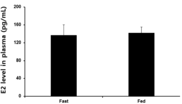
나일 틸라피아 암컷은 다회산란(multiple spawning)하 며 구중부화하는 어종(mouth breeders)이다. 구중부화하는 어종들은 수정된 알을 자신의 구강에서 부화시키기 때문 에 몇 주 동안 먹이 섭취를 거의 하지 못한다. 암컷은 구 중부화 후 체중이 감소하고, 난소주기(ovarian cycles)와 다음 산란시기가 지연된다(Smith and Wootton 1994, 1995; Tacon 1996). 그러나 본 연구에서 나일 틸라피아 암 컷의 경우 번식활동에 관여하는 유전자인 GnRH와 Kiss2 모두 절식에 의한 영향을 받지 않았다(Fig. 2). 두 유전자 모두 앞선 수컷과는 반대로 먹이를 섭취한 그룹보다 절식
한 그룹에서 약간 높은 발현양상을 나타내었지만 두 그룹 간의 유의한 차이는 없었다. 또한 암컷의 혈장 내 E
2의 농 도도 절식한 그룹에서 136.9 ± 23.27 pg/mL, 먹이를 섭취 한 그룹에서는 142 ± 13.1 pg/mL로 비슷한 수준을 나타내 면서 10일 동안의 절식에 영향을 받지 않았음을 확인하였 다(Fig. 4). 이와 같이 수컷과 다른 결과는 성별에 따른 차 이에 기인할 수도 있지만, 구중부화를 위해 수 주간 절식 을 하는 나일 틸라피아 암컷을 대상으로 절식에 의한 영 향을 확인하기에는 절식기간이 짧았을 가능성이 있다. 실 제로 구중부화를 하는 암컷 african cichlid fish(Astatotilapia burtoni)의 경우 4주 동안 절식 후에 GnRH 유전자 발현과 혈장 내 E
2의 농도가 감소되었다(Grone et al. 2012). 이는 암컷 나일 틸라피아도 절식이 번식관련 인자들에 영향을 줄 수 있음을 간접적으로 나타내는 결과이며, 절식기간 을 늘리는 등의 추가적인 실험을 통해 조사해 볼 필요가 있다.
앞선 결과들을 종합해 볼 때 비록 암컷에서는 절식에 의한 영향을 확인하지 못하였지만, 수컷의 경우 절식이 kisspeptin 을 포함한 번식관련 인자들에게 영향을 줄 수 있 음을 확인하였다. 또한 절식에 의해 동반 감소된 수컷의 kisspeptin과 GnRH 유전자 발현양상으로 미루어 볼 때, kisspeptin 이 나일 틸라피아 수컷의 먹이 섭취와 GnRH를 매개할 가능성이 있다. 그러나 두 유전자는 ghrelin, leptin, insulin 등 먹이 섭취와 관련한 인자들에게 각각 영향을 받을 가능성이 존재한다. 따라서 절식 → 시상하 부(kisspeptin →GnRH)→생식소(sex steroid)로 이어지는 연결 과정을 보다 명확히 밝히기 위해서는 먹이 섭취와 관련한 인자들과의 관련성을 조사하는 추가적인 연구가 요구된다.
Fig. 4. Effects of fasting on the plasma level of E
2in female Nile tilapia. E
2level was read at 450 nm and calculated using logistic (log-log) curve-fit standard curve (R
2> 0.99) which were generated by SOFTmax pro. Results are means ± S.E.M.
(n = 6)
사 사
이 논문은 2010년도 정부(교육부)의 재원으로 한국연구 재단의 지원을 받아 수행된 기초연구사업입니다(No.
2010-0024441).
참고문헌