Korean J Radiol 9(1), February 2008 91
Calcifying Aponeurotic Fibroma with Osseous Involvement of the Finger:
a Case Report with Radiologic and US Findings
Calcifying aponeurotic fibroma is a rare soft tissue tumor that occurs in the dis- tal extremities of children and adolescents. We report ultrasound and X-ray find- ings of a calcifying aponeurotic fibroma in the finger of a 36-year-old woman, associated with distal phalangeal bone involvement.
alcifying aponeurotic fibroma is a rare, locally aggressive fibroblastic lesion occurring primarily in the palms of the hands and soles of the feet in young children and adolescents under 20 years of age. Clinical presen- tation is a unique, hard, and painless palpable mass. This soft tissue tumor typically infiltrates into the surrounding fascia or muscle and has a predilection for recurrence after surgical removal. However, bone involvement in calcifying aponeurotic fibroma is a very rare condition and we found only three pediatric cases in the literature (1 3).
We present the ultrasound and radiographic findings of a calcifying aponeurotic fibroma in the finger of a 36-year-old woman, associated with erosive bone destruc- tion of the distal phalanx.
CASE REPORT
A 36-year-old woman with no history of any trauma exhibited a palpable mass on the volar ulnar aspect of her left middle finger at the level of the distal interphalangeal joint for a year. Physical examination revealed a 1 1.5-cm hard, non-tender, non- movable mass. Motion of the distal interphalangeal joint was normal. Plain radiographs of the finger revealed a calcifying soft tissue mass associated with large cortical scalloping on the volar ulnar side of the distal phalanx (Fig. 1). The distal interphalangeal joint was normal in appearance. On the high-resolution ultrasound (US) exam (HDI5000, Philips Medical System, Bothell, WA) with a high-frequency (15 7 MHz) compact linear transducer, a heterogeneously hyper-echoic mass with surface lobulation and extrinsic cortical erosion by the mass was clearly observable (Fig. 2A). The vascularity of the mass was not detected on color Doppler US (Fig. 2B).
Surgery revealed a flesh-colored lobulated mass was firmly attached to the periosteum of the distal phalanx and eroded it. However, the mass could be excised without any resection of the bone. Microscopic sections revealed a spindle-cell neoplasm with scattered calcifications and chondroid differentiation (Fig. 3). A pathologic diagnosis of calcifying aponeurotic fibroma was made. During the last nine months, the patient has been well with no signs of recurrence.
Soo-Jung Choi, MD1 Jae Hong Ahn, MD1 Gilhyun Kang, MD2 Jong Hyeog Lee, MD1 Man Soo Park, MD1 Dae Sik Ryu, MD1 Seung Moon Jung, MD1
Index terms :
Calcifying aponeurotic fibroma Soft tissue tumor
Ultrasound (US)
DOI:10.3348/kjr.2008.9.1.91
Korean J Radiol 2008 ; 9 : 91-93 Received November 13, 2006; accepted after revision November 30, 2006.
1Department of Radiology and
2Department of Pathology, Asan Foundation, GangNeung Asan Hospital, University of Ulsan College of Medicine, Gangneung 210-711, Korea
Address reprint requests to : Soo-Jung Choi, MD, Department of Radiology, Asan Foundation, GangNeung Asan Hospital, University of Ulsan, College of Medicine, 415 Bangdong-Li, Sachun-Myun, Gangneung 210-711, Korea.
Tel. (8233) 610-3485 Fax. (8233) 610-3111 e-mail: [email protected]
C
DISCUSSION
Calcifying aponeurotic fibroma usually appears in the first decade of life and most of the lesions manifest within
the first 2 decades of life. For this reason, this tumor is also referred to juvenile aponeurotic fibroma. It has a predilec- tion for the palms and soles. The tumor has since been reported in the fingers, wrist, forearm, elbow, upper arm, neck, abdominal wall, lumbar paravertebral area, leg, ankle, and thigh (4, 5). Male patients are twice as commonly affected as female patients (6).
Calcifying aponeurotic fibroma is considered to be a cartilage analogue of fibromatosis. Fibromatoses can be divided into two subdivisions, based on anatomic location and on age of presentation. One is the “juvenile
fibromatoses” including juvenile aponeurotic fibroma, congenital generalized fibromatosis, fibromatosis coli, Choi et al.
92 Korean J Radiol 9(1), February 2008
Fig. 2. A. High-resolution US demonstrates lobulated soft tissue mass (arrows) with internal punctate hyper-echoic foci (calcifications).
The mass is adhered and scalloped the cortex of the phalanx (arrowheads).
B. Color Doppler US shows hypo-vascularity of the mass.
A B
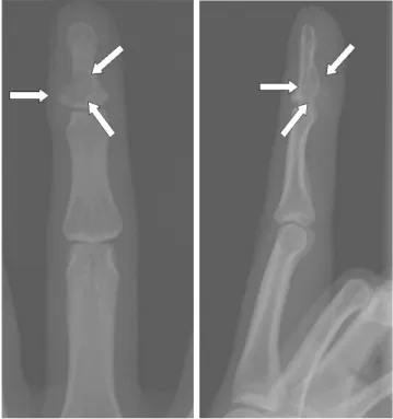
Fig. 1. Left middle finger AP (A) and lateral (B) views show eccentrically located well-defined osteolytic lesion in the base of the distal phalanx (arrows). Calcific foci are noted in the mass and soft tissue mass component is obvious. On these radiographs, soft tissue mass with large cortical erosion is indistinguishable with eccentrically locating osteolytic mass with soft tissue extension.
A B
Fig. 3. Histological section shows scattered calcifications (solid arrows) with surrounding chondroid differentiation (arrowheads) on the background of the fibrosis with interlacing bundles of spindle cells (open arrows) (Hematoxylin and Eosin staining, 40).
fibrous hamartoma of infancy, recurring digital fibrous tumor of Reye, juvenile hyaline fibromatosis, diffuse infantile fibromatosis, and hereditary gingival fibromatosis.
The second subdivision includes the aggressive
fibromatoses, plantar-palmar fibromatosis, and nodular fasciitis (1).
Radiologically, calcifying aponeurotic fibroma may show a soft tissue mass with no associated osseous lesions and a fine stippling of focal calcification (4). However, in
extremely rare cases, occasional scalloping of the cortex (1, 3) and thickening of the bone (2) have been reported in pediatric patients. To our knowledge, there has been no case with involvement of the distal digital phalanx. On MRI, Kwak et al. (7) reported that the signal intensity of the calcifying aponeurotic fibroma was lower than that of muscle on T1- and T2-weighted images. In our case, US also clearly demonstrated the extent and character of the soft tissue mass with internal calcific foci and cortical erosive changes of the adjacent bone. The recurrence rate after surgical excision of this tumor is high and generally reported at 50% (8). This necessitates an accurate diagno- sis and complete excision. Therefore, evaluation of the extent of the mass and its relationship with surrounding soft tissue or bone is important. US and MRI could be helpful modalities of the preoperative imaging.
The differential diagnosis of calcifying aponeurotic fibroma differs depending on the patient’s age, location of the lesion, presence of the calcification, and osseous involvement. Calcifying aponeurotic fibroma in adults should be differentiated from other fibrous tumors such as aggressive fibromatoses, plantar-palmar fibromatosis, and nodular fasciitis. However, in this case, considering distal phalangeal erosive soft tissue mass with calcification, the
differential diagnoses were parosteal/soft tissue chondroma, synovial sarcoma, and the calcified epider- moid.
In summary, calcifying aponeurotic fibroma is a rare soft tissue tumor that presents as a painless mass primarily on the volar surface of the hands and plantar aspects of the feet in juveniles, but this tumor should be also included in differential diagnoses of any mass with calcification and adjacent bone involvement in the distal phalanx of the finger. In addition, US could be useful for the preoperative evaluation of digital calcifying aponeurotic fibroma.
References
1. Karasick D, O’Hara AE. Juvenile aponeurotic fibroma. A review and report of a case with osseous involvement.
Radiology 1977;123:725-726
2. Robbin MR, Murphey MD, Temple HT, Kransdorf MJ, Choi JJ.
Imaging of musculoskeletal fibromatosis. Radiographics 2001;21:585-600
3. Rahmi M, Chakkouri K, Cohen D, Hassoun J, Trafeh M.
Juvenile aponeurotic fibroma. A case report with a review of the literature. Chir Main 2002;21:33-35
4. DeSimone RS, Zielinski CJ. Calcifying aponeurotic fibroma of the hand. J Bone Joint Surg Am 2001;83:586-588
5. Parker WL, Beckenbaugh RR, Amrami KK. Calcifying aponeu- rotic fibroma of the hand: radiologic differentiation from giant cell tumors of the tendon sheath. J Hand Surg (Am)
2006;31:1024-1028
6. Goldman RL. The cartilage analogue of fibromatosis (aponeu- rotic fibroma). Further observations based on 7 new cases.
Cancer 1970;26:1325-1331
7. Kwak HS, Lee SY, Kim JR, Lee KB. MR imaging of calcifying aponeurotic fibroma of the thigh. Pediatr Radiol 2004;34:438- 440
8. Carroll RE. Juvenile aponeurotic fibroma. Hand Clin 1987;3:219-224
Calcifying Aponeurotic Fibroma with Osseous Involvement of Finger
Korean J Radiol 9(1), February 2008 93