Pilomatricoma, or calcifying epithelioma of Malherbe, is a benign, usually asymptomatic tumor arising from hair follicular matrix cells. It is usually a solitary lesion and is most commonly found on the face and upper ex- tremities, appearing as a firm, dermal nodule covered by the normal skin. Although the tumor can occur in pa- tients of any age, about 60% of pilomatricomas are found in those aged less than twenty (1, 2). Most cases involve a single nodule, but multiplicity has been ob- served (3). Diagnosis is usually based on palpation of a superficial, hard mass and confirmed by histopathologic examination. The authors believe, however, that the ul- trasonographic findings of pilomatricoma facilitate accu- rate diagnosis, and if a calcified soft tissue mass is seen at ultrasonography, pilomatricoma should be included in the differential diagnosis.
Case Report
An otherwise healthy 22-year-old man presented with a two-month history of an expanding mass in the left in- terscapular area. He had no other associated symptom.
At initial physical examination, the tumor appeared as a superficial, bullous soft tissue mass with reddish discol- oration, and included a hard, internal nodule (Fig. 1A).
Possible clinical diagnoses at that time included der- moid, fibroma, and hemangioma. Plain radiography de- picted a well-defined bulging mass with tiny calcifica- tions in its central portion (Fig. 1B), and ultrasonography demonstrated a heterogeneously hypoechoic mass with multiple hyperechoic foci. There was no internal vascu- larity (Figs. 1C, D). The ultrasonic differential diagnoses were cartilaginous soft tissue mass, dermoid, heman- gioma and malignant soft tissue tumor. There was no detectable abnormality in complete blood count, blood chemistry (including serum calcium, phosphorous, and alkaline phosphatase)or urinalysis. The mass, about 3cm in diameter, was successfully excised with 1cm of safety margin. Grossly, its internal texture was wheel- like, a circular pattern of friable tissue was apparent,
J Korean Radiol Soc 2003;48:189-192
─ 189 ─
Pilomatricoma: Case Report1
Dong Hun Kim, M.D., Kyu Sung Cho, M.D.2, Do Hyun Kim, M.D.3
Pilomatricoma is a rare benign neoplasm arising from hair follicular matrix cells and exhibiting slow growth. The radiologic features of this neoplasm have rarely been de- scribed in the literature; in particular, the ultrasonographic findings have not been published in Korea. We report a case of pilomatricoma presenting as a well-marginated soft tissue mass with calcification in the dermis and with overlying bullous skin, a rare clinical variant.
Index words : Skin, neoplasms Skin, US
Pilomatricoma, neoplasms
1Department of Diagnostic Radiology, Armed Forces Kwang Ju Hospital
2Department of Plastic Surgery, Armed Forces Kwang Ju Hospital
3Department of Dermatology, Armed Forces Kwang Ju Hospital Received June 5, 2002 ; Accepted September 26, 2002
Address reprint requests to : Dong Hun Kim, M.D., Department of Radiology, Armed Forces Kwang Ju Hospital, 66 Hwajeong-dong, Seo-gu, Gwangju 502-240, Korea.
Tel. 82-61-390-5129 Fax. 82-62-381-3946
Dong Hun Kim, et al: Pilomatricoma
─ 190 ─
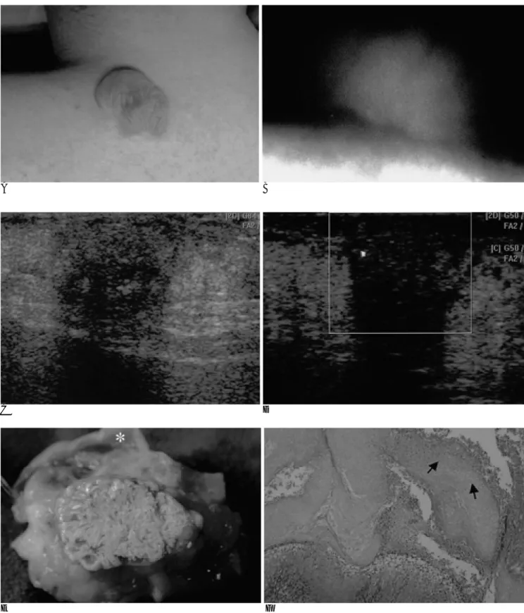
A B
C D
E F
Fig. 1. A 22-year-old man with pilomatricoma.
A. On physical examination, a bullous soft tissue mass with reddish discoloration is in the left interscapular area.
B. Plain radiograph shows the soft tissue mass with tiny calcifications.
C, D. Ultrasonograms demonstrate a heterogeneous hypoechoic mass located in the dermis. There is no vascularity. Multiple echogenic foci show posterior shadowings.
E. Cross section of the specimen shows calcified tissue arranged in a circular pattern. The mass is covered with the bullous skin (as- terisk).
F. Photomicrograph of histologic preparation (hematoxylin-eosin, original magnification ×100) shows the characteristic shadow cells (arrows). There is transition from nucleated basophilic cells to enucleated shadow cells.
and the mass was covered with bullous skin (Fig. 1E).
Pathologically, it consisted of pale, cornified cells, the nuclei of which had almost disappeared, and on the ba- sis of these and other pathologic findings was confirmed as a typical pilomatricoma (Fig. 1F).
Discussion
In 1880, Malherbe and Chenantais first described a certain benign tumor of the skin, thought to be derived from sebaceous glands and arising in the subcutis, and gave it the name, a calcifying epithelioma, wishing to correctly describe its origin in hair follicle matrix cells, Forbis and Helwig renamed the neoplasm ‘pilomatrixo- ma’(4).
Pilomatricoma has a stony consistency, which is its pathognomonic feature. The tumor slides freely over the underlying layer, and the overlying skin may have a reddish or bluish discoloration. It is well-circumscribed, somewhat friable when grasped firmly, and has a gray- ish-tan appearance. Patient are usually asymptomatic, but certain reports have mentioned that there may be associated pain during episodes of inflammation or ul- ceration (5).
Histopathologically, the tumor consists of irregularly shaped islands of epithelial cells arranged in a circular pattern. These islands are composed of two cell types:
(1) basaloid cells that are nucleated and arranged along the periphery of the tumoral islands, and (2) shadow (ghost) cells that are enucleated and located in the center of the islands. Calcification occurs in 70-85% of cases (5, 6). A malignant variety of the neoplasm, with distant metastasis to the lung, bone, brain, skin, and abdominal organs, has been reported (7).
Numerous studies have demonstrated that pilomatri- comas most commonly occur in the head and neck, fol- lowed by the upper extremity, trunk, and lower extrem- ity (8). A review of the literature has revealed female preponderance in those aged under 21. The neoplasm is mostly solitary, though synchronous multiplicity ac- counts for 2-3.5% of reported cases (3). A pilomatrico- ma can also be associated with Gardner syndrome, my- otonic muscular dystrophy, sarcoidosis, skull dysostosis, Rubinstein-Taybi syndrome, and Turner syndrome.
The tumor can usually be diagnosed solely on the ba- sis of its clinical features. However, various imaging modalities help the diagnostic process. Plain radi- ographs show nonspecific foci of calcification, while computed tomography depicts a well-marginated subcu-
taneous soft tissue mass adherent to the skin and with or without visible calcification (9). Magnetic resonance imaging demonstrates intermediate intensity at T2WI and slight enhancement at contrast-enhanced T1WI, mottled hypointense components corresponding to ar- eas of calcification (10). Ichikawa et al. (10) reported a case of giant pilomatricoma that was depicted at angiog- raphy as a hypervascular mass. Ultrasonography, a rela- tively fast and noninvasive technique, reveals the pres- ence of a well-defined, round, hyperechoic mass with a posterior dense acoustic shadow (6). A search of the lit- erature has failed to locate a description of the ultra- sonographic findings of this uncommon tumor in Korea, and this may well be the first such report to appear here.
In our case, ultrasonography demonstrated a well-mar- ginated heterogeneous, hypoechoic mass, unlike those described in the previous literature (6). The observed multiple echogenic foci with posterior acoustic shadow- ing indicated the presence of calcification. We believe that ultrasonographic findings mimicking a cartilaginous soft tissue mass, as in our case, are nonspecific, but that ultrasonographic findings supported by clinical informa- tion will help accurate diagnosis.
Since spontaneous regression of a pilomatricoma has never been observed, surgical excision is mandatory.
Occasionally, overlying skin may adhere to the tumor, requiring simultaneous excision of both, as in the pre- sent case, where overlying bullous skin was excised along with the tumor. Most recurrences arise due to in- completely excised neoplasms (6), and to minimize this risk, wide resection is recommended.
In conclusion, pilomatricoma is a rare, benign skin tu- mor derived from the matrix cells of the hair follicle. It has a characteristic clinical presentation, and the clinical diagnosis should thus be suspected at physical examina- tion. We suggest, however, that if ultrasonography re- veals a well-marginated, subcutaneous hypoechoic cal- cified mass adherent to the skin, and its nature has not been clinically proven, pilomatricoma should be includ- ed in the differential diagnosis when the tumor mimicks a cartilaginous soft tissue mass.
References
1. Yencha MW. Head and neck pilomatricoma in the pediatric age group: a retrospective study and literature review. Int J Pediatr Otorhinolaryngol 2001;57:123-128
2. Thomas RW, Perkins JA, Ruegemer JL, Munaretto JA. Surgical ex- cision of pilomatrixoma of the head and neck: a retrospective re- view of 26 cases. Ear Nose Throat J 1999;78(8):541, 544-546, 548 J Korean Radiol Soc 2003;48:189-192
─ 191 ─
3. Rotenberg M, Laccourreye O, Cauchois R, Laccourreye L, Putterman M, Brasnu D. Head and neck pilomatrixoma. Am J Otolaryngol 1996;17:133-135
4. Forbis R, Helwig EB. Pilomatrixoma (calcifying epithelioma). Arch Dermatol 1961;83:606-608
5. Duflo S, Nicollas R, Roman S, Magalon G, Triglia JM.
Pilomatrixoma of the head and neck in children: a study of 38 cas- es and a review of the literature. Arch Otolaryngol Head Neck Surg 1998;124(11):1239-1242
6. Boyd AS, Martin RW 3rd. Pathologic quiz case 1. Pilomatrixoma (calcified epithelioma of Malherbe) with secondary ossification.
Arch Otolaryngol Head Neck Surg 1992;118:212-215
7. Waxtein L, Vega E, Alvarez L, Cortes-Franco R, Hojyo T, Dominguez-Soto L. Malignant pilomatricoma: a case report. Int J Dermatol 1998;37(7):538-540
8. Domanski AM, Domanski HA. Cytology of pilomatrixoma (calci- fying epithelioma of Malherbe) in fine needle aspirates. Acta Cytol 1997;41:771-777
9. Lee KH, Kim HJ, Suh CH. Pilomatricoma in the head and neck:
CT findings in three patients. J Comput Assist Tomogr 2000;24(2):
332-335
10. Ichikawa T, Nakajima Y, Fujimoto H, et al. Giant calcifying ep- ithelioma of Malherbe (pilomatricoma): imaging features. Skeletal Radiol 1997;26:602-605
Dong Hun Kim, et al: Pilomatricoma
─ 192 ─
대한방사선의학회지 2003;48:189-192
모낭기질세포종: 증례 보고1
1국군광주병원 방사선과
2국군광주병원 성형외과
3국군광주병원 피부과 김동훈・조규성2・김도현3
모낭기질세포종은 모낭의 기질세포에서 기원하여 서서히 자라는 드문 양성종양이다. 이 종양에 대한 방사선학적 소 견은 드물게 보고되었고, 특히 초음파영상은 국내에 보고된적이 없다. 최근 저자들은 수포성 피부로 덮혀있으며 초음파 상 석회화가 동반된 경계가 좋은 저에코의 모낭기질세포종을 경험하였다. 따라서 저자들은 문헌고찰과 함께 드문 임상 양상의 모낭기질세포종을 보고하고자 한다.