Drug-induced liver injury (DILI), including herbal and dietary supplement hepatotoxicity, is often passed lightly; however, it can lead to the requirement of a liver transplant or may even cause death because of liver failure. Recently, the American College of Gastroenterology, Chinese Soci- ety of Hepatology and European Association for the Study of the Liver guidelines for the diagno- sis and treatment of DILI have been established, and they will be helpful for guiding clinical treatment decisions. Roussel Uclaf Causality Assessment Method scoring is the most commonly used method to diagnose DILI; however, it has some limitations, such as poor validity and repro- ducibility. Recently, studies on new biomarkers have been actively carried out, which will help di- agnose DILI and predict the prognosis of DILI. It is expected that the development of new thera- pies such as autophagy inducers and various other technologies of the fourth industrial revolu- tion will be applicable to DILI research.
Keywords: Diagnosis; Drug-induced liver injury; Herb; Management
Drug-induced liver injury
Jeong Ill Suh
Department of Internal Medicine, College of Medicine, Dongguk Unversity, Gyeongju, Korea
Introduction
In a narrow sense, drug-induced liver injury (DILI) deals with liv- er injury caused only by drugs. Herb-induced liver injury can be described as hepatotoxicity caused by herbal medicines other than drugs, but it is named herbal and dietary supplement (HDS) hepatotoxicity as a comprehensive concept that includes herbal medicine, health food, and folk remedies. In general, DILI is a liv- er injury caused by over-the-counter drugs, herbal medicines, health foods, folk remedies, and environmental hormones, as well as by a wide range of prescription drugs including HDS.
Although DILI is a concern in several clinical fields, it is often overlooked. DILI can progress into a chronic liver injury that lasts more than six months and can reach hepatic failure, which re- quires liver transplantation, or death [1-4].
Further, DILI not only hurts the health of the patient, but also has very serious personal, social, and national problems, including the financial burden of payment for treatment, loss of insurance fi- nances, distrust of prescription doctors, medical disputes sur-
Yeungnam Univ J Med 2020;37(1):2-12 https://doi.org/10.12701/yujm.2019.00297
Received: July 30, 2019 Revised: August 10, 2019 Accepted: August 22, 2019 Corresponding author:
Jeong Ill Suh
Department of Internal Medicine, College of Medicine, Dongguk University, 87 Dongdae-ro, Gyeongju 38067, Korea Tel: +82-54-770-8207 Fax: +82-54-770-8378 E-mail: [email protected]
rounding compensation issues, and the withdrawal of drugs de- veloped by investing enormous amounts of time and money from the market [5,6].
Therefore, basic knowledge and recent research trends about DILI are introduced.
Guidelines for drug-induced liver injury
In 2014, the American College of Gastroenterology (ACG) first established the ACG guideline, a diagnostic and treatment guide- line for the idiosyncratic DILI [7]. It presents an evidence-based approach for the diagnosis and management of DILI with special emphasis on DILI caused by HDS and DILI occurring in individ- uals with underlying liver disease. In 2017, the Chinese Society of Hepatology (CSH) established the CSH guideline, a diagnosis and treatment guideline for DILI that covers 16 evidence-based recommendations on diagnosis, differential diagnosis, treatment, and prevention of DILI [8]. Recently, the 2019 European Associ- ation for the Study of the Liver guideline presenting the available
Copyright© 2020 Yeungnam University College of Medicine
This is an Open Access article distributed under the terms of the Creative Commons Attribution Non-Commercial License (http://creativecommons.org/licenses/by-nc/4.0/) which permits unrestricted non-commercial use, distribution, and reproduction in any medium, provided the original work is properly cited.
evidence on risk factors, diagnosis, management, and risk minimi- zation strategies for DILI was also established [9].
Online information resource on drug- induced liver injury
The liver disease research branch of the National Institute of Dia- betes and Digestive and Kidney disease, in collaboration with the National Library of Medicine and Drug-Induced Liver Injury Network (DILIN), has developed an online resource for informa- tion on DILI resulting from prescription and over-the-counter drugs as well as from complementary and alternative medicines such as HDS. The web-based resource called “LiverTox,” provides up-to-date, accurate, and easy-to-access information about the di- agnosis, cause, prevalence, pattern, and management of DILI (http://livertox.nih.gov). China also provides a web-based re- source called “Hepatox” on DILI (http://hepatox.org); however, it is not very helpful as it is only in Mandarin.
Definition of drug-induced liver injury and categorization according to R-ratio
DILI is defined as a liver injury caused by various drugs, herbs, or other xenobiotics leading to abnormalities in liver function. Terms such as hepatitis, liver cirrhosis, and liver necrosis should be used only to support liver biopsy findings; the term liver injury should be used if biochemical abnormalities are present and a liver biopsy has not been conducted. Clinical chemistry criteria for DILI is de- fined as elevation of the alanine aminotransferase (ALT) or the as- partate aminotransferase (AST) >5 ×upper limit of normal (ULN) without symptoms, or rise of alkaline phosphate (ALP)
>2×ULN or rise of bilirubin >2×ULN with any rise of AST and ALT, or rise of AST or ALT <5 ULN with symptoms [10,11].
If liver injury recovers within 6 months, it is called acute liver in- jury (ALI), and if it persists for more than 6 months, it is said to be a chronic liver injury. ALI accounts for most cases of DILI and it is divided into hepatocellular, cholestatic, and mixed types based on the R-ratio. The R-ratio is calculated by dividing ALT by ALP, us- ing multiples of ULN for both values. R-ratios of > 5 define hepa- tocellular; < 2, cholestatic; and between 2 and 5, a mixed pattern of enzymes [12].
Spectrum of drug-induced liver injury
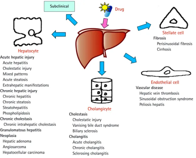
The DILI spectrum is variable and broad; in most cases, hepato- cytes are damaged, but cholangiocytes, stellate cells, and sinusoi- dal endothelial cells can also be damaged, and several other types
of cells can also be damaged simultaneously. DILI can manifest it- self as almost any kind of liver disease, from acute hepatitis to chronic hepatitis, fatty liver or steatohepatitis, vascular damage, liver cirrhosis, and even hepatic tumors (Fig. 1) [13,14].
Incidence of drug-induced liver injury
It is reported that the annual incidence of DILI is between 10 and 15 per 10,000 to 100,000 persons [15,16]. However, the actual incidence is estimated to be higher because diagnosis is not easy, and it is often disregarded unintentionally and is therefore not re- ported in the literature. A prospective study on DILI conducted in Korea estimated that 12 out of every 100,000 persons are admit- ted to university hospitals per year (data for 2005–2007) [17].
Diagnosis of drug-induced liver injury
1. History taking
The most important factor for the diagnosis of DILI is careful his- tory taking because DILI is a diagnosis of exclusion. The history of drug administration and the onset and progression of liver bio- chemical abnormalities must be accurate.
2. Roussel Uclaf Causality Assessment Method
The Roussel Uclaf Causality Assessment Method (RUCAM) is a diagnostic tool that makes a probabilistic decision using a score- card divided into 7 categories. The total scores ranges from less than 0 to 14, and the final score is interpreted as follows: highly probable (>8), probable (6–8), possible (3–5), unlikely (1–2), or excluded (<0) (Table 1) [18]. Currently, scores are conveniently calculated using the website (http://www.pmidcalc.org/?sid
=8229110&newtest=Y). Further, RUCAM can be easily used in clinical fields; however, in the case of liver transplantation for he- patic failure and HDS hepatotoxicity caused by Chinese medicine, health food, and folk remedies, the RUCAM scores may be low. In addition, the reproducibility of scoring is low [19].
3. Drug-Induced Liver Injury Network expert opinion DILIN is a network of US experts who have been conducting DI- LI-related research since 2004 [20]. The probability of DILI is di- vided into 5 categories: definite (95% or more), high likely (75%–
95%), probable (50%–74%), possibly (25%–49%), and unlikely (25% or less) (Table 2) [21]. Three DILIN experts take a decision based on data recorded for more than 6 months. There is a limita- tion where only DILIN experts can make decisions; however, there is a high reproducibility advantage over the RUCAM scoring.
Hepatocyte Acute hepatic injury
Acute hepatitis Cholestatic injury Mixed patterns Acute steatosis
Extrahepatic manifestations Chronic hepatic injury
Chronic hepatitis Chronic steatosis Steatohepatitis Phospholipidosis Chronic cholestasis
Chronic intrahepatic cholestasis Granulomatous hepatitis Neoplasia
Hepatic adenoma Angiosarcoma
Hepatocellular carcinoma
Cholangicyte Cholestasis
Cholestatic injury
Vanising bile duct syndrome Biliary sclerosis
Cholangitis Acute cholangitis Chronic cholangitis Sclerosing cholangitis
Endothelial cell Vascular disease
Hepatic vein thrombosis Sinusoidal obstruction syndrome Peliosis hepatis
Stellate cell Fibrosis
Perisinusoidal fibrosis Cirrhosis
Drug
Fig. 1. Spectrum of drug-induced liver injury. The spectrum of drug-induced liver injury is variable and broad from asymptomatic to liver failure. Drugs can damage not only hepatocytes but also cholangiocytes, stellate cells, sinusoidal endothelial cells, and they can cause acute hepatitis, chronic hepatitis, granulomatous hepatitis, neoplasia, cholestasis, cholangitis, vascular disease, and fibrosis.
Table 1. Roussel Uclaf Causality Assessment Method scale
Category Score Likelihood (%) Description
Highly probable > 8 > 75 Highly probable, including “highly likely” and “definite.” The evidence for the drug causing the injury is beyond a reasonable doubt, clear, and convincing
Probable 6–8 50–74 The preponderance of the evidence supports the link between the drug and the liver injury Possible 3–5 25–49 The evidence for the drug causing the injury is equivocal but present
Unlikely 1–3 < 25 There is evidence that an etiological factor other than a drug caused the injury
Excluded < 1 0 Causes could be excluded
Table 2. Drug-induced Liver Injury Network scale
Category Score Likelihood (%) Description
Definite 1 > 95 The evidence for the drug causing the injury is beyond a reasonable doubt Highly likely 2 75–95 The evidence for the drug causing the injury is clear and convincing but not definite Probable 3 50–74 The preponderance of the evidence supports the link between the drug and the liver injury Possible 4 25–49 The evidence for the drug causing the injury is equivocal but present
Unlikely 5 < 25 There is evidence that an etiological factor other than a drug caused the injury
Not applicable 0 0 Key elements of the drug exposure history, initial presentation, alternative diagnoses, and/or diagnostic evaluation prevent one from determining a causality score
Subclinical
4. Liver biopsy
Although there are no characteristic pathological indicators for DILI, sometimes characteristic pathological findings based on the drug may appear, and this can help identify other liver diseases and the severity of liver injury. According to the ACG guideline, a liver biopsy should be considered if autoimmune hepatitis re- mains a competing etiology and if immunosuppressive therapy is contemplated. Moreover, a liver biopsy may be considered if the liver biochemistries continues to increase or the liver function de- teriorates despite the interruption of a suspected drug; if the peak ALT level has not dropped by > 50% at 30–60 days after onset in cases of hepatocellular DILI; if the peak ALP level has not dropped by > 50% at 180 days in the cases of cholestatic DILI de- spite stopping the suspected offending agent; in cases of DILI where continued use or re-exposure to the implicated agent is ex- pected; or if liver biochemistry abnormalities persist beyond 180 days in the evaluation for the presence of chronic liver diseases and chronic DILI [7].
5. Biomarkers
Recently, there has been a growing interest in finding biomarkers that predict the occurrence of DILI, accurately diagnose it, and predict a poor prognosis. Biomarker candidates such as glutamate dehydrogenase, high-mobility group box 1, and keratin-18 have been found [22], and DILI studies using micro-ribonucleic acid, high throughput proteomics, genomics, and metabolomics have been attempted [23,24].
Mechanisms of drug-induced liver injury
Traditionally, DILI has been reported to be caused by intrinsic and idiosyncratic reactions, and sometimes, both may occur to- gether. Intrinsic liver injury is dose dependent and predictable.
On the other hand, idiosyncratic liver injury is divided into im- munoallergic and metabolic idiosyncratic reactions regardless of dose, and it is unpredictable.
However, Lammert et al. [25] reported the relationship be- tween the daily dose of oral medications and idiosyncratic DILI.
Higher daily doses (> 50 mg/day) were associated with serious hepatic events such as liver failure, liver transplantation, and death, but there was no association with lower daily doses (< 10 mg).
Thus, dosage also appears to play a role in idiosyncratic liver inju- ry. The traditional mechanism identified only the upstream of the mechanism of intrinsic and idiosyncratic reactions that cause liver injury. The current concepts of mechanisms in DILI focused not only on the upstream but also on the downstream. In other words, the hepatocyte injury mechanism is divided into 3 stages: hepato-
cyte injury (first stage), mitochondria permeability transition (second stage) belonging to downstream, and hepatocyte death (last stage) (Fig. 2) [26]. The initial hepatocyte injury caused by the drug does not uniformly progress to the third stage, but the in- jured hepatocyte may be recovered with the defense and regener- ation ability of the patient. In addition, various environmental and genetic factors are involved in each stage, and the degree of indi- vidual liver injury is different.
In addition to hepatocellular injury caused by drugs, drug-in- duced cholestasis is also important (Fig. 3). There are 3 triggering factors that induce cholestasis, including effects on drug trans- porters, various hepatocellular changes, and altered bile canaliculi dynamics [27]. The function of membrane drug transporters in- volved is inhibited, resulting in cholestasis. In addition, the drug metabolite that exits through the canalicular membrane damages the cholangiocyte, causing cholestasis. Liver injury may be exacer- bated as the accumulation of drugs in the liver via enterohepatic circulation or cholehepatic shunt.
In addition, liver injury may be further exacerbated by trans- mitting hepatic injury and inflammatory signals to neighboring cells through other gap junctions, and the liver injury may be tar- geted to non-parenchymal cells other than hepatocytes and chol- angiocytes [28].
Individual differences in drug-induced liver injury
Some people suffer liver injuries and others do not because every individual has different susceptibility to drugs. The risk factors for DILI can be divided into genetic factors and environmental fac- tors [29,30]. Genetic factors include mutations in the cyto- chromes P450 enzyme, the expression of transport proteins and nuclear receptors, and changes in the levels of immune compo- nents. Environmental factors include old age, female gender, drug combination, previous drug adverse reactions, nutritional status, pregnancy, alcohol consumption, inflammation, and existing dis- eases (Table 3). The important fact is that the different risk factors are involved in different three-step model concepts that describe the changed liver injury mechanism. Changes in gut microbiota affect the drug metabolism and immune system, and it is interest- ing that it is one of the risk factors of DILI [31].
Drug-induced liver injury in patients with pre-existing liver disease
There is much debate over whether DILI occurs more frequently in patients with pre-existing liver diseases than in normal people.
Fig. 2. Mechanism of drug-induced liver injury. The hepatocyte injury mechanism is divided into three stages: (1) first stage (initial hepatocellular injury); initial injury is exerted through direct cell stress, direct mitochondrial inhibition, and/or specific immune reactions;
(2) second stage (mitochondrial permeability transition); initial injury can lead to MPT. Direct cell stress causes MPT via the intrinsic pathway, and (3) final stage (hepatocyte death); MPT leads to necrosis or apoptosis depending on the availability of ATP, which is not uniformly progressive from the initial hepatocyte injury to the third stage, and damaged hepatocytes can be recovered depending on their defense and regeneration ability. Various environmental factors and genetic factors are involved in each step, and the degree of individual liver injury is different. MTP, mitochondrial permeability transition; ATP, adenosine triphosphate; CYP, cytochrome P450; NAt2, N-acetyltransferase 2; GST, glutathione S-transferase; UGT2B7, glycosyltransferase 2B7; BSEP, bile salt export pump; MDR3, multidrug resistance protein 3; MRP2, multidrug resistance-associated protein 2; OATP, organic-anion-transporting polypeptide; PXR, pregnane X receptor; CAR, constitutive androstane receptor; HLA, human leukocyte antigen; IL, interleukin; TNR-α, tumor necrosis factor-alpha;
SOD2, superoxide dismutase 2; Nrf2, nuclear factor erythroid 2-related factor 2; GSH, glutathione S-transferase; EtOH, ethanol.
There are several exceptions, but they are not more frequent.
However, it is known that DILI is more critical and has a higher mortality rate in patients with pre-existing liver diseases [32,33].
Predictive model of progression to acute liver failure
DILI is usually reversible and considered benign; however, it sometimes progresses to hepatic failure, requiring liver transplan-
tation or causing death. According to Hyman Zimmerman’s Hy’s law, the hepatocellular type of DILI is known to have a mortality rate of over 10% if accompanied by jaundice [34,35]. Hy’s law is known to have a high specificity (0.92) but a low sensitivity (0.68) for the prediction of acute liver failure (ALF) [36].
A new index predicting ALF in DILI has been recently pro- posed by Robles-Diaz et al. [37], which integrates Hy’s law with the new R-ratio (nR) and demonstrates a sensitivity of 90% and a specificity of 63%. The nR is calculated as (the highest AST or
· Inherited mitochondrial disease
· SOD2
· Acquired
· mitochondrial disease
· HLA
· IL-4, IL-6, IL-10
· TNF-α
· Oxidative stress
(SOD2, CYP2E1, GST, Nrf2)
· Infection, inflammation (danger signal)
· GSH depletion from EtOH
· Malnourishment
· Immunological sensitization
· CYP450, NAt2, GST UGT2B7, BSEP, MDR3 MRP2, OATP, PXR, CAR
· Inhibtion or induction of enzyme/transporter
· Caspase?
· Oxidative stress?
(SOD2, CYP2E1, GST)
· Preexisting liver disease
· Additional exposure to hepatotoxin
· ATP depletion Genetic factors
Initial injury Mitochondrial
permeability transition
2nd stage
1st stage 3rd stage
Hepatocyte death
Upstream Downstream
Progression of injury vs.
regeneration Apoptosis
Necrosis 1c. Immune reaction
1b. Direct mitochondrial inhibition 1a. Direct cell stress
Hapten
Inner Membrane
Outer Membrane
Cristae Matrix Pharmacokinetics
Metabolite formation Bioavailability
Cytokine
Environmental factors
Fig. 3. Different mechanism of drug-induced cholestasis. Drug-induced cholestasis is caused by the degradation of the expression and function of the transport protein due to the environmental factors that directly inhibit the function of the transport protein and the genetic variation of the transport protein. In addition, the drug metabolite that exits through the canalicular membrane damages cholangiocyte, causing cholestasis. A liver injury may be exacerbated by drug accumulation in the liver via drug reabsorption through enterohepatic or cholehepatic circulation. Directly inhibtion of BSEP function is cis-inhibtion, whereas indirectly inhibition of BSEP function from the canalicular lumen is trans-inhibtion. OATP, organic-anion-transporting polypeptide; MRP2, multidrug resistance- associated protein 2; BSEP, bile salt export pump; MDR3, multidrug resistance protein 3.
Table 3. Risk factors of drug-induced liver injury
Genetic factor Environmental factor
Phase 1 enzymes Age
CYP2C8, CYP2C9, CYP2C19, CYP2D6, CYP2E1 Sex
Phase 2 enzymes Race
NAT2, GSTM1, GSTT1, UGT2B7 Drug interaction
Phase 3 transporters Alcohol
BSEP, MRP2, MDR3 Inflammation
Phase 0 transporter Underlying disease
OATP
Nuclear receptors Pre-existing liver disease
PXR, CAR HIV
Immunologic Diabetes
HLA class antigen
Cytokines Pregnancy
IL-4, IL-10, TNF-α Nutrition
Mitochondrial mutation Previous history
DNA mutations (POLG), MnSOD Epigenetics
CYP, cytochrome P450; NAT2, N-acetyltransferase 2; GST, glutathione S-transferase; UGT2B7, glycosyltransferase 2B7; BSEP, bile salt export pump;
MRP2, multidrug resistance-associated protein 2; MDR3, multidrug resistance protein 3; OATP, organic-anion-transporting polypeptide; PXR, pregnane X receptor; CAR, constitutive androstane receptor; HIV, human immunodeficiency virus; HLA, human leukocyte antigen; IL, interleukin; TNR-α, tumor necrosis factor-alpha; DNA, deoxyribonucleic acid; POLG, polymerase gamma; MnSOD, manganese-dependent superoxide dismutase.
Trans-inhibition Vesicle transport
inhibition
Drug-drug interaction
Toxic
Hapten Continuous
drug exposure Cholehepatic circulation
Enterohepatic circulation
Covalent binding
Transporter retrieval
Mutant Mutant
Mutant
Mutant BSEP MDR2
MDR3 OATP
ALT/ULN)/(ALP/ULN); AST is substituted for ALT, if the AST yields a greater R-ratio.
Recently, a drug-induced liver toxicity ALF score (DrILTox ALF Score) was developed to predict the progression of liver fail- ure in DILI [36]. Scoring is based on platelet counts and total bili- rubin (TB) [DrILTox ALF Score= −0.00691292× platelet count +0.19091500× TB (per mg/dL)]. Although the specificity (0.76) was slightly lower, the sensitivity (0.91) was higher than those of Hy’s law criteria. The risk of liver failure becomes higher as the platelet count decreases and TB increases.
Herbal and dietary supplement hepatotoxicity
HDS include herbs or other plant materials, vitamins, and miner- als. The incidence of HDS hepatotoxicity is increasing compared to that in the past [38]. In general, it is estimated that HDS hepa- totoxicity is actually more likely to occur because it is not easy to diagnose, it is often missed if the symptoms are overlooked, and often, the cases are not reported in the literature in addition to in- adequate treatment for the patient. It is difficult to detect the toxic substances contained in the herb itself, unlike the commercial medicines whose causative substances are clearly defined, and it is difficult to prove causal relationships between the herb-specific components and the liver injury. Since the herb is a mixture of various substances, it is difficult to know which ingredient causes the liver injury. It can be contaminated with microorganisms or fungi during distribution or storage, or the liver injury may be caused by herb denaturation. It should be noted that there may also be liver injury caused by impurities, heavy metal contamina- tion, or illegal incorporation of drugs into the herb (Fig. 4).
Similar to that for the liver injury caused by commercial drugs, RUCAM scoring is applied to diagnose HDS hepatotoxicity.
However, it is necessary to adjust the RUCAM for HDS hepato- toxicity [39,40] because there are few reports of HDS hepatotox- icity, and thus, the results tend to be lower than the actual RU- CAM scores; further, the time between the termination of herb administration and symptom development is often long as low concentrations of plants are often taken over a long period of time.
Interestingly, Suh et al. [41] showed that psychological factors that present vulnerability to the temptation to use alternative medicines such as herbs and plant preparations are important for understanding toxic liver injury. Therefore, the treatment of toxic liver injury itself is important. However, to prevent toxic liver inju- ry and recurrence, it is necessary to implement an active strategy to understand and improve anxiety and depression faced by the patient.
Treatment of drug-induced liver injury
Stopping the suspected drugs is key to treatment. Other treat- ments involve the administration of N-acetyl cysteine (NAC) and steroids, and liver transplantation are considered when hepatic failure occurs.
1. Stopping the suspected drug
Follow-ups are necessary after stopping not only the suspected drug but also herbal medicines, plant preparations, and health food. Depending on when the drug is discontinued, the severity of the liver injury can vary; therefore, the drug should be stopped as early as possible. Since such liver injuries are reversible to nor- mal state, it is necessary to repeat liver function tests after stopping the medication. In some cases, even if the drug is discontinued, it may not immediately improve the liver condition and the liver in- jury may continue; therefore, careful follow-ups must be per- formed. It is very difficult to assess if the drug should be continued if there is a rise in hepatic enzymes in the liver function test during the course of the treatment. Further, if the suspected causative drug is important for the control of the underlying disease, the balance between the risk of progression of the underlying disease after drug withdrawal and the risk of exacerbation of liver damage due to the continued administration of potentially related drugs should be considered. According to the “stop rule” for new drugs developed by the US Food and Drug Administration (FDA) [42], the guidelines are based on AST, ALT, and TB, and the medica- tion being administered should be immediately stopped when any of the following results are obtained: (1) ALT or AST > 8× ULN;
(2) ALT or AST remains > 5× ULN over 2 weeks; (3) ALT or AST > 3× ULN & TB > 2× ULN or international normalized ra- tio (INR) > 1.5; (4) ALT or AST > 3 × ULN with symptoms (e.g., fatigue, nausea and vomiting, right upper quadrant pain, fe- ver, and rash) or eosinophilia. Therefore, it is acceptable to follow the FDA “stop rule” in clinical practice.
2. Specific treatment
Although no specific therapies are available for DILI, NAC (IV infusion, 50–150 mg/kg/day) may be administered for at least 3 days in patients with early or sub-ALF [43-45]; it is not recom- mended for children with severe DILI as it can lead to ALF. Sever- al studies have shown that steroids can prove effective; however, there have been some debates on the efficacy of corticosteroid in treating patients with DILI [46,47]. In the case of immunoallergic or autoimmune hepatitis-like DILI, the administration of gluco- corticoid may be considered.
3. Re-administration of suspected drugs
Although there is controversy regarding the unconditional re-ad- ministration of all suspected drugs that cause DILI, care should be taken because re-administration of immunoallergic reacting drugs may cause more serious liver damage than before.
4. Liver transplantation
Liver transplantation should be considered if the liver function deteriorates and is concomitant with coagulopathy and encepha- lopathy [48]. Survival is less than 20% if liver transplantation is not performed in drug-induced ALF caused by a hypersensitivity reaction. The recommendations of Kings’ College on indications for liver transplantation due to drug-induced liver failure as fol- lows: In case of acetaminophen-induced liver failure, liver trans- plantation is required when the arterial blood pH is < 7.3, regard- less of encephalopathy grade, or if grade III or IV encephalopathy
and an INR > 6.5 and a serum creatinine > 3.4 mg/dL. Liver transplantation is required for liver failure caused by non-acet- aminophen drugs as follows: patients with prothrombin time (PT) > 100 s (INR > 6.5) (with or without encephalopathy, re- gardless of grade) or who satisfy any 3 of the following criteria: (1) age < 10 or > 40 years of age; (2) etiology: non-A/non-B hepati- tis, drug-induced; (3) duration of jaundice to hepatic encephalop- athy > 7 days; (4) PT > 50 s (INR > 3.5); or (5) serum bilirubin level > 17 mg/dL (> 300 μmol/dL) [49,50].
5. New treatments for drug-induced liver injury
Many studies have been actively pursued to develop therapeutic agents aimed at nuclear receptors in DILI [51,52]. The activation of constitutive androstane receptor (CAR) and pregnane X recep- tor (PXR) exacerbates hepatotoxicity by acetaminophen [53,54].
Thus, compounds that inhibit CAR and PXR may be beneficial Fig. 4. Risk factors contributing to hepatotoxicity of herbal remedies. There are many causative factors for herb-induced liver injury such as the misidentification of the plant, mislabeling of the final product, unstandardized dose, plant-specific toxic substances, various ingredients, denaturalization during inadequate storage, illegal drug incorporation, contamination of the plant by various chemicals, heavy metals, microorganisms, individual difference due to genetic or environmental factors, and herb-drug interactions.
Misidentification of the plant
Unstandardized dose
Plant-specific toxic substances
Various ingredients
Mislabeling of the final product
Herb-drug interaction
Individual difference (genetic, environmental)
Contamination; pesticides, heavy metals (mercury, arsenic, lead), microorganism Illegal drug incorporation
Inadequate storage (denaturalization)
for the treatment of hepatic damage induced by acetaminophen.
Farnesoid X receptor (FXR) plays an important role in the regula- tion of bile acid synthesis and metabolism. Thus, FXR agonists such as obeticholic acid have been considered as promising targets for the treatment of cholestatic disorders involving drug-induced cholestasis.
Autophagy refers to the activity in which a cell obtains energy by dissolving its protein or removing unnecessary cell compo- nents when it becomes nutrient deficient. Recently, studies on au- tophagy have been actively conducted in various areas such as cancer, diabetes, infectious diseases, and Crohn’s disease. For DILI, a new therapeutic approach is being attempted to reduce the liver injury by controlling autophagy [55]. Acetaminophen overdose results in hepatic necrosis caused by mitochondrial damage. The activation of autophagy degrades the damaged cyto- plasmic proteins, which allows cells to survive without cell necro- sis. While liver injury is prevented by the administration of rapa- mycin (autophagy inducer), 3-methyladenine, or chloroquine (autophagy inhibitor), they have been shown to decrease liver in- jury. Further, it is expected that several new treatments, including autophagy induction, will be developed and applied to the treat- ment of DILI in clinical practice.
Drug-induced liver injury and the fourth industrial revolution
DILI has also been actively researched by applying various new technologies of the fourth industrial revolution. Recently, organ- on-a-chip (OOC) such as liver, lung, heart, nerve, and skin have been developed [56,57]. OOC is a technique that imitates the me- chanical and physiological cellular responses as well as the func- tions and characteristics of the organs by culturing cells that consti- tute a living OOC on which electronic circuits are placed. The OOC is worth using as a model for drug development and toxicity assessment. A liver-on-a-chip can be used to evaluate the toxicity of a drug without animal testing [58]. In addition, it is reported that deep learning using artificial intelligence can predict the occur- rence of DILI [59]. It is expected that research using various tech- nologies of the fourth industrial revolution will help predict the side effects and drug–drug interactions in advance through “In-Sil- ico,” a computer virtual test using biological big data [60,61].
Conclusion
DILI is a problem that can be encountered in clinical settings, and clinicians should therefore have appropriate knowledge and diag- nosis and treatment skills. We hope that various biomarkers to ac-
curately diagnose and predict the prognosis of DILI will be devel- oped and used conveniently, and various new technologies of the fourth industrial revolution will be developed and applied to DILI.
Acknowledgments
Conflicts of interest
No potential conflicts of interest relevant to this article was report- ed.
ORCID
Jeong Ill Suh, https://orcid.org/0000-0002-3040-8766
References
1. Chalasani N, Fontana RJ, Bonkovsky HL, Watkins PB, Davern T, Serrano J, et al. Causes, clinical features, and outcomes from a prospective study of drug-induced liver injury in the United States. Gastroenterology 2008;135:1924–34.
2. Hayashi PH, Bjornsson ES. Long-term outcomes after drug-in- duced liver injury. Curr Hepatol Rep 2018;17:292–9.
3. Zhu Y, Niu M, Chen J, Zou ZS, Ma ZJ, Liu SH, et al. Hepatobili- ary and pancreatic: comparison between Chinese herbal medi- cine and Western medicine-induced liver injury of 1985 pa- tients. J Gastroenterol Hepatol 2016;31:1476–82.
4. Lee WJ, Kim HW, Lee HY, Son CG. Systematic review on herb-induced liver injury in Korea. Food Chem Toxicol 2015;84:47–54.
5. Wysowski DK, Swartz L. Adverse drug event surveillance and drug withdrawals in the United States, 1969-2002: the impor- tance of reporting suspected reactions. Arch Intern Med 2005;165:1363–9.
6. Holt MP, Ju C. Mechanisms of drug-induced liver injury. AAPS J 2006;8:E48–54.
7. Chalasani NP, Hayashi PH, Bonkovsky HL, Navarro VJ, Lee WM, Fontana RJ, et al. ACG clinical guideline: the diagnosis and management of idiosyncratic drug-induced liver injury. Am J Gastroenterol 2014;109:950–66.
8. Yu YC, Mao YM, Chen CW, Chen JJ, Chen J, Cong WM, et al.
CSH guidelines for the diagnosis and treatment of drug-in- duced liver injury. Hepatol Int 2017;11:221–41.
9. European Association for the Study of the Liver. EASL clinical practice guidelines: drug-induced liver injury. J Hepatol 2019;
70:1222–61.
10. Fontana RJ, Seeff LB, Andrade RJ, Björnsson E, Day CP, Serra- no J, et al. Standardization of nomenclature and causality assess-
ment in drug-induced liver injury: summary of a clinical re- search workshop. Hepatology 2010;52:730–42.
11. Devarbhavi H. An update on drug-induced liver injury. J Clin Exp Hepatol 2012;2:247–59.
12. Bénichou C. Criteria of drug-induced liver disorders. Report of an international consensus meeting. J Hepatol 1990;11:272–6.
13. Visentin M, Lenggenhager D, Gai Z, Kullak-Ublick GA.
Drug-induced bile duct injury. Biochim Biophys Acta Mol Basis Dis 2018;1864:1498–506.
14. Wang T, Zhao X, Shao C, Ye L, Guo J, Peng N, et al. A proposed pathologic sub-classification of drug-induced liver injury. Hepa- tol Int 2019;13:339–51.
15. Ahmad J, Odin JA. Epidemiology and genetic risk factors of drug hepatotoxicity. Clin Liver Dis 2017;21:55–72.
16. Bell LN, Chalasani N. Epidemiology of idiosyncratic drug-in- duced liver injury. Semin Liver Dis 2009;29:337–47.
17. Suk KT, Kim DJ, Kim CH, Park SH, Yoon JH, Kim YS, et al. A prospective nationwide study of drug-induced liver injury in Korea. Am J Gastroenterol 2012;107:1380–7.
18. Danan G, Benichou C. Causality assessment of adverse reac- tions to drugs--I. A novel method based on the conclusions of international consensus meetings: application to drug-induced liver injuries. J Clin Epidemiol 1993;46:1323–30.
19. Hayashi PH. Overview of causality assessment in drug-induced liver injury. Clin Liver Dis (Hoboken) 2017;9:29–33.
20. Hoofnagle JH. Drug-induced liver injury network (DILIN).
Hepatology 2004;40:773.
21. Hayashi PH. Drug-induced liver injury network causality assess- ment: criteria and experience in the United States. Int J Mol Sci 2016;17:201.
22. Antoine DJ, Williams DP, Kipar A, Jenkins RE, Regan SL, Sathish JG, et al. High-mobility group box-1 protein and keratin-18, cir- culating serum proteins informative of acetaminophen-induced necrosis and apoptosis in vivo. Toxicol Sci 2009;112:521–31.
23. McGill MR, Jaeschke H. Biomarkers of drug-induced liver inju- ry. Adv Pharmacol 2019;85:221–39.
24. Neuman MG. Biomarkers of drug-induced liver toxicity. Ther Drug Monit 2019;41:227–34.
25. Lammert C, Einarsson S, Saha C, Niklasson A, Bjornsson E, Chalasani N. Relationship between daily dose of oral medica- tions and idiosyncratic drug-induced liver injury: search for sig- nals. Hepatology 2008;47:2003–9.
26. Russmann S, Kullak-Ublick GA, Grattagliano I. Current con- cepts of mechanisms in drug-induced hepatotoxicity. Curr Med Chem 2009;16:3041–53.
27. Gijbels E, Vinken M. Mechanisms of drug-induced cholestasis.
Methods Mol Biol 2019;1981:1–14.
28. Patel SJ, Milwid JM, King KR, Bohr S, Iracheta-Vellve A, Li M, et al. Gap junction inhibition prevents drug-induced liver toxici- ty and fulminant hepatic failure. Nat Biotechnol 2012;30:179–
83.
29. Chalasani N, Björnsson E. Risk factors for idiosyncratic drug-in- duced liver injury. Gastroenterology 2010;138:2246–59.
30. Björnsson ES. Epidemiology and risk factors for idiosyncratic drug-induced liver injury. Semin Liver Dis 2014;34:115–22.
31. Li H, He J, Jia W. The influence of gut microbiota on drug me- tabolism and toxicity. Expert Opin Drug Metab Toxicol 2016;
12:31–40.
32. Teschke R, Danan G. Diagnosis and management of drug-in- duced liver injury (DILI) in patients with pre-existing liver dis- ease. Drug Saf 2016;39:729–44.
33. Chalasani N, Regev A. Drug-induced liver injury in patients with preexisting chronic liver disease in drug development: how to identify and manage? Gastroenterology 2016;151:1046–51.
34. Zimmerman HJ. Hepatotoxicity: the adverse effects of drugs and other chemicals on the liver. 2nd ed. Philadelphia: Lippin- cott Williams & Wilkins; 1999.
35. Reuben A. Hy’s law. Hepatology 2004;39:574–8.
36. Lo Re V 3rd, Haynes K, Forde KA, Goldberg DS, Lewis JD, Car- bonari DM, et al. Risk of acute liver failure in patients with drug-induced liver injury: evaluation of Hy’s law and a new prog- nostic model. Clin Gastroenterol Hepatol 2015;13:2360–8.
37. Robles-Diaz M, Lucena MI, Kaplowitz N, Stephens C, Medi- na-Cáliz I, González-Jimenez A, et al. Use of Hy’s law and a new composite algorithm to predict acute liver failure in patients with drug-induced liver injury. Gastroenterology 2014;147:109–18.
38. de Boer YS, Sherker AH. Herbal and dietary supplement-in- duced liver injury. Clin Liver Dis 2017;21:135–49.
39. García-Cortés M, Stephens C, Lucena MI, Fernández-Castañer A, Andrade RJ. Causality assessment methods in drug induced liver injury: strengths and weaknesses. J Hepatol 2011;55:683–
91.
40. Teschke R, Wolff A, Frenzel C, Schwarzenboeck A, Schulze J, Eickhoff A. Drug and herb induced liver injury: Council for In- ternational Organizations of Medical Sciences scale for causality assessment. World J Hepatol 2014;6:17–32.
41. Suh JI, Sakong JK, Lee K, Lee YK, Park JB, Kim DJ, et al. Anxi- ety and depression propensities in patients with acute toxic liver injury. World J Gastroenterol 2013;19:9069–76.
42. Ford R, Schwartz L, Dancey J, Dodd LE, Eisenhauer EA, Gwyther S, et al. Lessons learned from independent central re- view. Eur J Cancer 2009;45:268–74.
43. Lee WM, Hynan LS, Rossaro L, Fontana RJ, Stravitz RT, Larson AM, et al. Intravenous N-acetylcysteine improves trans-
plant-free survival in early stage non-acetaminophen acute liver failure. Gastroenterology 2009;137:856–64.
44. Bateman DN, Dear JW, Thanacoody HK, Thomas SH, Ed- dleston M, Sandilands EA, et al. Reduction of adverse effects from intravenous acetylcysteine treatment for paracetamol poi- soning: a randomised controlled trial. Lancet 2014;383:697–
704.
45. Singh S, Hynan LS, Lee WM; Acute Liver Failure Study Group.
Improvements in hepatic serological biomarkers are associated with clinical benefit of intravenous N-acetylcysteine in early stage non-acetaminophen acute liver failure. Dig Dis Sci 2013;
58:1397–402.
46. Hu PF, Xie WF. Corticosteroid therapy in drug-induced liver in- jury: pros and cons. J Dig Dis 2019;20:122–6.
47. Hu PF, Wang PQ, Chen H, Hu XF, Xie QP, Shi J, et al. Beneficial effect of corticosteroids for patients with severe drug-induced liver injury. J Dig Dis 2016;17:618–27.
48. Reuben A, Koch DG, Lee WM; Acute Liver Failure Study Group. Drug-induced acute liver failure: results of a U.S. multi- center, prospective study. Hepatology 2010;52:2065–76.
49. Mindikoglu AL, Magder LS, Regev A. Outcome of liver trans- plantation for drug-induced acute liver failure in the United States: analysis of the United Network for Organ Sharing data- base. Liver Transpl 2009;15:719–29.
50. O’Grady JG, Alexander GJ, Hayllar KM, Williams R. Early indi- cators of prognosis in fulminant hepatic failure. Gastroenterolo- gy 1989;97:439–45.
51. Rudraiah S, Zhang X, Wang L. Nuclear receptors as therapeutic targets in liver disease: are we there yet? Annu Rev Pharmacol Toxicol 2016;56:605–26.
52. Suh JI. Role of PXR and CAR in cholestasis. Korean J Hepatol 2006;12:5–15.
53. Zhang J, Huang W, Chua SS, Wei P, Moore DD. Modulation of acetaminophen-induced hepatotoxicity by the xenobiotic re- ceptor CAR. Science 2002;298:422–4.
54. Cheng J, Ma X, Krausz KW, Idle JR, Gonzalez FJ. Rifampi- cin-activated human pregnane X receptor and CYP3A4 induc- tion enhance acetaminophen-induced toxicity. Drug Metab Dispos 2009;37:1611–21.
55. Ni HM, Jaeschke H, Ding WX. Targeting autophagy for drug-in- duced hepatotoxicity. Autophagy 2012;8:709–10.
56. Huh D, Torisawa YS, Hamilton GA, Kim HJ, Ingber DE. Micro- engineered physiological biomimicry: organs-on-chips. Lab Chip 2012;12:2156–64.
57. Kimura H, Sakai Y, Fujii T. Organ/body-on-a-chip based on mi- crofluidic technology for drug discovery. Drug Metab Pharma- cokinet 2018;33:43–8.
58. Beckwitt CH, Clark AM, Wheeler S, Taylor DL, Stolz DB, Grif- fith L, et al. Liver ‘organ on a chip’. Exp Cell Res 2018;363:15–
25.
59. Xu Y, Dai Z, Chen F, Gao S, Pei J, Lai L. Deep learning for drug-induced liver injury. J Chem Inf Model 2015;55:2085–93.
60. Fraser K, Bruckner DM, Dordick JS. Advancing predictive hep- atotoxicity at the intersection of experimental, in silico, and arti- ficial intelligence technologies. Chem Res Toxicol 2018;31:
412–30.
61. Suh JI. Establishment of tertiary hospital including southeast clini- cal research center in the era of the 4th industrial revolution. In:
Choi YJ, editor. The 58th annual meeting and international sym- posium of Korean Society of Life Science; 2017 Aug 3-4; Gyeo- ngju, Korea. Busan: Korean Society of Life Science; 2017. p. 71.