ISSN 2288-8128
Corresponding author : Byoung Kuk Jang
Division of Gastroenterology, Department of Internal Medicine, Keimyung University Dongsan Medical Center, 56 Dalseong-ro, Jung-gu, Daegu 41931, Korea
Tel. +82-53-250-7088, Fax. +82-53-250-7422 E-mail; [email protected]
간질환 병력이 없는 환자에게 처음 진단된 간세포암종에서 수술 후 조기 다발성 재발을 보인 1예
최왕용ㆍ장병국ㆍ정우진ㆍ황재석 계명대학교 동산의료원 소화기내과
A Case of Early Multiply Recurred Hepatocellular Carcinoma after
Surgical Resection in Patient Who Unprecedented Chronic Liver Disease
Wang Yong Choi, Byoung Kuk Jang, Woo Jin Chung, Jae Seok Hwang
Division of Gastroenterology, Department of Internal Medicine, Keimyung University Dongsan Medical Center, Daegu, Korea
Received Aug. 21, 2015 Revised Aug. 30, 2015 Accepted Sep. 15, 2015
Hepatocellular carcinoma (HCC) have relatively well known causative factors such as chronic hepatitis B, chronic hepatitis C, alcoholic liver disease, Non-alcoholic fatty liver disease (NAFLD), liver cirrhosis and so on. Recently, interesting reports that HCC in the absence of cirrhosis or other chronic liver disease and HCC associated with NAFLD and metabolic syndrome are increasing in USA. So far, there is no report about these issues in Korea. We present a 65 year- old obesity male who had no preceding chronic liver disease history. He was diagnosed as primary HCC and the mass was removed completely. However, HCC recurred shortly after operation. Multiple recurred HCC were treated with transcatheter arterial chemoembolization.
(J Liver Cancer 2015;15:112-117)
Keywords: Hepatocellular carcinoma; Resection; Recurrence; Chemoembolization, therapeutic
서 론
간세포암종의 주요 발생 위험 인자는 만성 B형 간염, 만 성 C형 간염, 알코올성 간질환, 비알코올성 지방간질환, 간 경변증 등으로 알려져 있다.
1국내의 경우에는 만성 B형 간 염, 만성 C형 간염, 알코올성 간질환이 대다수의 원인으로 보고된 바 있으며
2최근 미국에서 특별한 원인 질환 없이 간 경변증을 동반하지 않고 발생한 간세포암종 및 비알코올 성 지방간질환과 대사 증후군에 관련된 간세포암종이 증
가하고 있다는 보고도 나오고 있다.
3하지만 국내에서는 이 와 같이 만성 바이러스 간염과 간경변증 없이 발생한 간세 포암종에 대한 빈도와 추정되는 원인에 대한 보고는 아직 없다. 최근 저자들은 만성 간질환의 과거력이 없던 비만의 65세 남성에서 증상 없이 시행한 검진 영상으로 간세포암 종이 첫 진단되어 수술적 절제를 하였으나, 이후 빠른 시일 내에 다발성으로 재발하여 경동맥화학색전술(transcathe- ter arterial chemoembolization, TACE)을 시행한 사례를 경 험하였기에 저자들은 이에 대하여 보고하고자 한다.
증 례 1. 임상 소견
65세 남자로 검진 복부 전산화 단층 촬영(computed to-
mography, CT)에서 나타난 간 내 종괴를 주소로 본원 방문 하였다. 고혈압과 당뇨병으로 치료받고 있었으나 간질환
에 대한 과거력은 없었던 자로 주 1-2회 소주 한 병 정도의 음주력이 있었고 흡연은 하지 않았으며 수혈이나 문신 등
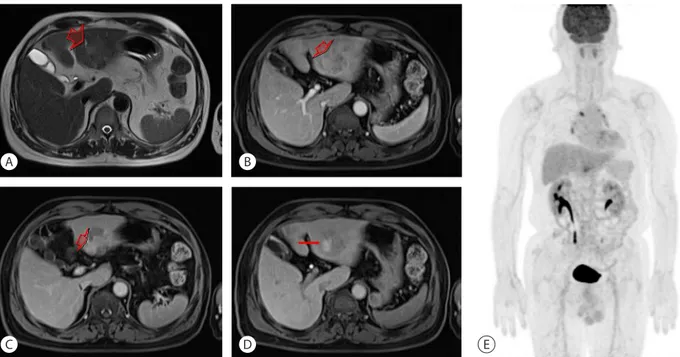
Figure 3. Liver primovist MRI and PET-CT. At S2-3, high signal intensity on T2-weighted image (A, arrow), enhancing on arterial phase (B, arrow), wash out on equilibrium phase (C, arrow) and defect on hepatobiliary phase (D, arrow) mass was shown and no distant metastasis (E). MRI, magnetic resonance image; PET-CT, positron emission tomography-computed tomography.
A
C
B
D E
A B C
Figure 2. Compared with previous CT scan (A, arrow), increased mass size was shown at follow up CT scan (B, arrow). Microscopic examination shows compatible with HCC (H&E, ×200) (C). CT, computed tomography; HCC, hepatocellular carcinoma .
A B C
Figure 1. Abdominal CT scan. About 3.0 cm sized rim-enhanced hepatic mass was shown at S2-3 in arterial phase (A, arrow) and non-visible in delayed phase (B). Microscopic finding shows fibrotic lesion with chronic inflammation (H&E, ×200) (C). CT, computed tomography.
의 과거력은 없었고 간 질환에 대한 가족력 또한 없었다. 내 원 당시 생체 징후는 안정적이었고 이학적 검사상 특이 소 견은 관찰되지 않았으며 특별하게 호소하는 증상 또한 없
었다. 신장 153.8 cm, 체중 63.5 kg, 체질량 지수(body mass index, BMI) 26.84 kg/m
2측정되었으며 검사실 소견은 말초 혈액 검사에서 백혈구 6,850/mm
3(호중구 51.8%, 호산구
Figure 4. Resected specimen (A), well-demarcated multi-lobulated single mass with fibrotic lesion. Glutamine synthetase (×100) (B), glypican 3 (×400) (C), heat shock protein 70 (×400) (D) were positive on immunochemistry test.
B A
C D
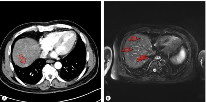
Figure 5. Newly developed arterial enhancing nodule on abdominal CT scan (A, arrow) and multiple recurred nodules on liver primovist MRI (B, arrows). CT, computed tomography; MRI, magnetic resonance image.
A B
2.1%), 혈색소 14.7 g/dL, 혈소판 246,000/mm
3, 생화학 검사 에서 총 단백질 7.3 g/dL, 알부민 4.6 g/dL, 식후 2시간 혈당 191 mg/dL, BUN 15 mg/dL, creatinine 0.78 mg/dL, AST/ALT 31/17 IU/L, gamma glutamyl transpeptidase 58 IU/L, alka- line phosphatase 183 IU/L, 총 콜레스테롤 188 mg/dL, 총 빌 리루빈 1.05 mg/dL (직접 빌리루빈 0.28 mg/dL), prothrom- bine time 10.6초(international normalized ratio, INR 1.02),
바이러스간염표지자 검사상 HBs Ag (-), anti-HBs Ab (+), anti-HCV Ab (-)였고 종양표지자 검사상 alpha-fetoprotein (AFP) 4.9 ng/mL, CA 19-9 17.27 IU/mL로 측정되었다.
2. 영상 및 병리 소견
복부 CT상 S2-3에 3.0 cm 크기의 병변이 보였다(Fig. 1A,
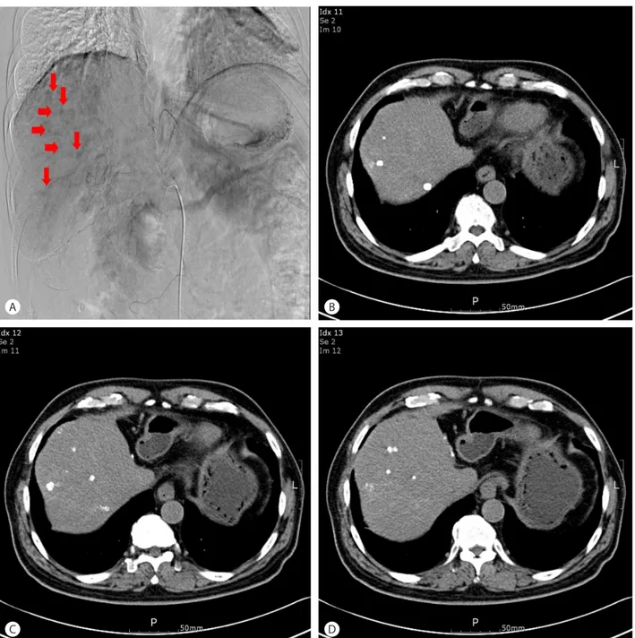
Figure 6. Hepatic angiography shows multiple tumor staining (A, arrows). After TACE, compact lipiodol uptake was shown on follow up non- enhancing CT scan (B, C, D). TACE, transcatheter arterial chemoembolization; CT, computed tomography.
A
C
B
D
B). 정확한 진단을 위해 조직 검사를 시행하였으며 조직 검 사 결과에서 만성 염증을 동반한 섬유화의 소견을 보여 (Fig. 1C) 추적 관찰하기로 하였다. 3개월 후 시행한 복부 CT에서 4 cm로 크기가 증가하는 양상을 보이고(Fig. 2A, B) AFP 6.1 ng/mL, prothrombin induced by vitamin K absence- II (PIVKA-II) 552 mAU/mL 소견을 보여 조직 검사를 다시 시행하였으며, 조직 검사 결과 상에 HCC에 합당한 소견을 보였다(Fig. 2C). 이어 시행한 자기공명영상(magnetic resonance image, MRI)에서 S2-3에 지방 변성을 동반한 명 백한 HCC 소견을 보였고(Fig. 3A, B, C, D) 그 외 간내 다른 병변은 관찰되지 않았으며 양전자방출단층촬영(Positive emission tomography, PET)에서 원격 전이는 관찰되지 않 았다(Fig. 3E).
3. 진단 및 치료 경과
영상검사에 따른 병기상 Modified Union for Internation- al Cancer Control (UICC) stage II (T2N0M0)로 수술을 시 행하였고(Lateral sectionectomy) 별다른 합병증 없이 병변 은 성공적으로 절제되었다(Fig. 4A). 절제된 종양 조직의 면 역 조직 화학 검사상 Glutamine synthetase, Glypican3, Heat shock protein 70에서 모두 양성 소견을 보였고(Fig. 4B, C, D) 주변 조직에는 간경변증의 소견은 보이지 않았으며, 25% 이상의 지방간 소견이 관찰되었다. 수술 5개월째 추적 관찰 위해 시행한 복부 CT (Fig. 5A)에서 S7에 새롭게 생긴 동맥기 조영 증강을 보이는 결절이 관찰되었고 MRI상 다 발성 소결절들이 관찰되었으며(Fig. 5B) 종양표지자검사상 AFP 7.2 ng/mL, PIVKA-II 81 mAU/mL로 측정되었다. 다발 성 간 내 전이 의심하에 혈관 조영술을 시행하였다. 혈관 조 영술 상 과혈관성의 작은 다발성 종양들이 관찰되어 TACE 를 시행하였고 이후 별다른 합병증은 보이지 않았다(Fig.
6A). TACE 시행 3주 후 시행한 복부 CT에서 조밀한 리피오 돌 섭취 상태를 보였으나(Fig. 6B, C, D) AFP 7.4 ng/mL, PIVKA-II 1,071 mAU/mL으로 측정되었다. 이후 환자는 본 인이 원하여 추가 치료를 위해 타 병원으로 전원되었다.
고 찰
최근 보고된 간경변증을 동반하지 않은 간세포암 환자에 대한 연구에 따르면 1,500명의 간세포암종 환자들 중 약 13%의 환자에서 간경변증을 동반하지 않았으며, 간경변증
을 동반하지 않은 간세포암종의 주된 위험 인자는 비알코 올성 지방간질환과 대사 증후군이었다.
3또 다른 보고에 따 르면 최근 미국에서 2004년에서 2009년 동안 비알코올성 지방간질환과 대사 증후군에 관련된 간세포암종이 매년 9%씩 증가하고 있으며
4이러한 간세포암종에서 생존 기간 이 더 짧고 빠르게 진행하는 경과를 보이며 간 이식 가능성 이 낮아서 위험군에서 HCC에 대한 선별 검사의 중요성에 대한 인식이 지금보다 더 높아져야 한다는 연구가 보고되 었다.
5하지만 아직 우리나라에서는 간경변증을 동반하지 않은 간세포암종 환자들의 발생 빈도와 특징 등에 대한 보 고는 없으며, 또한 비알코올성 지방간질환과 연관된 간세 포암조 환자들에 대한 발생 빈도도 보고된 바 없다. 본 증례 에서 환자는 바이러스표지자검사상 음성이었고, 음주력이 없었으나 당뇨병, 고혈압 및 비만 등과 같은 대사 증후군을 동반한 상태로 영상 검사와 수술 후 종양 주변 조직에서도 간경변증 소견은 보이지 않았다. 이에 이 환자에서 간세포 암종의 원인으로 비알코올성 지방간질환 또는 대사 증후 군일 가능성이 높다고 생각된다. 최근 국내에서도 비만 인 구가 증가하고 대사 증후군 및 비알코올성 지방간질환의 유병률이 서구와 같이 증가하고 있어 이와 같은 환자들의 발생도 점차 증가할 것으로 생각된다. 따라서 추후 임상적 으로 비알코올성 지방간질환이 있거나 대사 증후군을 갖 고 있는 환자들에 대해서 간세포암종 발생 가능성과 감시 검사에 대해서 보다 많은 관심과 심도 있는 연구가 필요하 겠다.
중심 단어: 간세포암종; 절제술; 재발; 항암색전술
Conflicts of Interest
The authors have no conflicts to disclose.
REFERENCES
1. El-Serag HB. Hepatocellular carcinoma. N Engl J Med 2011;365:
1118-1127.
2. Korean Liver Cancer Study Group (KLCSG); National Cancer Center, Korea (NCC). 2014 KLCSG-NCC Korea Practice Guideline for the Management of Hepatocellular Carcinoma. Gut Liver. 2015;9:267- 317.
3. Mittal S, El-Serag HB, Sada YH, Kanwal F, Duan Z, Temple S, et al. Hepatocellular carcinoma in the absence of cirrhosis in United States Veterans is associatted with nonalcoholic fatty liver disease.
Clin Gastroenterol Hepatol 2015. [Epub ahead of print]
4. Perumpail RB, Wong RJ, Ahmed A, Harrison SA. Hepatocellular Carcinoma in the Setting of Non-cirrhotic Nonalcoholic Fatty Liver Disease and the Metabolic Syndrome: US Experience. Dig Dis Sci 2015;60:3142-3148.
5. Younossi ZM, Otgonsuren M, Henry L, Venkatesan C, Mishra A, Erario M, et al. Association of Non-alcoholic Fatty Liver Disease (NAFLD) with hepatocellular carcinoma (HCC) in the United States from 2004-2009. Hepatology 2015. [Epub ahead of print]