INTRODUCTION
Drug-induced liver injury (DILI) is an important liver dis- ease with a worldwide incidence of 14 to 19 cases per 100,000 persons [1,2]. Approximately 3%–5% of hospital admissions for jaundice and 10% of acute hepatitis cas- es are associated with DILI [3]. DILI is the most common cause of acute liver failure (ALF) in Western society [4,5].
Azithromycin, a semisynthetic macrolides derived from erythromycin, was approved in 1994 [6]. It is extensively prescribed for treating otitis media, upper respiratory tract infections, bronchitis, and community-acquired pneumonia [7]. Azithromycin is frequently selected to first line antibi- otics in patients with respiratory infection in Korea. The
prescription rate of macrolide including azithromycin was higher (46.8%–49.0%) than that of penicillin (2.1%–2.5%) or third-generation cephalosporin (18.2%–19.5%) during 2011–2015 [8]. Common adverse effects of azithromycin include nausea, vomiting, abdominal pain and diarrhea [9].
Azithromycin-induced liver failure needed to liver transplantation has rarely been reported. In the United States, a 60-year-old woman with underlying nonalcohol- ic steatohepatitis was treated for a respiratory tract infec- tion with azithromycin for 3 days and developed fatigue, anorexia and nausea 4 days later, followed by jaundice and deterioration of mental status. She underwent liver transplantation 1 month after the initial presentation, and recovered from progressive liver failure [10]. Herein, we
Liver transplantation for azithromycin-induced severe liver injury
Hyun Joon Park
1, Kwang Il Seo
1, Young Il Choi
21 Department of Internal Medicine, Kosin University Gospel Hospital, Kosin University College of Medicine, Busan, Korea
2Department of Surgery, Kosin University Gospel Hospital, Kosin University College of Medicine, Busan, Korea
Drug-induced liver injury is the most common cause of acute liver failure in Western countries by prescription drugs and herbal medications. Liver injury due to azithromycin has rarely been reported. This is a brief report of a patient administered azithromycin and who developed acute liver failure leading to liver transplantation. We report the case of a 68-year-old woman who developed jaundice 1 week after she started taking a azith- romycin. On the 3rd day of hospitalization, her hepatic function rapidly deteriorated and level of consciousness decreased to drowsiness. The model for end-stage liver disease score was confirmed to be 33, and liver transplantation was considered. On the 8th day of hospitalization, she underwent emergency living donor liver transplantation, receiving a right lobe liver graft from a 35-year-old male donor, the patient’s son. Currently, she is alive with good liver function after 25 months of transplant. This case suggests that azithromycin may cause rare hepatitis with liver failure. Therefore, at the beginning of the azithromycin treatment, patients should visit the hospital immediately if symptoms such as jaundice and abdominal pain are experienced.
Keywords: Drug induced liver injury; Liver failure; Liver transplantation
Received May 19, 2020 Revised June 30, 2020 Accepted August 10, 2020 Corresponding author: Young Il Choi Department of Surgery, Kosin University College of Medicine, 262 Gamcheon-ro, Seo-gu, Busan 49267, Korea
Tel: +82-51-990-6462 Fax: +82-51-246-6093 E-mail: [email protected]
© The Korean Society for Transplantation This is an Open Access article distributed under the terms of the Creative Commons Attribution Non-Commercial License (http://creativecommons.org/licenses/
by-nc/4.0/) which permits unrestricted non-commercial use, distribution, and reproduction in any medium, provided the original work is properly cited.
pISSN 2671-8790
eISSN 2671-8804
report a first case of liver transplantation for azithromy- cin-induced liver failure in a patient without previous liver disease in Korea.
CASE REPORT
This study was approved by the Institutional Review Board of Kosin University Gospel Hospital (IRB No. 2020- 05-014). Informed consent was waived by the Review Board due the retrospective case report. This study was conducted in compliance with the principles of the Decla- ration of Helsinki.
A 68-year-old woman with jaundice was admitted to our hospital. Jaundice had developed 3 days before the visit. She manifested general weakness, fatigue, and de- creased appetite. Although icteric sclera was observed, no other abnormal findings were noted on physical exam- ination. On hospitalization, all her vital signs were normal range. Computed tomography (CT) and bronchoscopy revealed acute bronchitis, and she was administered tranexamic acid (250 mg), N-acetylcysteine (200 mg), dihydrocodeine, and azithromycin (250 mg) for a week.
She had no history of allergic diseases and denied alcohol consumption and smoking.
Initial laboratory test results were as follows: white blood cell count, 9,100/mm
3; hemoglobin level, 13.7 g/dL; platelet count, 149,000/mm
3; aspartate aminotransferase level, 1,631 IU/L; alanine aminotransferase (ALT) level, 1,711 IU/L; total bilirubin level, 24.20 mg/dL; direct bilirubin level, 14.99 mg/dL;
alkaline phosphatase (ALP) level, 226 IU/L; gamma-glutam- yl transferase level, 232 U/L; total protein concentration, 5.3 g/dL; albumin level, 3.1 g/dL; blood urea nitrogen level, 6.4 mg/dL; creatinine level, 0.5 mg/dL; sodium concentra- tion, 142 mmol/L; potassium concentration, 3.7 mmol/L;
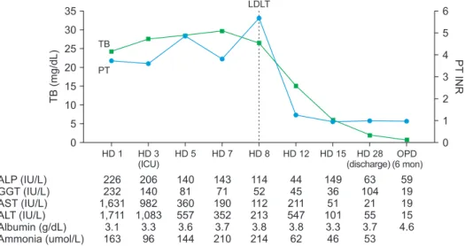
amylase level, 29 IU/L; lipase level, 53 IU/L; and ammonia concentration, 163 µMol/L. Prothrombin time was extend- ed to 37.5 seconds and partial thromboplastin time was extended to 55.9 seconds. The high sensitivity C-reactive protein level was 0.932 mg/dL. Fig. 1 summarizes the time course of blood tests for liver function. Hepatitis A virus immunoglobulin M (IgM) antibody, hepatitis B surface antigen, anti-HCV, Epstein-Barr virus (EBV) viral capsid antigen-IgM, EBV early anti gen-diffuse restrict IgM, cyto- megalovirus (CMV) IgM, and CMV real-time polymerase chain reaction tests were all negative. Herpes simplex virus (HSV)-IgM (enzyme immunoassay [ELISA]) was positive at 1.4 and HSV-immunoglobulin G (IgG; ELISA) was also posi- tive at 23.5. Toxoplasma IgM and human immunodeficiency virus serum tests were found to be negative. In addition, an- tinuclear antibody, anti-mitochondrial antibody, anti-smooth muscle antibody and liver kidney microsomal antibody tests were all negative. The IgG serum level was normal at 774.6 mg/dL.
The CT image obtained of the liver in the week before HIGHLIGHTS
• This case report is the first published report to proceed to liver transplantation after taking azithromycin in pa- tients who have had no previous liver disease.
HD 1 HD 3 (ICU) 35
30 25 20 15 10 5
HD 7 HD 8 HD 12 HD 15 HD 28 (discharge)
OPD (6 mon)
TB(mg/dL)
ALP (IU/L) GGT (IU/L) AST (IU/L) ALT (IU/L) Albumin (g/dL) Ammonia (umol/L)
0
PTINR
6 5 4 3 2 1 0 HD 5
226 232 1,631 1,711 3.1 163
206 140 982 1,083
3.3 96
140 81 360 557 3.6 144
143 71 190 352 3.7 210
114 52 112 213 3.8 214
44 45 211 547 3.8 62
149 36 51 101
3.3 46
63 104
21 55 3.7 53
59 19 19 15 4.6 TB
PT
LDLT
Fig. 1. Trends in liver function test results
of the case patient during hospitalization period and follow-up visit. TB, total bilirubin;
PT, prothrombin time; LDLT, living donor liver
transplantation; INR, international normal-
ized ratio; HD, hospital day; ICU, intensive
care unit; OPD, outpatient department; ALP,
alkaline phosphatase; GGT, gamma-glutamyl
transferase; AST, aspartate aminotransfer-
ase; ALT, alanine aminotransferase.
initiating azithromycin treatment showed normal findings (Fig. 2A). However, on the day of admission, periportal edema, gallbladder wall edema, and small amount of asci- tes were observed in the abdominal CT scan (Fig. 2B).
She was clinically suspected of having azithromycin-in- duced liver injury and all medications were discontinued.
The Roussel Uclaf Causality Assessment Method (RUCAM) scores for tranexamic acid, N-acetylcysteine, and dihydro- codeine were all 4 (possible), and the RUCAM score for azithromycin was 7 (probable) (Table 1) [11]. The R value for phenotype assessment was more than 5 and was con- sidered to be a hepatocellular type [10].The diagnosis of azithromycin-induced liver injury and ALF was made. The patient received medical treatment. However, laboratory test values had worsened (Fig. 1). On the 3rd day of hospi- talization, flapping tremor was observed, and the level of consciousness decreased to drowsiness. Hepatic enceph- alopathy persisted and the consciousness deteriorated to semi-coma. On the 8th day, she underwent emergency liv- ing donor liver transplantation, receiving a 950 g right lobe liver graft from a 35-year-old male donor, the patient’s son.
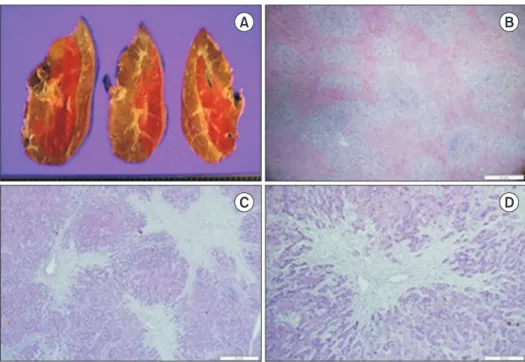
Gradual recovery of consciousness and liver function was observed after the transplant. The patient was discharged from the hospital after 20 days of transplant. Currently, she is alive, asymptomatic, and with adequate liver graft function after 25 months of transplant (Fig. 2C and D). His- tological examination of the explanted liver revealed fulmi- nant hepatitis, zone 3 necrosis, and extensive hepatocyte
death (Fig. 3).
DISCUSSION
DILI is typically classified as direct or idiosyncratic. Di- rect hepatotoxicity is common, predictable, and dose–
dependent. However, idiosyncratic hepatotoxicity can rarely occur due to agents with little or no intrinsic toxic- ity. It develops independently of drug route or duration of administration or drug dose [12]. Azithromycin, a culprit drug in this study, induced hepatotoxicity that is typically classified to idiosyncratic mechanism [10].
Based on the R value, idiosyncratic hepatotoxicity is classified into three phenotypes: hepatocellular (R ≥5), cholestatic (R ≤2), and mixed (2≤ R ≤5) [13]. The R value is the ratio of ALT to ALP relative to their respective normal upper limits—[serum (AST/ALT the upper limit of normal [ULN])÷(ALP/ALP ULN)] [10]. Hepatocellular phenotype is the most common and is characterized by a marked in- crease in ALT and a slight increase in ALP [12,14]. Accord- ing to Hy's law by Hyman J. Zimmerman, the mortality rate of cholestatic phenotype with jaundice is low; how- ever, the mortality rate of hepatocellular phenotype with jaundice is high, usually 10% or higher [15]. We reported a case of woman without previous liver disease who devel- oped azithromycin-induced ALF. This case represents a
A B
C D
Fig. 2. Axial computed tomography scan
images of the case patient. (A) Before
initiating azithromycin therapy. (B) On the
admission day, periportal edema (black
arrows) is observed. (C) After 6 days of liver
transplantation, mild periportal edema (black
arrow) is observed. (D) After 1 year of dis-
charge.
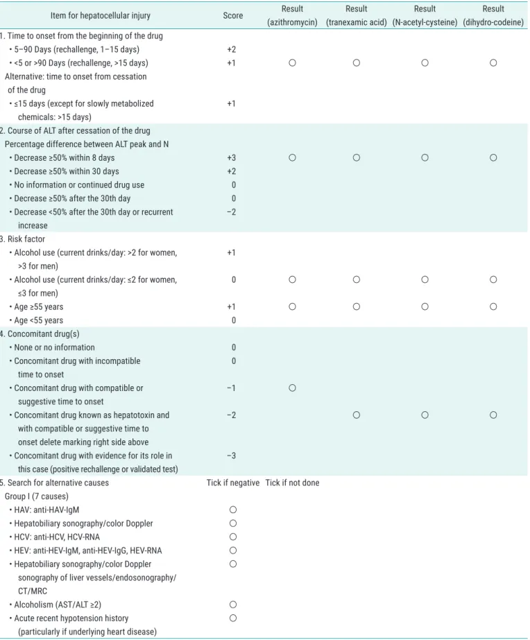
Table 1. RUCAM for the hepatocellular injury of DILI
Item for hepatocellular injury Score Result
(azithromycin)
Result (tranexamic acid)
Result (N-acetyl-cysteine)
Result (dihydro-codeine) 1. Time to onset from the beginning of the drug
• 5–90 Days (rechallenge, 1–15 days) +2
• <5 or >90 Days (rechallenge, >15 days) +1
○ ○ ○ ○Alternative: time to onset from cessation of the drug
• ≤15 days (except for slowly metabolized chemicals: >15 days)
+1 2. Course of ALT after cessation of the drug
Percentage difference between ALT peak and N
• Decrease ≥50% within 8 days +3
○ ○ ○ ○• Decrease ≥50% within 30 days +2
• No information or continued drug use 0
• Decrease ≥50% after the 30th day 0
• Decrease <50% after the 30th day or recurrent increase
–2 3. Risk factor
• Alcohol use (current drinks/day: >2 for women,
>3 for men)
+1
• Alcohol use (current drinks/day: ≤2 for women,
≤3 for men)
0
○ ○ ○ ○• Age ≥55 years +1
○ ○ ○ ○• Age <55 years 0
4. Concomitant drug(s)
• None or no information 0
• Concomitant drug with incompatible time to onset
0
• Concomitant drug with compatible or suggestive time to onset
–1
○• Concomitant drug known as hepatotoxin and with compatible or suggestive time to onset delete marking right side above
–2
○ ○ ○• Concomitant drug with evidence for its role in this case (positive rechallenge or validated test)
–3
5. Search for alternative causes Tick if negative Tick if not done Group I (7 causes)
• HAV: anti-HAV-IgM
○• Hepatobiliary sonography/color Doppler
○• HCV: anti-HCV, HCV-RNA
○• HEV: anti-HEV-IgM, anti-HEV-IgG, HEV-RNA
○• Hepatobiliary sonography/color Doppler sonography of liver vessels/endosonography/
CT/MRC
○
• Alcoholism (AST/ALT ≥2)
○• Acute recent hypotension history (particularly if underlying heart disease)
○
fatal clinical course with icteric hepatocellular injury.
Azithromycin-induced idiosyncratic hepatotoxicity is predominantly present as cholestatic or mixed phenotype.
It has been reported that most of the patients exhibit be- nign clinical features, rarely patients exhibit fatal or chron- ic liver injury; however, the exact mechanism of injury is unknown [10]. Herein, we reviewed 22 cases of azithromy- cin-induced liver injury that were previously reported and classified according to the three phenotypes as follows:
hepatocellular, 10 patients (45.5%); cholestatic, 8 patients
(36.3%); and mixed, 4 patients (18.2%) (Table 1). The me- dian (range of quartiles) age of all the patients was 46 years (19.8–66), and the proportion of women was 77.3%.
In our case, the patient was a 68-year-old woman, older than the median age of all the aforementioned patients as well as the hepatocellular group (53.5 years [36.8–60]).
Jaundice developed 4 days after azithromycin prescrip- tion, faster than the median (range of quartiles) days of all the cases (17.5 days [8.5–30.8]) as well as the hepato- cellular group (20.5 days [15.3–32.5]). The median (range
Table 1. ContinuedItems for Hepatocellular Injury Score Result
(Azithromycin)
Result (Tranexamic acid)
Result (N-acetyl-cysteine)
Result (dihydro-codeine) Group II (5 causes)
• Complications of underlying disease(s) such as sepsis, metastatic malignancy, autoimmune hepatitis, chronic hepatitis B or C, primary biliary cholangitis or sclerosing cholangitis, genetic liver diseases
○
• Infection suggested by PCR and titer change for
• CMV (anti-CMV-IgM, anti-CMV-IgG) ○
• EBV (anti-EBV-IgM, anti-EBV-IgG) ○
• HSV (anti-HSV-IgM, anti-HSV-IgG) ○
• VZV (anti-VZV-IgM, anti-VZV-IgG) ○
Evaluation of groups I and II
• All causes-groups I and II—reasonably ruled out +2
• 7 causes of group I ruled out +1 ○ ○ ○ ○
• 6 or 5 causes of group I ruled out 0
• Less than 5 causes of group I ruled out –2
• Alternative cause highly probable –3
6. Previous hepatotoxicity of the drug
• Reaction labelled in the product characteristics +2 ○
• Reaction published but unlabelled +1
• Reaction unknown 0 ○ ○
7. Response to unintentional re-exposure
• Doubling of ALT with the drug alone, provided ALT below 5N before re-exposure
+3
• Doubling of ALT with the drug(s) already given at the time of first reaction
+1
• Increase of ALT but less than N in the same conditions as for the first administration
–2
• Other situations 0 ○ ○ ○ ○
Total score for the case 7 4 4
RUCAM, Roussel Uclaf Causality Assessment Method; DILI, drug-induced liver injury; ALT, alanine aminotransferase; N, upper limit of the normal range;
HAV, hepatitis A virus; IgM, immunoglobulin M; HCV, hepatitis C virus; HEV, hepatitis E virus; CT, computer tomography; MRC, magnetic resonance
cholangiography; AST, aspartate aminotransferase; PCR, polymerase chain reaction; CMV, cytomegalovirus; EBV, Epstein-Barr virus; HSV, herpes simplex
virus; VZV, varicella zoster virus.
of quartiles) peak serum levels of ALT and ALP in the he- patocellular group were 1,271.5 IU/L (923.5–3,201.3) and 224.5 IU/L (159.5–444), respectively. In our case, peak ALT and ALP levels were 1,711 IU/L and 226 IU/L, respec- tively, close to those in the hepatocellular group. The me- dian (interquartile range) peak total bilirubin level in the hepatocellular group was 17 mg/dL (10.8–21.5), whereas in our case it was 29.67 mg/dL. As described earlier, it is thought to be highly related to the fatal clinical course of liver transplantation. Pathologic findings were described in 11 of the 22 patients. Of the 11 patients, four cases showed hepatitis with no visible cholestasis, two cases showed acute cholestasis. Three other cases reported cholestatic hepatitis. Of the remaining two cases, one showed zone 3 necrosis and one showed complex find- ings. A total of 13 (59.1%) of 19 patients available for fol- low-up fully recovered, and the recovery period was 4–60 days after drug discontinuation. Four patients (18.2%) showed chronic progression. One patient (4.5%) recov- ered after liver transplantation, one patient (4.5%) died.
To summarize, the latency in our case was much shorter than that of previous cases, and our case patient did not have underlying liver disease compared to patients with previous cases.
In conclusion, we report a case of azithromycin-in- duced liver failure treated liver transplantation in the pa- tient without underlying liver diseases in Korea. Because azithromycin-induced liver injury is unpredictable, it is
important to suspect DILI when a patient manifests acute jaundice and abdominal pain after administering azithro- mycin.
ACKNOWLEDGMENTS Conflict of Interest
No potential conflict of interest relevant to this article was reported.
Funding/Support
This study was supported by research grant from the Kore- an Society for Transplantation (2020-00-03002-003).
ORCID
Hyun Joon Park https://orcid.org/0000-0001-5443-4873 Kwang Il Seo https://orcid.org/0000-0001-8854-5205 Young Il Choi https://orcid.org/0000-0002-9630-6287 Author Contributions
Conceptualization: YIC. Data curation: YIC, KIS. Formal analysis & Funding acquisition: YIC. Investigation: HJP, KIS.
Methodology: KIS. Project administration & Resources:
YIC. Software: HJP, KIS. Supervision: YIC, KIS. Validation
& Visualization: YIC. Writing–original draft: HJP. Writing–
review & editing: YIC.
A B
C D
Fig. 3. Pathological finding of the explanted