대한소화기학회지 2007;49:400-404
접수: 2006년 10월 4일, 승인: 2007년 5월 7일
연락처: 천재희, 120-752, 서울시 서대문구 신촌동 134 연세대학교 의과대학 내과학교실
Tel: (02) 2228-1990, Fax: (02) 393-6884 E-mail: [email protected]
본 연구는 2006년도 연세대학교 학술연구비의 부분적인 지 원에 의하여 이루어진 것임.
회결장동맥의 동맥류 파열에 의한 대량 장출혈을 동반한 베체트 장염
연세대학교 의과대학 내과학교실, 소화기병연구소, 진단영상의학교실*, 외과학교실†, 병리학교실‡
김승업ㆍ천재희ㆍ임준석*ㆍ백승혁†ㆍ김상겸‡ㆍ이상길ㆍ이용찬ㆍ김원호
Massive Gastrointestinal Bleeding due to Aneurysmal Rupture of Ileo-colic Artery in a Patient with Behcet's Disease
Seung Up Kim, M.D., Jae Hee Cheon, M.D., Joon Seok Lim, M.D.*, Seung Hyuk Paik, M.D.†, Sang Kyum Kim, M.D.‡, Sang Kil Lee, M.D., Yong Chan Lee, M.D., and Won Ho Kim, M.D.
Department of Internal Medicine and Institute of Gastroenterology, Radiology*, Surgery†, and Pathology‡, Yonsei University College of Medicine, Seoul, Korea
Behcet's disease has been recognized as a systemic vasculitis characterized by the involvement of multiple organs such as orogenital ulcers, eye lesions including uveitis and optic neuritis, and skin lesions including folliculitis and erythema nodosum. Vascular involvement occurs occasionally and is classified into thrombosis and aneurysm.
However, massive gastrointestinal bleeding from arterial aneurysm is a rare manifestation of intestinal Behcet's disease. Recently, we experienced a case of intestinal Behcet's disease presenting with massive gastrointestinal bleeding due to aneurysmal rupture of ileo-colic artery. A 30-year-old male with Behcet's disease was admitted because of massive gastrointestinal bleeding. A large ileo-cecal ulcer was revealed as a bleeding focus on colonoscopic examination. Celiac angiography showed aneurysm and stenosis of ileo-colic artery. After the failure of hemostasis with arterial embolization, ileocecectomy was performed. After the resection hematochezia was completely stopped. (Korean J Gastroenterol 2007;49:400-404)
Key Words: Behcet's disease; Gastrointestinal bleeding; Ileocecal ulcer; Aneurysm; Aneurysmal rupture
Correspondence to: Jae Hee Cheon, M.D.
Department of Internal Medicine, Yonsei University College of Medicine, 134, Sinchon-dong, Seodaemun-gu, Seoul, Korea Tel: +82-2-2228-1990, Fax: +82-2-393-6884
E-mail: [email protected]
서 론
베체트 병은 만성적으로 나타나는 전신 질환으로 반복적 으로 재발하는 구강 및 음부 궤양과 홍채염, 포도막염, 망막 혈관염 등의 안과 질환을 주 증상으로 하는 원인 불명의 염 증 질환이다. 1937년 Behcet1가 처음 소개한 이후에 최근에 는 많은 장기를 침범하는 전신 질환으로서 심혈관계, 중추 신경계, 호흡기계, 비뇨기계, 장관, 피부 및 관절 등을 침범
하여 임상 소견이 매우 다양한 질환으로 알려져 있다.
베체트 병은 7-35%에서 혈관을 침범하며,2 약 7%에서 동 맥을 침범한다.3 동맥 침범은 혈전증이나 동맥류로 나타날 수 있다. 베체트 병의 동맥류는 모든 크기의 혈관을 침범할 수 있으나, 흉부와 복부대동맥, 경동맥, 쇄골하동맥, 총장골 동맥, 폐동맥 그리고 말초동맥 등을 주로 침범한다.4,5 동맥 류는 파열이나 국소 압박 등에 의한 합병증을 일으킬 수 있 으며, 동맥류 파열은 베체트 병에서 주요한 사망원인 중 하
김승업 외 7인. 동맥류 파열에 의한 장출혈을 동반한 베체트 장염 401
Fig. 1. Colonoscopy. It reveals current bleeding from an ulcer (arrow) in the ascending colon just below the ileocecal valve.
A B
Fig. 3. Celiac angiography. (A) It shows an arterial aneurysm (white arrow) and stenosis (black arrow) in ileo-colic artery. (B) Selection and embolization of the artery were done with gelform. The artery was embolized successfully (arrow).
Fig. 2. Abdominal CT scan. It shows a small, contrast-enhancing aneurysm (arrow) located in the wall of ileocecal area.
나이다.
베체트 병의 위장관 침범은 1940년에 Bechguard이 처음 기술하였으며 베체트 병 환자의 40-50%에서 복부 팽만감, 복통, 연하장애, 식욕부진, 구토, 설사, 출혈 등의 증상을 일 으킨다. 이처럼 위장관 증상은 흔하지만 서양에서는 실제 장관 궤양이나 출혈 및 천공은 베체트 병 환자의 1% 미만 으로 보고되고 있다.
그러나 우리나라를 비롯한 극동 아시아 국가에서는 베체 트 병의 장 침범이 서양보다는 흔한 편이며 궤양에 의한 출 혈이 드물지 않게 발생한다. 하지만 장간막 혈관의 동맥류 파열에 의한 대량 장출혈은 매우 드물고,6 국내에서 장관 침 범을 보이는 베체트 병 환자에서 회결장 동맥류 및 동맥류 파열에 의한 대량 출혈의 보고는 아직 없었다.
최근 저자들은 회맹판 직하부의 상행결장 궤양과 회결장 동맥류 출혈을 동반한 베체트 병을 경험하여서 문헌 고찰과 함께 보고한다.
증 례
30세 남자 환자가 내원 1시간 전부터 시작된 대량의 혈변 을 주소로 내원하였다. 환자는 14년 전부터 구강 궤양, 포도 막염 그리고 피부병변이 있어 베체트 병을 진단받고 5-ami- nosalicylic acid (5-ASA)와 azathioprine을 복용하면서 외래에 서 경과 관찰 중이었다.
내원 시 혈압 90/60 mmHg, 맥박 102회/분, 호흡수 26회/
분, 체온 36.7oC였고, 급성병색을 보였으며, 의식은 명료하 였다. 구강 점막, 설상연 및 성기 부위에 궤양은 관찰되지 않았다. 결막이나 공막에 빈혈이나 황달 소견은 없었으며 경부 림프절 비대는 없었다. 심음 및 호흡음도 정상이었다.
복부 소견에서 우하복부에 국한된 경직과 압통이 있었고 장 음은 정상이었다.
말초혈액검사에서 혈색소 12.3 g/dL, 헤마토크리트 36.4%
(MCV 83.7 fL, MCH 28.2 pg, MCHC 33.7 g/dL, RDW 39.7%),
402 The Korean Journal of Gastroenterology: Vol. 49, No. 6, 2007
A B
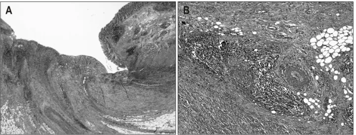
Fig. 5. Microscopic findings. (A) Enlarged ulcer undermines surrounding tissues. Edema-like swelling with crater formation around the ulcer margins produces a characteristic “collar-stud” appearance. The ulcer contains nonspecific chronic inflammation (H&E stain, ×100). (B) Mononuclear cells infiltrate around capillaries and venules accompanied by intimal thickening, which demonstrates non-necrotizing lymphocytic vasculitis (H&E stain, ×400).
Fig. 4. Gross finding. The specimen is a product of ileocecetomy, including cecum attached to ileum and appendix. It shows a deep penetrating ulcer (arrow) in the ascending colon just below the ileocecal valve.
백혈구 14,310/mm3, 중성구 83.6%, 혈소판 462,000/ mm3, 혈 액침강속도 22 mm/hr이었고, 혈액응고검사와 소변검사는 정상이었다. 생화학검사에서 나트륨 140.0 mEq/L, 칼륨 4.0 mEq/L, 염소 100.0 mEq/L, 총 이산화탄소 26.0 mEq/L, 혈액 요소질소 13.3 mg/dL, 크레아티닌 0.9 mg/dL, 혈당 132 mg/dL, 칼슘 8.7 mg/dL, 총 빌리루빈 0.7 mg/dL, ALT 58 IU/L, AST 98 IU/L, 아밀라아제 35 U/L, 리파아제 21 U/L, 총 단백 6.7 g/dL, 알부민 3.6 g/dL, 요산 4.4 mg/dL이었고, 혈 청 검사에서 CRP는 3.8 mg/dL이었다.
흉부 X-선과 단순복부 촬영에서 이상 소견은 없었다. 위 장관 출혈을 확인하기 위해 시행한 상부위장관내시경 검사
에서 위전정부와 십이지장 구부에 얕은 미란이 있었으나 출 혈의 증거는 없었다. 대장내시경 검사에서는 회맹판 직하부 의 상행결장 부위에 주변의 부종을 동반한 궤양과 궤양저에 서 출혈을 관찰할 수 있었다(Fig. 1). 복부 전산화단층촬영 검사에서 회맹판 직하부의 상행결장벽 비후가 관찰되었으 며, 장벽에 조영 증강되는 동맥류가 관찰되었다(Fig. 2). 입 원 이후 스테로이드 정맥 투여를 포함한 내과 치료에도 불 구하고 지속적으로 혈변 증상이 있어 혈관조영술을 시행하 였다. 혈관조영술에서 회맹판 직하부의 회결장 동맥류 출혈 소견을 보여 색전술을 시행하였다(Fig. 3). 하지만 색전술 시 행 다음날 다시 대량의 혈변이 지속되어 혈압이 80/60 mmHg까지 감소하고, 당시 시행한 혈액검사에서 혈색소가 6.5 g/dL까지 감소하였으며, 의식 상태가 혼미한 수준까지 악화되는 등 저혈량에 의한 쇼크가 악화되는 소견을 보여 응급 회맹장절제술 및 회장-상행결장문합술을 시행하였다 (Fig. 4). 조직 소견에서 회맹판 직하부의 상행결장에 주변의 정상 점막으로 둘러싸인 깊은 함요 궤양을 보였으며 주변 혈관에 혈관염 소견이 관찰되어 베체트 병을 시사하였다 (Fig. 5). 수술 이후에 출혈은 없었고 5-ASA, colchicine, azathioprine을 복용하며 퇴원하였으며 현재 더 이상의 출혈 없이 외래에서 추척 관찰 중이다.
고 찰
이번 증례는 베체트 병을 진단 받은 30세 남자 환자에서 회맹판 직하부의 상행결장 동맥류 파열에 의한 대량 출혈이 있었으며, 그에 따른 치료로 혈관조영술을 통한 동맥 색전
Kim SU, et al. Behcet's Disease with Gastrointestinal Bleeding due to Aneurysmal Rupture 403
술을 시행하였으나 실패하여 회맹장절제술 및 회장-상행결 장문합술로 호전된 예이다.
베체트 병에서 혈관염은 혈전증 또는 동맥류의 형태로 나 타나며 소혈관 침범이 대부분이지만 대혈관 병변도 약 25%
에서 나타난다.7,8 여러 혈관이 동시에 침범되기도 하고 대 혈관 병변이 최초 증상으로 나타나기도 한다. 혈관 침범은 환자의 7-35%에서 발생하며, 동맥보다는 정맥을 흔히 침범 한다.2 동맥 침범은 베체트 병 환자의 약 7%에서 발생하며,3 혈전증 또는 동맥류 형성이 특징적인 소견이다. 대부분의 동맥류는 팽창된 종괴의 형태로 발견되지만, 국소적인 압박 에 의한 합병증을 보일 수도 있고, 이번 증례에서와 같이 동 맥류 파열과 같은 심각한 합병증이 발생할 수 있다.5,7 일반 적으로 장관을 침범한 베체트 병에서 장출혈에 대한 치료는 스테로이드나 면역억제제를 통한 내과 치료, 대장내시경을 통한 지혈, 혈관조영술을 이용한 색전술, 또는 수술 요법이 있다.9 일반적인 베체트 병의 장침범에서 보이는 궤양 출혈 은 대부분 스테로이드 등의 항염증제와 면역억제제를 통한 보존 치료로 호전을 보인다. 만약 내과 치료에 반응하지 않 거나 대량 출혈일 경우에는 선택적인 장간막 혈관조영술 및 색전술을 시행할 수 있다. 혈관조영술은 장출혈의 진단과 치료에 모두 효과적이며 이번 증례과 같이 혈관조영술을 통 해 동맥류를 진단하고 색전술8,10 또는 혈관 내 vasopressin 투여11가 시도된 증례들이 보고된 바 있다. 대부분의 장출혈 은 내과 치료와 색전술로 호전을 보이나 이번 증례처럼 이 런 치료에 효과가 없거나 천공이 발생한 경우에는 수술이 치료 원칙이다. 다만 수술 이후 문합부에서 가성 동맥류의 재발을 유념해야 하며, 수술 후 약물 치료가 재발 문합부 동 맥류 또는 다른 부위의 동맥류 형성을 막는 데 도움이 된 다.3 국내에는 비슷한 증례가 보고되지 않았지만 국외에서 Smith와 Siddiqui12는 베체트 병을 가진 환자의 전 결장에서 관찰되는 많은 수의 궤양과 그에 따르는 대량 장출혈로 우 측 대장절제술을 시행한 증례를 보고하였고, Fujita 등9은 말 단 회장과 전 결장에서 궤양의 소견을 보이는 환자에서 상 장간막 동맥을 통한 혈관조영술을 시도하여 말단 회장 부위 의 출혈을 관찰하여 코일로 색전술을 시도하였으나 실패하 여 동맥내 스테로이드 투여 및 회장절제술을 통해 치료한 증례를 보고하였다.
베체트 병에서 동맥류가 형성되는 기전은 정확히 알려져 있지 않으나, 현재까지 인정받고 있는 가설을 살펴보면 혈 관벽에 면역복합체의 침착으로 보체와 다형핵 호중구를 활 성화시키고 활성화된 호중구는 산소 유리 라디칼을 분비하 여 vasa vasorum의 손상을 일으킨다.13 Vasa vasorum의 손상 과 그에 따르는 폐쇄는 동맥 혈관벽의 괴사를 일으키게 되 고 이러한 혈관벽의 퇴행성 변화로 결국에는 혈관벽의 파열 및 가성 동맥류가 형성된다.5,13,14 그러나 이번 증례와 같이
기존의 장 궤양이 심해 오랫동안 염증을 앓고 있는 부위에 서 혈관벽이 노출되어 있을 가능성이 높은 경우 췌장염 환 자에서 발생하는 혈관벽 주위 염증에 의한 가성 동맥류와 같은 기전으로 동맥류가 생겼을 가능성도 배제할 수 없다.
즉 이번 증례의 동맥류는 위와 같은 혈관염의 기전으로 발 생한 동맥류인지 아니면 심한 궤양과 만성 염증으로 인해 이차적인 혈관벽의 염증으로 생긴 가성 동맥류인지는 알 수 없다.
저자들은 국내에서 아직 보고된 바가 없는 베체트 병을 진단 받은 30세 남자 환자에서 발생한 대량의 장출혈을 혈 관조영술을 통해 회맹판 직하부의 상행결장 동맥류로 진단 하고 회맹장절제술 및 회장-상행결장문합술을 통해 치료한 예를 경험하여서 문헌 고찰과 함께 보고한다. 결론으로 베 체트 병에서 흔하지 않으나 장 출혈이 있을 경우 병태생리 학 관점에서 동맥류 발생 가능성이 있으므로 이를 염두에 두어 빠른 진단과 치료가 가능할 수 있도록 해야 한다.
참고문헌
1. Behcet H. Uber rezidivierende, aphthos durch ein virus erusachate Geschwure am Mund, am Auge und Genitalian.
Dermat Wehnschr 1973;105:1152.
2. Shimizu T, Ehrlich GE, Inaba G, Hayashi K. Behcet disease.
Semin Arthritis Rheum 1979;8:223-260.
3. Bartlett ST, McCarthy WJ 3rd, Palmer AS, Flinn WR, Bergan JJ, Yao JS. Multiple aneurysms in Behcet's disease.
Arch Surg 1988;123:1004-1008.
4. Behcet's Disease Research Committee of Japan. Behcet's disease: guide to diagnosis of Behcet's disease. Jpn J Op- hthalmol 1974;18:291-294.
5. Hamza M. Large artery involvement in Behcet's disease. J Rheumatol 1987;14:554-559.
6. Men S, Ozmen MN. Superior mesenteric artery aneurysm in Behcet's disease. Abdom Imaging 1994;19:333-334.
7. Koc Y, Gullu I, Akpek G, et al. Vascular involvement in Behcet's disease. J Rheumatol 1992;19:402-410.
8. Park JH, Lee CB, Han MC, Choi SJ. Radiologic findings of vascular involvement in Behcet's disease. Korean Circ J 1984;14:309-314.
9. Fujita H, Kiriyama M, Kawamura T, et al. Massive hemorrhage in a patient with intestinal Behcet's disease:
report of a case. Surg Today 2002;32:378-382.
10. Zuckerman GR, Prakash C. Acute lower intestinal bleeding.
Part II: etiology, therapy, and outcomes. Gastrointest Endosc 1999;49:228-238.
11. Baba S, Hiramatsu K. Selective angiography for diagnosis and
404 대한소화기학회지: 제49권 제6호, 2007
treatment for intestinal bleeding. Gastroenterol Jpn 1991;
26(suppl 3):116S-120S.
12. Smith JA, Siddiqui D. Intestinal Behcet's disease presenting as a massive acute lower gastrointestinal bleed. Dig Dis Sci 2002;47:517-521.
13. Niwa Y, Miyake S, Sakane T, Shingu M, Yokoyama M.
Auto-oxidative damage in Behcet's disease: endothelial cell damage following the elevated oxygen radicals generated by stimulated neutrophils. Clin Exp Immunol 1982;49:247-255.
14. Akiyama K, Hirota J, Ohkado A, Shiina Y. Multivarious clinical manifestations of multiple pseudoaneurysms in Be- hcet's disease. J Cardiovasc Surg 1998;39:175-178.