ORIGINAL ARTICLE
한국인 비알코올성 지방간 질환 환자에서 P2/MS 및 기타 비침습적 섬유화 지표의 유효성
유수종1, 김동희2, 이정훈1, 정고은2, 임정윤2, 박민정2, 김윤준1, 윤정환1, 장자준3, 이효석1
서울대학교 의과대학 내과학교실, 간연구소1, 서울대학교병원 헬스케어시스템 강남센터 내과, 헬스케어연구소2, 서울대학교 의과대학 병리학교실3
Validation of P2/MS and Other Noninvasive Fibrosis Scoring Systems in the Korean Population with Nonalcoholic Fatty Liver Disease
Su Jong Yu1, Donghee Kim2, Jeong-Hoon Lee1, Goh Eun Chung2, Jeong Yoon Yim2, Min Jung Park2, Yoon Jun Kim1, Jung-Hwan Yoon1, Ja-June Jang3 and Hyo-Suk Lee1
Department of Internal Medicine and Liver Research Institute, Seoul National University College of Medicine1, Department of Internal Medicine and Healthcare Research Institute, Seoul National University Hospital, Healthcare System Gangnam Center2, Department of Pathology, Seoul National University College of Medicine3, Seoul, Korea
Background/Aims: P2/MS is a noninvasive marker for detecting hepatic fibrosis in patients with viral hepatitis. However, the applicability of P2/MS in patients with nonalcoholic fatty liver disease (NAFLD) has not yet been validated. This study aimed to validate P2/MS and compare it to other noninvasive fibrosis scoring systems in Korean patients with NAFLD.
Methods: Consecutive patients who underwent liver biopsy between January 2002 and December 2009 at Seoul National University Hospital, Seoul, Korea were enrolled in this study. Fibrosis stage was determined using the METAVIR scoring system.
Results: A total of 235 patients were included in the study: advanced fibrosis (METAVIR F3-F4) was present in 7 patients. No patient was over-staged among 162 patients with a P2/MS score above the high cut-off (95), resulting in a high negative predictive value (NPV) of 100% (95% confidence interval, 97.1-100). There was no significant difference between the area under the receiver-operating characteristic curve (AUROC) of the FIB-4 (0.964) and the AUROC of the NAFLD fibrosis score (0.964) or P2/MS (0.940) for detecting advanced fibrosis. If P2/MS was implemented in the Korean patients with NAFLD, 68.9% of liver biopsies might be avoided.
Conclusions: P2/MS has a high NPV for excluding advanced fibrosis in Korean patients with NAFLD, and can reduce the burden of liver biopsy in the majority of cases. Since there were few patients with advanced fibrosis, further studies are warranted in a cohort including more patients with advanced fibrosis to validate the low cut-off value. (Korean J Gastroenterol 2011;57:19-27)
Key Words: Fatty liver; Nonalcoholic fatty liver disease; Liver fibrosis
Received November 25, 2010. Revised December 22, 2010. Accepted December 23, 2010.
CC This is an open access article distributed under the terms of the Creative Commons Attribution Non-Commercial License (http://creativecommons.org/
licenses/by-nc/3.0) which permits unrestricted non-commercial use, distribution, and reproduction in any medium, provided the original work is properly cited.
교신저자: 김동희, 135-984, 서울시 강남구 역삼동 737, 강남파이낸스센터, 서울대학교병원 헬스케어시스템 강남센터 내과, 헬스케어연구소
Correspondence to: Donghee Kim, Department of Internal Medicine and Healthcare Research Institute, Seoul National University Hospital, Healthcare System Gangnam Center, 39th FL Gangnam Finance Center, 737 Yeoksam-dong, Gangnam-gu, Seoul 135-984, Korea. Tel: +82- 2-2112-5574, Fax: +82-2-2112-5635, E-mail: [email protected]
Financial support: None. Conflicts of interest: None.
INTRODUCTION
The prevalence of nonalcoholic fatty liver disease (NAFLD) is increasing and becoming one of the most common chronic liver diseases (CLD).1 NAFLD is known to be related to predictors of cardiovascular disease such as dyslipidemia, central obesity, insulin resistance, and metabolic syndrome.2 Several studies have suggested a possible role of NAFLD in the development of car- diovascular disease and that NAFLD increases overall mortality.1,2 The spectrum of NAFLD ranges from simple steatosis through nonalcoholic steatohepatitis (NASH), to fibrosis and cirrhosis and hepatocellular carcinoma (HCC).3,4 Patients with simple steatosis on presentation usually have a benign prognosis, while patients with NASH may develop the progression of fibrosis leading to cirrhosis and cirrhosis-related complications including HCC.5 In NASH, cirrhosis development occurs at an old- er age than in other liver diseases, even though once cirrhosis develops in patients with NASH, their clinical outcome is similar to patients with other causes of cirrhosis.5 Approximately 5% and 15% percent of pa- tients with NAFLD and NASH will progress to cirrhosis, respectively.6,7 Thus, the identification of the patients with fibrosis among those with NAFLD is important for predicting prognosis.4
In the absence of decompensated cirrhosis, liver biop- sy is the gold standard for determining the prognosis of NAFLD based on fibrosis severity.8 However, liver biopsy has several limitations such as invasiveness, cost, intra- and interobserver variability, sampling error, and the risk of complications.8-10 In addition, it is likely that most physicians are more reluctant to perform a liver biopsy in patients with NAFLD than in patients with viral-re- lated CLD.11 These considerations underscore efforts to develop noninvasive methods for the assessment of liver fibrosis.
Several clinical noninvasive fibrosis scoring systems based on simple clinical or laboratory parameters have been developed to identify advanced fibrosis in patients with NAFLD and other liver diseases.12 These include the aspartate aminotransferase (AST)/alanine amino- transferase (ALT) ratio (AAR),13 AST/platelet ratio (APR),14 the AST-to-platelet ratio index (APRI),15 the
BARD score,16 the Goteburg University Cirrhosis Index (GUCI),17 cirrhosis determinant score (CDS),18 the simple panel,4 the NAFLD fibrosis score,8 and the FIB-4.19 However, many of these tests were derived and vali- dated in Caucasian.8 In Asian patients, NASH and other metabolic complications have a propensity to occur at a lower body mass index (BMI).20 It is uncertain whether ethnicity affects the accuracy of these simple non- invasive fibrosis scoring systems.1 Therefore, these sys- tems should be validated in Asian patients prior to their global use. In addition, P2/MS, a simple and non- invasive test that was developed and has been con- firmed for the detection of hepatic fibrosis in Korean pa- tients with virus-related CLD.21-23 Although P2/MS was known to be accurate, the applicability of P2/MS in pa- tients with NAFLD has not yet been validated. In this study, we aimed to validate the diagnostic performance of P2/MS and to compare it with other noninvasive fib- rosis scoring systems in terms of identifying the pres- ence of advanced fibrosis in Korean patients with NAFLD.
SUBJECTS AND METHODS
1. Patients
We performed a retrospective cohort study. We en- rolled consecutive patients who underwent liver biopsy for the evaluation of an abnormal liver function or a liver donor work-up for liver transplantation at Seoul National University Hospital, Seoul, Korea between January 2002 and December 2009. Patients with the following were excluded: coexistent liver disease including chronic viral hepatitis, drug-induced liver disease, autoimmune hep- atitis, primary biliary cirrhosis, primary sclerosing chol- angitis, hemochromatosis, Wilson’s disease, α1-anti- trypsin deficiency, or biliary obstruction. Patients that consumed more than 20 g of alcohol per day were also excluded. In addition, patients that met any of the fol- lowing conditions were also excluded: prior or current malignancy; a concomitant serious medical illness such as chronic kidney disease, congestive heart failure, or hematological disease; or active infection. If the fasting plasma glucose was more than 126 mg/dL or symptoms of diabetes plus casual plasma glucose concentration
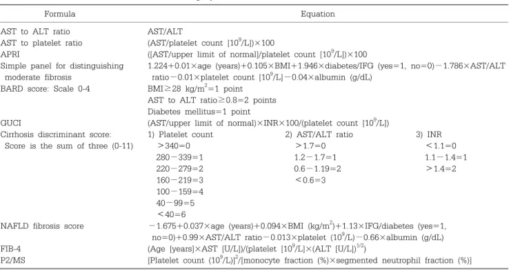
Table 1. Formulae of Noninvasive Fibrosis Scoring Systems
Formula Equation
AST to ALT ratio AST/ALT
AST to platelet ratio (AST/platelet count [109/L])×100
APRI ([AST/upper limit of normal]/platelet count [109/L])×100
Simple panel for distinguishing 1.224+0.01×age (years)+0.105×BMI+1.946×diabetes/IFG (yes=1, no=0)−1.786×AST/ALT moderate fibrosis ratio−0.01×platelet count [109/L]−0.04×albumin (g/dL)
BARD score: Scale 0-4 BMI≥28 kg/m2=1 point AST to ALT ratio≥0.8=2 points Diabetes mellitus=1 point
GUCI (AST/upper limit of normal)×INR×100/(platelet count [109/L])
Cirrhosis discriminant score: 1) Platelet count 2) AST/ALT ratio 3) INR
Score is the sum of three (0-11) >340=0 >1.7=0 <1.1=0
280−339=1 1.2−1.7=1 1.1−1.4=1
220−279=2 0.6−1.19=2 >1.4=2
160−219=3 <0.6=3
100−159=4 40−99=5 <40=6
NAFLD fibrosis score −1.675+0.037×age (years)+0.094×BMI (kg/m2)+1.13×IFG/diabetes (yes=1, no=0)+0.99×AST/ALT ratio−0.013×platelet (109/L)−0.66×albumin (g/dL) FIB-4 (Age [years]×AST [U/L])/(platelet [109/L]×(ALT [U/L])1/2)
P2/MS [Platelet count (109/L)]2/[monocyte fraction (%)×segmented neutrophil fraction (%)]
AST, aspartate transaminase; ALT, alanine transaminase; APRI, AST to platelet ratio index; BMI, body mass index; IFG, impaired fasting glucose; INR, international normalization ratio; GUCI, Goteborg University Cirrhosis Index; NALFD, nonalcoholic fatty liver disease.
was more than 200 mg/dL, a patient was diagnosed to have diabetes mellitus. Impaired fasting glucose was defined as a fasting glucose between 110 and 125 mg/dL.1 Pediatric subjects were excluded due to the var- iable patterns of fibrosis observed in children.24 This study was reviewed and approved by the Institutional Review Board of Seoul National University Hospital.
2. Noninvasive fibrosis scoring systems
The following noninvasive fibrosis scoring systems were calculated for each patient: AAR, APR, APRI, the BARD score, GUCI, CDS, the simple panel, the NAFLD fibrosis score, the FIB-4 score, and P2/MS. The specific formulae used to determine these markers are listed in Table 1.
3. Liver biopsy specimen examination
Liver biopsy specimens were fixed in formalin and embedded in paraffin. Hematoxylin and eosin (H&E) and Masson’s trichrome staining were performed. Histologi- cal examinations were carried out by an experienced pathologist (J.J.J., with over 20 years of experience) who
was blinded to any clinical information. Liver tissues that contained <11 portal tracts or with a length of
<20 mm were also excluded.21,25 Fatty liver was defined as the presence of more than 5% steatosis.26 Fibrosis stage was reviewed and assessed according to the METAVIR scoring system.25 Fibrosis was staged on a 0-4 scale as following: F0=no fibrosis, F1=portal fibrosis without septa, F2=few septa, F3=numerous septa with- out cirrhosis, F4=cirrhosis. Advanced fibrosis was de- fined as F3 or F4.
4. Statistical analysis
Receiver operating characteristic (ROC) curves were constructed and area under the ROC curves (AUROCs) were calculated to assess the diagnostic accuracies of ten noninvasive fibrosis scoring systems. The AUROCs of these noninvasive fibrosis scoring systems for detect- ing advanced fibrosis were calculated. The method sug- gested by Hanley and McNeil was applied for the com- parison of AUROC values among these noninvasive fib- rosis scoring systems.27 The sensitivity, specificity, pos- itive predictive value (PPV), negative predictive value
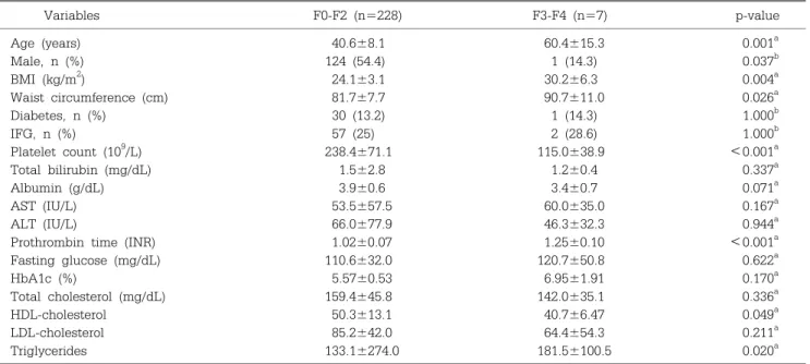
Table 2. Baseline Characteristics of All Patients
Variables F0-F2 (n=228) F3-F4 (n=7) p-value
Age (years) 40.6±8.1 60.4±15.3 0.001a
Male, n (%) 124 (54.4) 1 (14.3) 0.037b
BMI (kg/m2) 24.1±3.1 30.2±6.3 0.004a
Waist circumference (cm) 81.7±7.7 90.7±11.0 0.026a
Diabetes, n (%) 30 (13.2) 1 (14.3) 1.000b
IFG, n (%) 57 (25) 2 (28.6) 1.000b
Platelet count (109/L) 238.4±71.1 115.0±38.9 <0.001a
Total bilirubin (mg/dL) 1.5±2.8 1.2±0.4 0.337a
Albumin (g/dL) 3.9±0.6 3.4±0.7 0.071a
AST (IU/L) 53.5±57.5 60.0±35.0 0.167a
ALT (IU/L) 66.0±77.9 46.3±32.3 0.944a
Prothrombin time (INR) 1.02±0.07 1.25±0.10 <0.001a
Fasting glucose (mg/dL) 110.6±32.0 120.7±50.8 0.622a
HbA1c (%) 5.57±0.53 6.95±1.91 0.170a
Total cholesterol (mg/dL) 159.4±45.8 142.0±35.1 0.336a
HDL-cholesterol 50.3±13.1 40.7±6.47 0.049a
LDL-cholesterol 85.2±42.0 64.4±54.3 0.211a
Triglycerides 133.1±274.0 181.5±100.5 0.020a
Values are expressed as mean±SD.
BMI, body mass index; IFG, impaired fasting glucose; AST, aspartate transaminase; ALT, alanine transaminase; INR, interna- tional normalization ratio; HbA1c, hemoglobin A1c; HDL, high density lipoproteins; LDL, low density lipoproteins.
aMann-Whitney test. bTwo-sided Fisher’s exact test.
(NPV), positive likelihood ratio, and negative likelihood ratio were then calculated for each of the scoring sys- tems using the previously published cut-off values. All analyses were conducted using SPSS version 17.0 (SPSS Inc., Chicago, IL, USA) and p-values of <0.05 were con- sidered significant.
RESULTS
1. Patient characteristics
Of the 297 patients initially included, 62 patients with at least one potential cause of chronic liver disease were excluded: 23 with hepatitis B, 10 with hepatitis C, 12 with autoimmune hepatitis, 7 with excessive alcohol consumption, and 10 with a prior or a current malignancy. A total of 235 patients finally included in this study. Patient characteristics are summarized in Table 2. There was no evidence of liver decompensation among the included patients. Fibrosis stage was de- termined in all 235 patients: 212 patients with METAVIR F0, 11 with F1, 5 with F2, 1 with F3, and 6 with F4.
2. Comparison of P2/MS with the other noninvasive fibrosis scoring systems
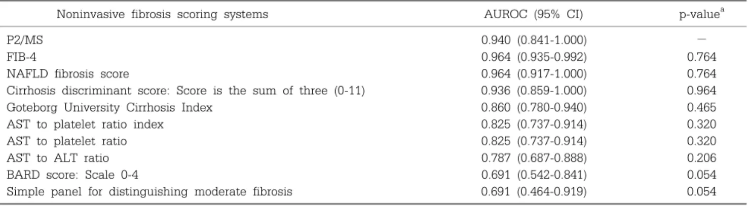
To determine the clinical utility for detecting ad- vanced fibrosis, i.e. METAVIR F3-F4, ROC curves were plotted for each of the ten noninvasive fibrosis scoring systems. The AUROC was greatest for FIB-4 (0.964; 95%
CI, 0.935-0.992) and NAFLD fibrosis score (0.964; 95%
CI, 0.917-1.000) (Fig. 1A) and then followed by P2/MS (0.940; 95% CI, 0.841-1.000) (Fig. 1B), CDS (0.936; 95%
CI, 0.859-1.000), GUCI (0.860; 95% CI, 0.780-0.940), APRI (0.825; 95% CI, 0.737-0.914), APR (0.825; 95% CI, 0.737-0.914), AAR (0.787; 95% CI, 0.687-0.888), BARD score (0.691, 95% CI, 0.542-0.841), and the simple panel (0.691, 95% CI, 0.464-0.919). There was no significant difference between the AUROC for P2/MS and the AUROC for FIB-4, NAFLD fibrosis score, P2/MS, CDS, GUCI, APRI, APR, or AAR (Table 3).
3. Determination of an optimal cut-off values of P2/
MS for reflection of advanced fibrosis in patients with NAFLD
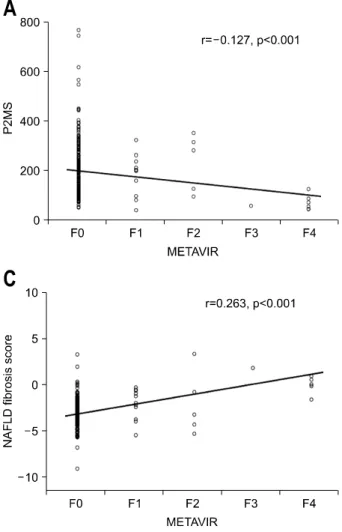
P2/MS scores ranged from 0.620 to 764.35 and its mean value decreased as a function of the METAVIR
Fig. 1. Comparisons of the ROC of P2/MS and other noninvasive fibrosis scoring systems for the diagnosis of advanced fibrosis (defined as METAVIR F3-F4). (A) P2/MS versus FIB-4 and (B) P2/MS versus NAFLD fibrosis score.
Table 3. AUROC of Noninvasive Fibrosis Scoring Systems for Predicting Advanced Fibrosis (METAVIR F3-F4)
Noninvasive fibrosis scoring systems AUROC (95% CI) p-valuea
P2/MS 0.940 (0.841-1.000) −
FIB-4 0.964 (0.935-0.992) 0.764
NAFLD fibrosis score 0.964 (0.917-1.000) 0.764
Cirrhosis discriminant score: Score is the sum of three (0-11) 0.936 (0.859-1.000) 0.964
Goteborg University Cirrhosis Index 0.860 (0.780-0.940) 0.465
AST to platelet ratio index 0.825 (0.737-0.914) 0.320
AST to platelet ratio 0.825 (0.737-0.914) 0.320
AST to ALT ratio 0.787 (0.687-0.888) 0.206
BARD score: Scale 0-4 0.691 (0.542-0.841) 0.054
Simple panel for distinguishing moderate fibrosis 0.691 (0.464-0.919) 0.054
AUROC, area under the receiver operating characteristic curve; CI, confidence interval; NAFLD, nonalcoholic fatty liver disease;
P2/MS, [Platelet count (109/l)]2/[monocyte fraction (%)×segmented neutrophil fraction (%)].
aCompared to AUROC of P2/MS.
fibrosis stage, ranging from 171.4±144.8 in F0 cases to 33.5±32.7 in F4 cases. Furthermore, P2/MS showed a significant inverse correlation with the METAVIR fib- rosis stage (Spearman’s correlation coefficient=−0.127, p<0.001) (Fig. 2A).
Based on our ROC curve analysis, we suggested opti- mized cut-off values of P2/MS for the reflection of ad- vanced fibrosis in patients with NAFLD. At a cut-off val- ue of 49, P2/MS detected advanced fibrosis with a spe- cificity of 92.1% (95% confidence interval [CI], 87.6-95.1) and a PPV of 21.7% (95% CI, 8.3-44.2); and at a cut-off value of 95, P2/MS ruled out advanced fibrosis with a sensitivity 100% (95% CI, 56.1-100) and a NPV of 100%
(95% CI, 97.1-100). If liver biopsies were only performed in patients with a P2/MS <95, 162 (68.9%) of 235 biop-
sies could have been avoided (Table 4).
4. Clinical utility of FIB-4 and NAFLD fibrosis score for detecting advanced fibrosis
FIB-4 scores ranged from 0.24 to 13.15 and its mean value increased as a function of the METAVIR fibrosis stage, ranging from 1.24±1.00 in F0 cases to 4.87±2.38 in F4 cases. FIB-4 showed a significant positive correla- tion with the METAVIR fibrosis stage (Spearman’s cor- relation coefficient=0.235, p<0.001) (Fig. 2B). NAFLD fibrosis score ranged from −7.85 to 8.50 and its mean value increased as a function of the METAVIR fibrosis stage, ranging from −2.30±1.38 in F0 cases to 1.93±
3.32 in F4 cases. NAFLD fibrosis score showed a sig- nificant positive correlation with the METAVIR fibrosis
Fig. 2. Scatter plot of noninvasive fibrosis scoring systems versus pathologically determined fibrosis stage. Fibrosis stage was determined using the METAVIR scoring system.25 Circles represent the values of each noninvasive fibrosis scoring systems. The solid line represents a trend-line. (A) P2/MS values showed a significant inverse correlation with the METAVIR fibrosis stage (Spearman’s correlation coefficient=
−0.127, p<0.001), (B) FIB-4 scores showed a significant in- verse correlation with the METAVIR fibrosis stage (Spearman’s correlation coefficient=0.235, p<0.001), (C) NAFLD fibrosis score values showed a significant inverse correlation with the METAVIR fibrosis stage (Spearman’s correlation coefficient=
0.263, p<0.001).
Table 4. Predictive Values of P2/MS for Advanced Fibrosis (METAVIR F3-F4)
Low
(<49) Intermediate High (>95) Total
Total 23 50 162 235
No advanced fibrosis 18 48 162 228
Advanced fibrosis 5 2 0 7
Sensitivity 71.4% 100%
Specificity 92.1% 71.1%
PPV 21.7% 9.60%
NPV 99.1% 100%
PLR 9.05 3.45
NLR 0.31 0
PPV, positive predictive value; NPV, negative predictive value; PLR, positive likelihood ratio; NLR, negative likelihood ratio.
stage (Spearman’s correlation coefficient=0.263, p<
0.001) (Fig. 2C).
We evaluated the clinical utilities of FIB-4 and NAFLD fibrosis score based on the results of AUROC
comparisons of the noninvasive fibrosis scoring systems.
At a lower cut-off value of 1.45, FIB-4 ruled out ad- vanced fibrosis with a sensitivity of 100% (95% CI, 56.1-100) and a NPV of 100% (95% CI, 97.2-100); and at a cut-off value of 3.25, FIB-4 detected advanced fibrosis with a specificity of 95.6% (95% CI, 91.8-97.8) and a PPV of 33.3% (95% CI, 13.0-61.3). If liver biopsies were only performed in patients with an FIB-4 above the low cut-off value (1.45), 168 (71.5%) of 235 biopsies could have been avoided.
At a cut-off value of −1.455, NAFLD fibrosis score ruled out advanced fibrosis with a sensitivity of 100%
(95% CI, 56.1-100) and a NPV of 100% (95% CI, 97.2- 100); and at a cut-off value of 0.676, NAFLD fibrosis score detected advanced fibrosis with a specificity of 99.1% (95% CI, 96.5-99.8) and a PPV of 33.3% (95% CI, 1.8-87.5). If liver biopsies were only performed in pa- tients with a NAFLD fibrosis score above the low cut-off value −1.455, 170 (72.3%) of 235 biopsies could have
been avoided.
DISCUSSION
The principal findings of this study relate to the de- tection of severe hepatic fibrosis in patients with NAFLD using P2/MS. P2/MS was showed a statistically significant inverse correlation with the METAVIR fib- rosis stage and diagnostic accuracy for detecting ad- vanced fibrosis (METAVIR F3-F4) in patients with NAFLD. In addition, FIB-4 and NAFLD fibrosis score were found to have greater diagnostic accuracy than the other eight noninvasive fibrosis scoring systems for de- tecting advanced fibrosis in Korean patients with NAFLD, though P2/MS was found to have a diagnostic accuracy similar to FIB-4 or NAFLD fibrosis score. To our knowledge, this is the first study validating the diag- nostic accuracy of P2/MS for detecting advanced fibrosis in patients with NAFLD.
In this study, we focused on patients with advanced fibrosis (METAVIR F3-F4).4,28 The underlying rationale was that this approach would help identify those who should or not undergo a biopsy because the detection of advanced fibrosis would lead to closer screening for HCC,28 as patients with CLD tend to show a gradual progression from hepatic fibrosis to liver cirrhosis and HCC.29 In our cohort, 68.9% of the liver biopsies might have been avoided if the procedure was only performed in patients with a P2/MS score below the high cut-off value of 95. Therefore, P2/MS would be particularly use- ful to reduce unnecessary liver biopsies in Korean pa- tients with NAFLD.
It is well known that a proportion of patients with NAFLD, even those with advanced fibrosis, have normal liver enzyme levels and low AST levels until the late dis- ease stage.1,30 P2/MS which is based on a complete blood cell count will work well in such patients and our findings that the AUROC of P2/MS is similar to or better than those of the other tests, which require liver enzyme information, support the implementation of P2/MS for the detection of advanced fibrosis in patients with NAFLD. Furthermore, P2/MS has advantages over other noninvasive fibrosis scoring systems based on BMI, be- cause P2/MS is not based on BMI. Although there was
a significant difference in the mean value of BMI be- tween patients with advanced fibrosis and patients with F0-F2 in this study, the mean value of BMI of Asian pa- tients with advanced fibrosis was lower than those of Caucasian patients with advanced fibrosis (30.2±6.3 vs.
35±6.0).28 Indeed, 62 patients were classified as inter- mediate according to NAFLD fibrosis score, whereas 50 patients were classified as intermediate according to P2/MS (p=0.06).
In addition, we also investigated other noninvasive fibrosis scoring systems, which were mainly derived in Caucasians. Of these, FIB-4 and NAFLD fibrosis score were found to have greatest diagnostic accuracy for the detection of advanced fibrosis. In our cohort, 71.5% and 72.3% of the liver biopsies might have been avoided if the procedure was only performed in patients with an FIB-4 <1.45 and an NAFLD fibrosis score <−1.455, respectively. These findings are in agreement with those of previous studies, which reported that 70% and 79%
of liver biopsies could be replaced by FIB-4 and NAFLD fibrosis score, respectively.1,31 Therefore, it appears that FIB-4 and NAFLD fibrosis score could be used to reduce unnecessary liver biopsies in the Korean population where advanced fibrosis is uncommon.
This study has several limitations that bear considera- tion. The prevalence of advanced fibrosis in our cohort was only 2.98%. This proportion is smaller than that re- ported by other Asian studies,32,33 which would tend to increase the NPV of the noninvasive fibrosis scoring sys- tems examined in the present study. This divergence may have been caused by the low mean BMI of our pa- tients, which was <25 kg/m2 in 67.2% of our cohort.
Thus, in further study, the NPV of P2/MS could be de- creased after statistical adjustment of the number of pa- tients with F0-F2. Although patients were recruited at a single tertiary referral medical center, our patients may have represent NAFLD patients in the community.
However, potential applications of P2/MS and other non- invasive fibrosis scoring systems as a screening method for silent liver disease with advanced fibrosis in the gen- eral population require validation in properly designed studies.28 Although the prevalence of NAFLD was re- ported to be high in male patients in previous studies, female patients were predominant in advanced fibrosis
group in this study. This is similar to previous studies in which 59-71% of patients with advanced fibrosis were female patients.12,28 In this study, this might be asso- ciated with the significant difference of mean age be- tween female and male patients (43.5 years vs. 39.1 years, p<0.001). Indeed, the mean age of female pa- tients with advanced fibrosis was 64.5 years in this study and postmenopausal status was thought to be re- lated with high prevalence of advanced fibrosis. There- fore, further prospective studies including more patients with advanced fibrosis are needed to validate the high cut-off values of FIB-4 and NAFLD fibrosis score and the low cut-off value of P2/MS. As in all validation studies of noninvasive fibrosis scoring systems for hepatic fib- rosis, in the present study, liver biopsy was used as the gold standard, which introduces potential sampling errors.1 We tried to minimize these errors by excluding liver biopsy samples with inadequate tissue length and numbers of portal tracts.21,25 In addition, fibrosis stage was assessed according to the METAVIR scoring system which was developed for viral hepatitis. However, the METAVIR system was also used in NAFLD and was thought to be a better reference for noninvasive panels based on blood tests.34 Another major issue relates to whether changes in P2/MS scores correspond to changes in hepatic fibrosis over time. In the present study, the sensitivity of the P2/MS to changes in hepatic fibrosis was not examined, and thus, further studies should be undertaken to determine if changes in P2/MS can be utilized to monitor hepatic fibrosis in patients with NAFLD. Finally, since we performed a retrospective analysis with data from a single medical center, an in- dependent external validation of P2/MS in patients with NAFLD is also required.
In conclusion, P2/MS as well as FIB-4 and NAFLD fibrosis score were found to have good negative pre- dictive values for excluding the possibility of advanced fibrosis in Korean patients with NAFLD, and thus, could be used to reduce the burden of liver biopsies. A further larger-scale prospective study containing more patients with advanced fibrosis is warranted.
REFERENCES
1. Wong VW, Wong GL, Chim AM, et al. Validation of the NAFLD fibrosis score in a Chinese population with low prevalence of advanced fibrosis. Am J Gastroenterol 2008;103:1682-1688.
2. Targher G, Bertolini L, Padovani R, Zenari L, Zoppini G, Falezza G. Relation of nonalcoholic hepatic steatosis to early carotid atherosclerosis in healthy men: role of vis- ceral fat accumulation. Diabetes Care 2004;27:2498- 2500.
3. Ko JS. Nonalcoholic fatty liver disease. Korean J Gastro- enterol 2010;56:6-14.
4. Guha IN, Parkes J, Roderick P, et al. Noninvasive mark- ers of fibrosis in nonalcoholic fatty liver disease:
Validating the European Liver Fibrosis Panel and explor- ing simple markers. Hepatology 2008;47:455-460.
5. Starley BQ, Calcagno CJ, Harrison SA. Nonalcoholic fatty liver disease and hepatocellular carcinoma: a weighty connection. Hepatology 2010;51:1820-1832.
6. Adams LA, Sanderson S, Lindor KD, Angulo P. The histo- logical course of nonalcoholic fatty liver disease: a longi- tudinal study of 103 patients with sequential liver biopsies. J Hepatol 2005;42:132-138.
7. Ekstedt M, Franzén LE, Mathiesen UL, et al. Long-term follow-up of patients with NAFLD and elevated liver enzymes. Hepatology 2006;44:865-873.
8. Angulo P, Hui JM, Marchesini G, et al. The NAFLD fib- rosis score: a noninvasive system that identifies liver fib- rosis in patients with NAFLD. Hepatology 2007;45:846- 854.
9. Bedossa P, Dargère D, Paradis V. Sampling variability of liver fibrosis in chronic hepatitis C. Hepatology 2003;38:
1449-1457.
10. Ratziu V, Charlotte F, Heurtier A, et al. Sampling varia- bility of liver biopsy in nonalcoholic fatty liver disease.
Gastroenterology 2005;128:1898-1906.
11. Grattagliano I, D'Ambrosio G, Palmieri VO, Moschetta A, Palasciano G, Portincasa P; "Steatostop Project"
Group. Improving nonalcoholic fatty liver disease man- agement by general practitioners: a critical evaluation and impact of an educational training program. J Gastrointestin Liver Dis 2008;17:389-394.
12. McPherson S, Stewart SF, Henderson E, Burt AD, Day CP. Simple non-invasive fibrosis scoring systems can reli- ably exclude advanced fibrosis in patients with non-alco- holic fatty liver disease. Gut 2010;59:1265-1269.
13. Williams AL, Hoofnagle JH. Ratio of serum aspartate to alanine aminotransferase in chronic hepatitis. Relation- ship to cirrhosis. Gastroenterology 1988;95:734-739.
14. Bourliere M, Penaranda G, Renou C, et al. Validation and comparison of indexes for fibrosis and cirrhosis pre-
diction in chronic hepatitis C patients: proposal for a pragmatic approach classification without liver biopsies.
J Viral Hepat 2006;13:659-670.
15. Borroni G, Ceriani R, Cazzaniga M, et al. Comparison of simple tests for the non-invasive diagnosis of clinically silent cirrhosis in chronic hepatitis C. Aliment Pharmacol Ther 2006;24:797-804.
16. Blonsky JJ, Harrison SA. Review article: nonalcoholic fat- ty liver disease and hepatitis C virus--partners in crime.
Aliment Pharmacol Ther 2008;27:855-865.
17. Islam S, Antonsson L, Westin J, Lagging M. Cirrhosis in hepatitis C virus-infected patients can be excluded using an index of standard biochemical serum markers. Scand J Gastroenterol 2005;40:867-872.
18. Bonacini M, Hadi G, Govindarajan S, Lindsay KL. Utility of a discriminant score for diagnosing advanced fibrosis or cirrhosis in patients with chronic hepatitis C virus infection. Am J Gastroenterol 1997;92:1302-1304.
19. Sterling RK, Lissen E, Clumeck N, et al. Development of a simple noninvasive index to predict significant fibrosis in patients with HIV/HCV coinfection. Hepatology 2006;
43:1317-1325.
20. Wong VW, Hui AY, Tsang SW, et al. Metabolic and adi- pokine profile of Chinese patients with nonalcoholic fatty liver disease. Clin Gastroenterol Hepatol 2006;4:1154- 1161.
21. Lee JH, Yoon JH, Lee CH, et al. Complete blood count reflects the degree of oesophageal varices and liver fib- rosis in virus-related chronic liver disease patients. J Viral Hepat 2009;16:444-452.
22. Kim BK, Han KH, Park JY, et al. External validation of P2/MS and comparison with other simple non-invasive indices for predicting liver fibrosis in HBV-infected patients. Dig Dis Sci 2010;55:2636-2643.
23. Kim BK, Han KH, Park JY, et al. Prospective validation of P2/MS noninvasive index using complete blood counts for detecting oesophageal varices in B-viral cirrhosis. Liver Int 2010;30:860-866.
24. Schwimmer JB, Behling C, Newbury R, et al. Histopath- ology of pediatric nonalcoholic fatty liver disease. Hepa-
tology 2005;42:641-649.
25. Bedossa P, Poynard T. An algorithm for the grading of activity in chronic hepatitis C. The METAVIR Coopera- tive Study Group. Hepatology 1996;24:289-293.
26. Kleiner DE, Brunt EM, Van Natta M, et al. Design and validation of a histological scoring system for non- alcoholic fatty liver disease. Hepatology 2005;41:1313- 1321.
27. Hanley JA, McNeil BJ. A method of comparing the areas under receiver operating characteristic curves derived from the same cases. Radiology 1983;148:839-843.
28. Shah AG, Lydecker A, Murray K, Tetri BN, Contos MJ, Sanyal AJ; Nash Clinical Research Network. Comparison of noninvasive markers of fibrosis in patients with non- alcoholic fatty liver disease. Clin Gastroenterol Hepatol 2009;7:1104-1112.
29. Matsumura H, Moriyama M, Goto I, Tanaka N, Okubo H, Arakawa Y. Natural course of progression of liver fib- rosis in Japanese patients with chronic liver disease type C--a study of 527 patients at one establishment. J Viral Hepat 2000;7:268-275.
30. Mofrad P, Contos MJ, Haque M, et al. Clinical and histo- logic spectrum of nonalcoholic fatty liver disease asso- ciated with normal ALT values. Hepatology 2003;37:
1286-1292.
31. Vallet-Pichard A, Mallet V, Nalpas B, et al. FIB-4: an in- expensive and accurate marker of fibrosis in HCV infection. comparison with liver biopsy and fibrotest.
Hepatology 2007;46:32-36.
32. Malik A, Cheah PL, Hilmi IN, Chan SP, Goh KL. Non-al- coholic fatty liver disease in Malaysia: a demographic, anthropometric, metabolic and histological study. J Dig Dis 2007;8:58-64.
33. Tsang SW, Ng WF, Wu BP, Chow DA, Li ET, Wong TC.
Predictors of fibrosis in Asian patients with non-alcoholic steatohepatitis. J Gastroenterol Hepatol 2006;21:116- 121.
34. Calès P, Boursier J, Chaigneau J, et al. Diagnosis of differ- ent liver fibrosis characteristics by blood tests in non-al- coholic fatty liver disease. Liver Int 2010;30:1346-1354.