Internal Fixation Using Clavicle Hook Plates for Distal Clavicle Fractures
Kwang-Yul Kim, Hyung-Chun Kim , Sung-Jun Cho, Su-Han Ahn, Dong-Seon Kim
Department of Orthopedic Surgery, Wallace Memorial Baptist Hospital, Busan, Korea
Background: To report the radiological and clinical outcomes of internal fixation using distal clavicle hook plates for distal clavicle frac- tures.
Methods: From April 2008 to December 2012, 32 patients with distal clavicle fractures underwent surgery using an AO hook plate. The reduction was qualified and evaluated according to the radiological findings. The evaluation of the clinical outcomes was performed with the University of California at Los Angeles (UCLA) score, the Korean Shoulder score, and the visual analogue scale (VAS) pain score.
Results: By radiological evaluation, we found that 31 of 32 patients showed anatomical reduction and solid bone union. Although we obtained satisfactory UCLA scores, Korean Shoulder Scale scores, and VAS pain scores, 12 cases of complications were present. We found 4 cases of osteolysis of the acromion, 1 case of nonunion, 3 cases of periprosthetic fractures, 3 cases of subacromial pain, and 1 case of skin irritation. We performed re-operations in 2 patients.
Conclusions: To avoid complications associated with clavicle hook plates, choosing the appropriate hook size and bending of the hook according to the slope of the acromion undersurface is critical. Also, we believe that early removal of clavicle plates may help reduce complications.
(Clin Shoulder Elbow 2015;18(1):21-27)
Key Words: Clavicle; Distal clavicle fracture; Hook plate; Complication Clinics in Shoulder and Elbow Vol. 18, No. 1, March, 2015
http://dx.doi.org/10.5397/cise.2015.18.1.21
Received August 6, 2014. Revised January 11, 2015. Accepted January 17, 2015.
Correspondence to: Hyung-Chun Kim
Department of Orthopedic Surgery, Wallace Memorial Baptist Hospital, 200 Geumdan-ro, Geumjeong-gu, Busan 609-728, Korea Tel: +82-51-580-1422, Fax: +82-55-583-2568, E-mail: [email protected]
Financial support: None. Conflict of interests: None.
Introduction
While fractures of clavicle shafts manage to fuse well after conservative treatment, distal clavicle fractures are often unsuc- cessfully treated by conservative means and require surgical treatments.1) For instance, when unstable Neer Type II distal clavicle fractures separated from the coraco-clavicular ligament were treated through conservative management by Edwards et al.,2) approximately 30% of fractures showed non-union. Many others have presented with similar results and argued for the ne- cessity of surgical treatments for distal clavicle fractures.3-6)
Fixation using clavicle hook plates allows anatomical reduc- tion of clavicle fractures and displaced acromioclavicular joints,
and also merits from early return to previous mobility and func- tion after surgery. A downside of fixation using clavicle hook plates is the high incidence of complications such as subacromial pain at abduction, skin irritation around acromioclavicular joints, subacromial bone erosion, and reduction loss by non-union.3,7,8)
The purpose of this study was to analyze the treatment and complications of Neer Type II distal clavicle fractures by internal fixation using AO Hook plates. Especially, we wanted to address the possibility that the complications may arise due to the hook plate that is left inside the patient’s body even after bone union is achieved and the hook inserted into the subacromial space.
And if so, what alternative protocols may be devised to mini- mize the hook plate-associated complications.
Methods
Subjects of Study
We enrolled 32 patients with Neer Type II distal clavicle frac- tures who underwent the clavicle hook plate fixation between April 2008 and December 2012 and were able to participate in at least a 6-month follow-up. The mean period of follow-up was 14.7 months (range, 8 to 39 months). The ratio of sex was 24 males to 8 females. The injury was on the right arm in 19 patients and on left in 13 patients. The mean age of the patients at the time of surgery was 46.2 years (range, 21 to 81 years) of age, and the mean duration from hospitalization to surgery was 2.4 days. The etiology of the distal clavicle fractures were car ac- cidents in 11 cases, misstep in 15 cases, sports-related injury in 5 cases, and by falling in 2 cases. Other concomittant injuries were 3 rib fractures, 1 ipsilateral distal radial fracture and 1 ipsilateral scapular body fracture. No nerve injuries were seen in all cases.
When the fractures were classified according to the Neer’s clas- sification, we found 14 cases of Type IIA fractures where the medial coracoclavicular ligament is fractured but not torn and18 cases of Type IIB fractures where the conoid ligament is torn with only the trapezoidal ligament attached on the distal bone.
Surgical Methods
Under general anesthesia, we made a 6- to 8-cm skin inci- sion along the Langer’s line starting from the lateral third-half of the clavicle. The patient was placed in a semifowler position on a beach-chair. We exposed the fracture site by dissecting the deltoid-trapezius border. After temporarily reducing the fractures using Kirschner (K)-wires or bone reduction forceps, hooks of the clavicle hook plates were inserted into the subacromial space, and the proximal ends were positioned by the clavicle. We used a signal intensifier to evaluate the amount of direct contact be- tween the hook and the bone at the subacromion and to direct anatomical reduction. We used leverage to fix the acromion into its anatomic position and inserted cortical screws to the medial screw holes to fix the fracture medially. After suturing the deltoid and trapezius to the fascia, the skin was sutured. We did not carry out further treatments such as suturing the coracoclavicular ligaments.
After surgery, all patients wore an arm sling for 2 weeks. Pa- tients were begun shoulder range of motion exercises and pas- sive forward flexion from the 4th postoperative day and active joint exercises from the 2nd postoperative week.
The removal of internal fixatives was conducted after on average 4.8 months of confirmed bone union. When removing the fixatives, the acromioclavicular joint was not exposed but, to minimize the damages to the regenerated ligaments, only two thirds of the inner incision line was cut to remove the fixation screws and the plate.
Assessment Methods
We assessed the patients through radiography at the follow- ing postoperative follow-ups; 2 weeks, 4 weeks, 2 months, and every month thereafter until bone union was achieved. We considered bone union as when callus that is superior and in- ferior to the fracture site became connected or when no callus formed, when the fracture line disappears and the trabecular bone passes through the fracture. For radiological examination, the antero-posterior and the cephalic tilt radiographies of the clavicle were viewed to evaluate plate position, bone union, and bone erosion. Further radiological examination was performed in patients who received plate removal and were able to attend follow-ups to assess complications such as re-fracture. For the clinical assessment, we used the University of California at Los Angeles (UCLA) score, Korean Shoulder Scale (KSS) score, and visual analogue scale (VAS) score for pain, which were measured at the postoperative 6 month follow-up. We compared the val- ues of the clinical tests using the Student’s t-test, and statistical significance was set to a p-value of less than 0.05. All statistical analyses were performed using SPSS software ver. 12.0 (SPSS Inc., Chicago, IL, USA).
Results
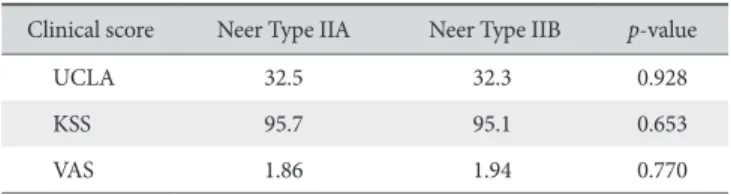
In the 32 patients who received internal fixation of fractures using clavicle hook plates, the plate depth of the plates used was 15 mm in 25 patients and 18 mm in 7 patients, and the plate size was a 4-hole plate in 2 patients, 5-hole plate in 19, 6-hole plate in 10, and a 7-hole plate in 1. Except in one patient with nonunion of the bone, through radiological assessment we found a mean bone union time of 11.6 weeks post-operation in 31 patients. We found that the mean UCLA scores were 32.5 and 32.3 in patients who had Neer Type IIA fractures (12 excel- lent/good and 2 fair/poor) and Neer Type IIB fractures (16 excel- lent/good and 2 fair/poor), respectively (p=0.928). We found that the respective mean KSS scoreswere 95.7 and 95.1, and the respective VAS scores were 1.86 and 1.94 (p=0.653, 0.770;
Table 1). None of these scores between the two fracture groups showed a statistically significant difference. Further, we did not find a significant difference between the groups in terms of the
Table 1. Summary of Clinical Results
Clinical score Neer Type IIA Neer Type IIB p-value
UCLA 32.5 32.3 0.928
KSS 95.7 95.1 0.653
VAS 1.86 1.94 0.770
UCLA: University of California at Los Angeles, KSS: Korean Shoulder Scale, VAS: visual analogue scale.
shoulder range of motion (ROM) after affirmation of bone union (p=0.734, 0.825, 0.781; Table 2). We performed plate removal surgery in 28 out of 32 patients at a mean 4.8 months after con- firmation of bone union by radiography.
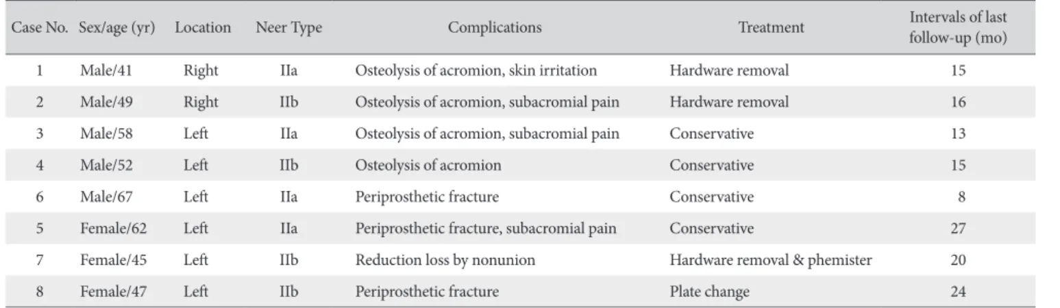
The number of complications in 8 patients totaled to 12, showing a prevalence of 25%. We observed 4 cases of subacro- mial erosion, 1 case of bone nonunion-induced reduction loss, 3 cases of secondary fractures around the plate, 3 cases of mod- erate subacromial pain at abduction, and 1 case of skin irritation around the clavicle fracture (Table 3). We performed re-fixation after removing the fixation devices in a patient that showed non- union of the bone and in another who showed secondary frac- tures around the plate fixation site. They saw bone-union and return of shoulder ROM to normal levels after the operation. Ac- cording to the Neer classification, we found that the prevalence of complications was 28.6% in Neer Type IIA fracture patients (7 complications in 4 patients) and 22.2% in Neer Type IIB frac- ture patients (5 complications in 4 patients). Despite a higher prevalence of complications in patients with Type IIA fractures, we found that it was difficult to directly compare the prevalence rates due to a small number of case reports that were compared and the fact that Neer classification does not take into account communition fracture. Thus, we did not delve further into the difference in the prevalence of complications according to frac- ture type.
Discussion
Dislocated unstable distal clavicle fractures often require sur-
gical treatment,1) but an optimal or standardized method is not yet in place. Especially, Neer Type II fractures are commonly as- sociated with injury to the coracoclavicular ligament or the me- dial ligament, which increases instability of the fracture site and the likelihood of bone nonunion after conservative treatment.3-6) Many types of surgical interventions exist, the choice of which depends on the size and positon of spicules and the presence or absence of coracoclavicular ligament tears in patients. The types of treatment of clavicle fractures include fixation of clavicle frac- tures using screws,9) polydioxanone sutures,6) or Dacron tape,10) fixation of clavicle fractures along with acromioclavicular joints using K-wires,1,11) Steinmann pins,2) or Knowles pins,12) and lastly, fixation of clavicle fractures through the plating system using the tension band wires,13) Wolter plates,14) or the Hook plates.15)
In this study, for the fixation of AO Hook plates, we used a 3.5-mm locking screw, a 3.5-mm cortical screw, or a 4.0-mm cancellous screw. Depending on the plate size, we were able to screw up to 8 holes. At the distal end of the plate where it is fixed to the clavicle, there is a 12° curvature of the plate. Further, a hook step stretching between the plate and the subacromial space makes a hook depth of 12 mm, 15 mm, or 18 mm. Thus, hook size was chosen according to the patient build and acro- mion size.
Internal fixation of distal clavicle fractures using hook plates provides the mechanical strength to retain reduction, and a relatively small bone-plate contact region allows abundant vas- cular supply at the clavicle. Not only do hook plate systems have rotational and horizontal stability, it is associated with early reha- bilitation after surgery.16-18) However, limitations to this method include a requirement for a secondary surgery to remove internal fixation devices, broad skin incision, subacromial oste- olysis, plate-induced pain, subacromial impingement-induced pain, and reduced ROM.3,7,8) Renger et al.18) found that in pa- tients who receive clavicle hook plating, 68% had subacromial impingement-induced pain, reduced shoulder ROM, and sub- acromial erosion. They later found that these symptoms resolved in 90% of patients whose plates were removed postoperatively.
Table 2. Summary of Range of Motion at the 6 Month Follow-up Range of motion (o) Neer Type IIA Neer Type IIB p-value
Forward flexion 168.6 165.4 0.734
External rotation 65.1 63.9 0.825
Abduction 158.2 155.7 0.781
Table 3. Summary of Complications
Case No. Sex/age (yr) Location Neer Type Complications Treatment Intervals of last
follow-up (mo)
1 Male/41 Right IIa Osteolysis of acromion, skin irritation Hardware removal 15
2 Male/49 Right IIb Osteolysis of acromion, subacromial pain Hardware removal 16
3 Male/58 Left IIa Osteolysis of acromion, subacromial pain Conservative 13
4 Male/52 Left IIb Osteolysis of acromion Conservative 15
6 Male/67 Left IIa Periprosthetic fracture Conservative 8
5 Female/62 Left IIa Periprosthetic fracture, subacromial pain Conservative 27
7 Female/45 Left IIb Reduction loss by nonunion Hardware removal & phemister 20
8 Female/47 Left IIb Periprosthetic fracture Plate change 24
Similarly, Meda et al.19) found that the complications, 6 cases of impingement syndrome and 5 cases of subacromial osteolysis, observed in patients who underwent internal fixation with plates were resolved after plate removal. Further, Muramatsu et al.17) found that in patients whose fixative plates were not removed even when bone union was attained there was subacromial dis- placement of the plate hook and proposed that plate removal is a necessity. In line with previous findings, we found that when we removed the internal fixatives in 28 patients, excluding the patient with reduction loss and the 3 patients with secondary fractures, from a mean 4.8 months of bone union, no complica- tions were seen.
In this study, we observed 4 cases of subacromial osteolysis of which in 2 cases, we carried out removal of the internal fixa- tives at 4 months of surveillance when we saw that subacromial osteolysis and clavicle joint pain worsened. Pain resolved after surgery and further deformities were not detected (Fig. 1). In the other 2 cases of subacromial osteolysis, the patients complained only of mild symptoms without additional progression of symp- toms. These patients were monitored by active surveillance.
After taking into consideration the acromial slope, we made the ends of the hooks to bend in to increase the hook-bone surface area in 9 cases. At 12-month follow-up, we could that we could not observe complications such as bone osteolysis or erosion in these 9 cases. However, due to a small sample, a comparative study with a greater patient sample is needed to confirm our re- sults.
In this study, we found other complications such as 3 cases of secondary clavicle shaft fractures. In one instance, we found a secondary fracture around the region where the screw was fixed to the plate at 3-month postoperation without other obvious in- juries. Currently, we are actively surveying the secondary fracture at follow-ups due to this patient’s non-compliance towards ad- ditional operation (Fig. 2). In another example, a female patient with severe osteoporosis exhibited with a secondary clavicle B
A C
Fig. 1. Radiographs of a 52-year-old male. (A) Postoperative radiograph of the right shoulder shows a Neer Type II distal clavicle fracture and fixation using a hook plate. (B) At 2-month follow-up, the radiograph shows osteolysis of the acromion near the hook plate. (C) At 4-month follow-up, the hook plate was re- moved.
A
B
C
Fig. 2. Radiographs of a 67-year-old male. (A) A preoperative radiograph shows a left distal clavicle fracture. (B) Postoperative radiograph. The fracture was reduced and stabilized with a hook plate. (C) A postoperative radiograph at 2-month follow-up shows a clavicle shaft fracture (arrow).
shaft fracture where a screw was fixed to the plate at 2 months postoperation. Describing a similar case, Bottlang et al.20) found that in bone shaft fractures in patients with severe osteoporosis, the use of locking screws on either sides of the locking compres- sion plate led to an increase in pressure at the interface of the screw and bone, thereby increasing the likelihood of a second- ary fracture. They showed that using pre-used screws rather than locking screws at the distal ends of the locking plate reduces the likelihood of secondary fractures around the plate region. How- ever, if at the discretion of the surgeon a strong pull out strength is needed, locking screws may be used with the distance be- tween the screws and the fracture line in mind, and additional cortical screws if necessary. However, this particular patient refused further surgical operation, thus we carried out conserva- tive treatment using a clavicle brace and actively surveyed the patient. At 12-week postoperation, a radiological bone union was observed after which shoulder ROM was begun (Fig. 3).
Lastly, we found a patient with a secondary clavicle shaft fracture around the plate at one month post-operation. We deduced that this secondary fracture was probably due to an insufficient plate length and subsequent failure of fixation. Re-fixation by exchanging the plate to a 6-hole plate led to a successful bone union at 10-week post re-operation without complications.
In the case of plate-induced loss in reduction, we did not see bone union even at the postoperative 9-month follow-up but saw loss in reduction at subsequent follow-ups. At the time of surgery, making an anatomical reduction in this patient was challenging due to a communition fracture. We hypothesize
that because we were unable to fix the locking screws parallel to the fracture line, micro-movements of the fracture led to an in- complete internal fixation and bone nonunion. For this patient, we carried out plate removal operation, then a bone graft and Phemister surgery. When we assessed the radiological outcome at the 12-week post-removal follow-up we assessed a radiologic bone union and restored shoulder ROM (Fig. 4).
In our study, 3 patients with subacromial impingement syn- drome complained of a locking sensation of the plate during exercise accompanied by pain and a reduced shoulder ROM.
These complications disappeared once the internal fixation de- vices were removed in these patients.
Altogether, we propose that in order to avoid complications after internal fixation of distal clavicle fractures using hook plates such as subacromial impingement syndrome, and improve post- operative ROM, prophylactic removal of plate fixatives as soon as bone union is affirmed is a critical step.
Limitations to our study include having only a small number of case reports and a short duration of follow-up after plate removal. A further prospective study using a greater number of cases to compare the clinical and radiological outcomes is needed.
Conclusion
We found that although using clavicle hook plates for distal clavicle fractures give satisfactory clinical and radiological out- comes, surgeons and clinicians need to be aware of and reduce Fig. 3. Radiographs of a 62-year-old female. (A) A preoperative radiograph of the left distal clavicle fracture. (B) Postoperative radiograph. The fracture was reduced and stabilized with a hook plate. (C) A postoperative radiograph at 4-week follow-up shows a clavicle shaft fracture. (D) A postoperative radiograph at 12-month follow-up shows bony union without any complications.
A B
C D
complications associated with this procedure. Choosing the ap- propriate hook plate depths on a patient by patient basis and bending the hook plate sufficiently when inserting it into the subacromial space are important considerations to make. Fur- ther, once bone union is confirmed by radiologic assessment, we believe that taking the internal fixatives out at the earliest pos- sible stage helps to minimize complications and gives the best clinical results.
References
1. NEER CS 2nd. Nonunion of the clavicle. J Am Med Assoc.
1960;172:1006-11.
2. Edwards DJ, Kavanagh TG, Flannery MC. Fractures of the distal clavicle: a case for fixation. Injury. 1992;23(1):44-6.
3. Charity RM, Haidar SG, Ghosh S, Tillu AB. Fixation failure of the clavicular hook plate: a report of three cases. J Orthop Surg (Hong Kong). 2006;14(3):333-5.
4. Levy O. Simple, minimally invasive surgical technique for treatment of type 2 fractures of the distal clavicle. J Shoulder Elbow Surg. 2003;12(1):24-8.
5. Chen CH, Chen WJ, Shih CH. Surgical treatment for distal
clavicle fracture with coracoclavicular ligament disruption. J Trauma. 2002;52(1):72-8.
6. Mall JW, Jacobi CA, Philipp AW, Peter FJ. Surgical treatment of fractures of the distal clavicle with polydioxanone suture ten- sion band wiring: an alternative osteosynthesis. J Orthop Sci.
2002;7(5):535-7.
7. Salem KH, Schmelz A. Treatment of Tossy III acromioclavicular joint injuries using hook plates and ligament suture. J Orthop Trauma. 2009;23(8):565-9.
8. Faraj AA, Ketzer B. The use of a hook-plate in the manage- ment of acromioclavicular injuries. Report of ten cases. Acta Orthop Belg. 2001;67(5):448-51.
9. Habernek H, Weinstabl R, Schmid L, Fialka C. A crook plate for treatment of acromioclavicular joint separation:
indication, technique, and results after one year. J Trauma.
1993;35(6):893-901.
10. Goldberg JA, Bruce WJ, Sonnabend DH, Walsh WR. Type 2 fractures of the distal clavicle: a new surgical technique. J Shoulder Elbow Surg. 1997;6(4):380-2.
11. Kona J, Bosse MJ, Staeheli JW, Rosseau RL. Type II distal clavicle fractures: a retrospective review of surgical treatment. J Orthop Trauma. 1990;4(2):115-20.
Fig. 4. Radiographs of a 45-year-old female. (A) A preoperative radiograph shows a left distal clavicle fracture. (B) Postoperative radiograph. The fracture was reduced and fixed with a hook plate. (C) A postoperative radiograph at 6 month follow-up shows proximal screw loosening and plate migration without bony union. (D) A postoperative radiograph at 12-month follow-up shows a removed hook plate, internal fixation with Kirschner wires, and bone grafts. (E) A postoperative radiograph at 16-month follow-up shows bony union.
C D
E
A B
12. Craig EV. Fracture of the clavicle. In: Rockwood CA Jr, Green DP, Bucholz RW, Heckman JD, eds. Fracture in adult. 4th ed.
Philadelphia: Lippincort-Raven; 1996. 1109-61.
13. Kao FC, Chao EK, Chen CH, Yu SW, Chen CY, Yen CY. Treat- ment of distal clavicle fracture using Kirschner wires and tension-band wires. J Trauma. 2001;51(3):522-5.
14. Mizue F, Shirai Y, Ito H. Surgical treatment of comminuted fractures of the distal clavicle using Wolter clavicular plates. J Nippon Med Sch. 2000;67(1):32-4.
15. Flinkkilä T, Ristiniemi J, Lakovaara M, Hyvönen P, Leppilahti J.
Hook-plate fixation of unstable lateral clavicle fractures: a re- port on 63 patients. Acta Orthop. 2006;77(4):644-9.
16. Haidar SG, Krishnan KM, Deshmukh SC. Hook plate fixation for type II fractures of the lateral end of the clavicle. J Shoulder Elbow Surg. 2006;15(4):419-23.
17. Muramatsu K, Shigetomi M, Matsunaga T, Murata Y, Tagu- chi T. Use of the AO hook-plate for treatment of unstable fractures of the distal clavicle. Arch Orthop Trauma Surg.
2007;127(3):191-4.
18. Renger RJ, Roukema GR, Reurings JC, Raams PM, Font J, Verleisdonk EJ. The clavicle hook plate for Neer type II lateral clavicle fractures. J Orthop Trauma. 2009;23(8):570-4.
19. Meda PV, Machani B, Sinopidis C, Braithwaite I, Brownson P, Frostick SP. Clavicular hook plate for lateral end fractures:- a prospective study. Injury. 2006;37(3):277-83.
20. Bottlang M, Doornink J, Byrd GD, Fitzpatrick DC, Madey SM.
A nonlocking end screw can decrease fracture risk caused by locked plating in the osteoporotic diaphysis. J Bone Joint Surg Am. 2009;91(3):620-7.