J. Exp. Biomed. Sci. 13 (2007) 161–168
Effects of Solvent-extracted Fractions from Salicornia herbacea on Anti-oxidative Activity and Lipopolysaccharide-induced NO
Production in Murine Macrophage RAW264.7 Cells
Whi Min Lee1, Hye Jin Sung1, Jae Chan Song1, Jae Youl Cho2, Hwa Jin Park3, Suk Kim4 and Man Hee Rhee1,†
1Department of Veterinary Medicine, College of Veterinary Medicine, Kyungpook National University, Daegu 702-701, 2School of Biotechnology and Bioengineering, Kangwon National University,
Chuncheon 200-701, 3Department of Biomedical Laboratory Science, College of Biomedical Science and Engineering, Inje University, Gimhae 602-749, 4Department of Veterinary Medicine,
College of Veterinary Medicine, Gyeongsang National University, Jinju 660-701, Korea
Salicornia herbacea L. (Chenopodiaceae: S. herbacea) is a salt marsh plant, which has long been prescribed in traditional medicines for the treatment of intestinal ailments, nephropathy, and hepatitis in Oriental countries. In order to elucidate the mechanisms of this herb, we conducted an anti-oxidative activity, the inhibition of nitric oxide (NO) production, and the suppression of the pro-inflammatory cytokine genes, with the solvent-extracts of S. herbacea. We found that both ethyl acetate and n-butanol fractions showed potent anti-oxidative effects in comparison to other fractions using xanthine oxidase assay with IC50 values of 66.0±0.5 µg/ml and 82.5±3.8 µg/ml, respectively. In addition, both ethyl acetate and n-butanol fractions showed more electron donating activity (EDA) than other fractions, according to DPPH (2, 2-Diphenyl-lpicrylhydrazyl radical) assay. The EDA of ethyl acetate fraction (IC50 values of 117.5±3.8 µg/ml) is more significant than that of n-butanol fraction (IC50 values of 375.0±12.5 µg/ml). Among potential anti-oxidative fractions, ethyl acetate fraction dose-dependently suppressed lipopolysaccharide (LPS, 0.1 µg/ml)-induced nitric oxide (NO) production in RAW264.7 cell, while n-butanol did not. As expected, ethyl acetate fraction suppressed the expression of inducible NO synthase (iNOS) in RAW264.7 cell stimulated by 0.1 µg/ml of LPS. Moreover, the ethyl acetate fraction suppressed the expression of interleukin-1 (IL)-1β and granulocyte/macrophage colony-stimulating factor (GM-CSF) mRNA in LPS-stimulated RAW264.7 cells. Therefore, these results suggest that S. herbacea may have anti-oxidative and anti-inflammatory activities by modulating radical-induced toxicity and various pro-inflammatory responses.
Key Words: Salicornia herbacea, Anti-oxidative activity, Nitric oxide, Inflammatory cytokines
INTRODUCTION
S. herbacea is a salt marsh plant, which is commonly known as 'Tungtungmadi' in Korea and grows well in tidelands due to its anti-salt characteristics (Lee et al., 2004;
Chung et al., 2005). Several halophytes such as S. herbacea, S. asparagoides, and Calystegia soldanella, are widely di-
stributed in Korea estuaries (Ihm and Lee, 1986; Tori et al., 2000; Kim et al., 2004; Lee et al., 2004; Min, 2005).
Among them, the pharmacological and chemical characteri- zation of S. herbacea have recently been examined (Lee et al., 2004; Lee et al., 2005). The polysaccharide fraction of this plant has previously shown to stimulate nitric oxide (NO) production and inducible NO synthase (iNOS), and cytokine production, such as tumor necrosis factor (TNF)-α and interleukin (IL)-1β (Im et al., 2006; Lee et al., 2006;
Im et al., 2007). As described above, some constituents of this plant had been studied in specific animal model or on immunological potency, but systemic analysis of this plant's solvent extracts regarding on the plant's anti-oxidative and
*Received: August 20, 2007
Accepted after revision: September 6, 2007
†Corresponding author: Man Hee Rhee, Department of Veterinary Medicine, College of Veterinary Medicine, Kyungpook National University, Daegu 702-701, Korea.
Tel: +82-53-950-5967, Fax: +82-53-950-5955 e-mail: [email protected]
anti-inflammatory activities using in vitro model system had not been studied. In addition, although the S. herbacea plant is traditionally employed medicinal remedy for illnesses such as intestinal ailments, nephropathy, and hepatitis, the mechanisms of its pharmacological activity have not been fully elucidated.
Free radicals such as ROS (e.g. superoxide anion radical, hydroxyl radical, singlet oxygen, hydrogen peroxide) and peroxynitrite are highly reactive molecules, which are gene- rated predominantly during cellular respiration and normal metabolism. An imbalance between the cellular production of free radicals and the ability of cells to defend against them is referred as oxidative stress, which is implicated as a potential contributor to lipid peroxidation. Oxidative stress, however, can damage many targets other than lipids, in- cluding proteins, DNA and small molecules. An antioxidant is defined as 'any substance that, when present at low con- centrations compared to those of an oxidizable substrate, significantly delays or prevents oxidation of that substrate' (Halliwell et al., 1995; Wiseman et al., 1997; Mates et al., 1999). Antioxidants are of interest to biologists and clini- cians because they help to protect the human body against damage by reactive free radicals found in cancer, athero- sclerosis, and aging (Halliwell et al., 1995; Mates et al., 1999). Synthetic antioxidants (e.g. tert-butylhydroxytoluene [BHT] and tert-butylhydroxyanisol (Shankar et al., 2006) have been developed. The clinical use of these antioxidants, however, is restricted due to their toxicity, low potency and limitations. Therefore, now interest is in screening safer and more potent antioxidants, which are from natural products, rather than synthetic antioxidants. There are many reports that natural products and their derivatives have efficient antioxidative characteristics, such as possessing anti-cancer, hypolipidemic, anti-aging, and anti-inflammatory activity (Halliwell et al., 1995; Wiseman et al., 1997; Hogg, 1998;
Mates et al., 1999; Aruoma, 2003; Cho et al., 2006).
Macrophages play a central role in managing many different immunopathological phenomena such as the over- production of pro-inflammatory cytokines and inflammatory mediators (i.e. ROS and NO) (Lundberg, 2003; Walsh, 2003). In the case of oxidative stress, NO and ROS affect virtually every step of the development of inflammation.
Macrophages mediate the inflammatory process through the release of chemokines (e.g. MIP-1α and MCP-1) and cytokines (e.g. TNF-α, IL-1β and IL-6).
Indeed, a number of inflammatory stimuli such as LPS and pro-inflammatory cytokines activate immune cells to up-regulate such inflammatory states (Gallucci et al., 1998) and therefore, the stimuli are often used in the development of both developing new anti-inflammatory drugs and for determining the potential of exploring molecular anti- inflammatory mechanisms.
Therefore, in this study, first we determined whether the solvent-extracted fractions of S. herbacea, used ethnophar- macologically for long time, displayed antioxidant activity and if they inhibited LPS-induced NO production and cyto- kine expression in RAW264.7 cells.
MATERIALS AND METHODS
1. Materials
Ascorbate, dimethylsulfoxide (DMSO), DPPH, LPS, Griess's reagent, and xanthine oxidase were obtained from Sigma Co (St. Louis, MO). Xanthine was from Merck Co.
(Milwaukee, WI). All other reagents were of reagent grade.
2. Solvent extraction
S. herbacea was collected from the province of Boryung (Korea) in August, 2002. The voucher specimen with number PLSA-2200 is deposited in the herbarium of our laboratory.
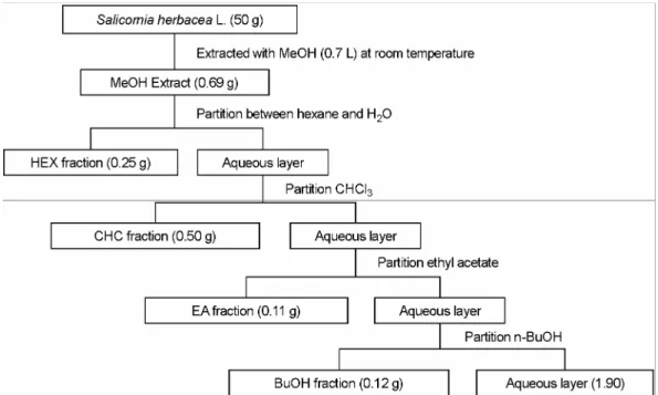
One-hundred g of powder, from the leaves of S. herbacea, were extracted with 0.7 L of methanol overnight (Fig. 1).
The methanol extract was subsequently filtered through filter paper (Whatman No. 3) and centrifuged at 5,000 g for 10 min. The filtrate was evaporated in a Rotavapor (yield 3.57 g [3.6%]). The methanol extract was successively extracted except for 0.69 g, which is suitable for a methanol fraction.
The extraction was conducted using hexane, chloroform, ethyl acetate, n-butanol and water and the yield amounts were 8.7%, 17.4%, 3.7%, 4.2%, and 66.0%, respectively.
The crude extracts were stored in -20℃ until use.
3. DPPH Radical Scavenging Activity
The DPPH assay measured hydrogen atom (or one
electron) donating activity and hence provided an evaluation of antioxidant activity due to free radical scavenging. The DPPH, a purple-colored stable free radical, is reduced into yellow-colored diphenylpicryl hydrazine. The Blios method was used in this experiment with slight modifications (Blois, 1958). A fresh batch of a radical stock solution was prepared daily. The EDA described the difference in the absorbance, between the mixture and the control solution, in terms of a percentage: EDA (%) = (the absorbance of the control - the absorbance of the mixture) / the absorbance of the control × 100.
4. Assay for inhibition of xanthine oxidase activity
The activity of xanthine oxidase with xanthine, as a sub- strate, was measured spectrophotometrically by using the procedures of Noro et al. (Noro et al., 1983), with the follo- wing modifications. The final concentration of xanthine oxidase was 250 µU/ml in a 0.1 mM phosphate buffer (pH 7.4). Xanthine and xanthine oxidase were mixed in a cuvette with either compound being tested or vehicles. The difference of the absorbance was measured at 295 nm for 3 min and enzyme activity was calculated with references: (the activity of control - the activity of the mixture) / (the activity of control) × 100.
5. Cell culture
RAW264.7 cells were maintained in RPMI supplemented with 100 U/ml of penicillin and 100 µg/ml of streptomycin and a 5% FBS. Cells were grown at 37℃ and in 5% CO2
in humidified air.
6. Measurement of nitrite
In order to determine the NO concentration, nitrite (NO2-) was measured using Griess reagent (1% sulfanilamide, 0.1% naphthylethylenediamine dihydrochloride, and 2%
phosphoric acid), as described previously (Hong et al., 2003; Cho et al., 2006). The concentrations of nitrite were calculated from regression analysis, using serial dilutions of sodium nitrite as a standard. The percentage inhibition was calculated based upon the ability of the extracts to inhibit NO formation by cells, as compared with the control (cells in media without extracts containing triggering agents and DMSO), which was considered as no inhibition (0%).
7. Extraction of total RNA
Total RNAs from LPS treated-RAW264.7 cells were prepared by adding Easy blue Reagent (InTron Biotechno- logy Co. Seoul), according to manufacturer's instructions.
Fig. 1. Schematic diagram of extraction and fractionation of S. herbacea. MeOH, methanol; HEX, hexane; CHC, chloroform; EA, ethyl acetate; BuOH, n-butanol.
The total RNA solution was stored at -70℃ until use.
8. Semiquantitative RT-PCR amplification
Semiquantitative RT reactions were carried using RT premix (Bioneer Co. Daejeon). Briefly, total RNAs (2 µg) were incubated with oligo-dT18 at 70℃ for 5 minutes and cooled on ice for 3 minutes. The reaction mixture were incubated for 90 minutes after the addition of RT premix at 42.5℃. The reactions were terminated at 95℃ for 5 minutes, for the inactivation of the reverse transcriptase. The PCR reaction was further conducted using a PCR premix (Bio- neer Co. Daejeon), with an appropriate sense and antisense primer, under the following incubation conditions: a 45- second denaturation time at 94℃, an annealing time of 45 seconds at 55 to 60℃, an extension time of 45 seconds at 72℃, and final extension of 10 minutes at 72℃ at the end of cycles. The primers (Bioneer Co. Daejeon) used in this experiment are described in earlier reports (Cho et al., 2006).
9. Statistical analysis
The one-way and the two-way ANOVA were used to determine the statistical significance of differences between values for the various experimental and control groups.
Data are expressed as means ± standard errors (SEM) in triplicates. P values of 0.05 or less were considered to be statistically significant.
RESULTS AND DISCUSSION
1. S. herbacea displays anti-oxidative effects
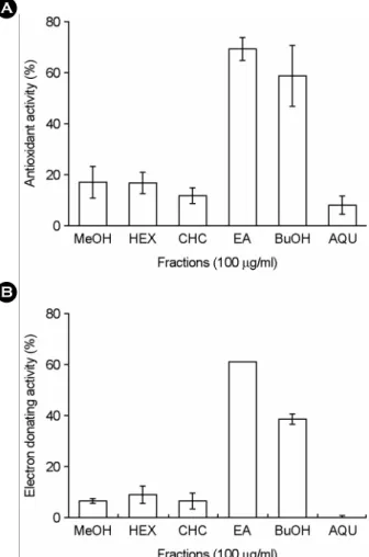
In order to compare the antioxidant capacity of methanol, hexane, chloroform, ethyl acetate, n-butanol, and aqueous fractions of S. herbacea, first, we measured the antioxidant activity of each extract (100 µg/ml) of the plant, by using a xanthine oxidase assay and a DPPH assay. In a xanthine oxidase assay, 100 µg/ml of each extract show antioxidant activity with a significant difference between the samples tested (Fig. 2A). The antioxidant activity of the ethyl acetate and n-butanol fractions of S. herbacea was much higher than those of methanol, hexane, chloroform, and the aqueous fractions of the plant. On the other hand, in a DPPH assay, the radical scavenging activity of ethyl acetate fractions of
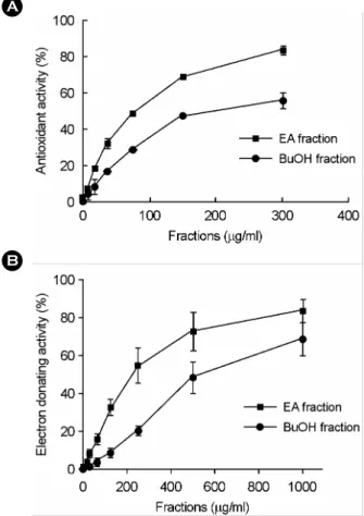
S. asparagoides was much stronger than those of the other fractions of the plant (Fig. 2B). Of various solvent-extracted fractions, we chosen both ethyl acetate and n-butanol frac- tions of the plant due to relatively higher potency at 100 µg/ml. Next, we examined the potency of the antioxidant activity of ethyl acetate and n-butanol fractions, by using a xanthine oxidase and DPPH assay. As shown in Fig. 3A, the IC50 of the ethyl acetate fraction (66.0 µg/ml) is less than those of the n-butanol fraction (82.5 µg/ml). The standard compound, ascorbic acid, also displayed scaven- ging activity with IC50 values of 180 µM (data not shown).
In order to confirm the radical scavenging effect of S.
Fig. 2. Antioxidant activities of various solvent extract from S.
herbacea in xanthine oxidase assay (A) and DPPH assay (B).
Either phosphate buffer (0.1 mM, pH 7.4, for xanthine oxidase assay) or acetate buffer (10 mM, pH 5.5, for DPPH assay) and methanol (MeOH), chloroform (CHC), hexane (HEX), ethyl acetate (EA), n-butanol (BuOH) or aqueous (AQU) extracts (100 µg/ml) were mixed and assay was carried out as described in 'Materials and Methods'. Each value is the mean ± SEM of three determin- tions, performed in triplicate.
B A
herbacea, a DPPH assay was employed. As shown in Fig.
3B, the ethyl acetate extract of S. herbacea also highly scavenged the radical generation, with an IC50 value of 117.5 µg/ml. The radical scavenging activity of the n-butanol fractions was less than that of ethyl acetate fraction from S.
herbacea (i.e. IC50 values of 375.0 µg/ml).
In this study, we found that ethyl acetate fraction of S.
herbacea showed anti-oxidative activity by using xanthine oxidase assay and DPPH assay and n-butanol fraction of the plant did less potential. These different assay systems for anti-oxidative screening showed similar profiles of the
radical scavenging activity of the followings: The ethyl acetate fractions (probably including more lipophilic com- ponents) of the herbs had a more potent antioxidant effect than the n-butanol fraction (probably including hydrophilic components) in xanthine oxidase assay and DPPH assay (see Fig. 2 and Fig. 3). This seems to imply that lipophilic compounds of the antioxidative characteristics of the plant play an important role in protecting against various oxidative stresses such as radical generation and radical scavenging activity. At present, however, which ingredients of these solvent extracts display strong anti-oxidative effect is not exactly investigated yet. Therefore, further study should be followed in the next study to identify the active principles.
B A
Fig. 3. The antioxidant activity of ethyl acetate and n-butanol extracts of S. herbacea in xanthine oxidase assay (A) and DPPH assay (B). A. Phosphate buffer and various concentrations of ethyl acetate (EA) or n-butanol (BuOH) extract were mixed. After adding xanthine and xanthine oxidase, the difference of the absorbance at 295 nm was monitored for 3 min and the enzyme activity was calculated as described in 'Materials and Methods'. B. The acetate buffer and various concentrations of ethyl acetate or n-butanol extracts of S. herbacea were mixed. After adding an ethanolic DPPH solution, the absorbance was monitored at 595 nm and enzyme activity was calculated as described in Materials and Methods. Means ± SEM was calculated from three independent experiments that were performed in triplicate.
A
B
Fig. 4. The effects of ethyl acetate fraction of S. herbacea on the production of NO and on the mRNA expression of iNOS in LPS-activated RAW264.7 cells (1×10⁶ cells/ml). RAW264.7 cells (1×10⁶ cells/ml) were stimulated by a LPS (0.1 µg/ml) and incubated with ethyl acetate (EA) fraction of S. herbacea. A.
Supernatants were collected after 18 h and nitrite formation was determined using Griess' reagent. Means ± SEM was calculated from three independent experiments that were performed in tripli- cate. B. The preparation of total RNA and RT-PCR were performed as described in Materials & Methods. The figures represent the results of three independent experiments. RAW264.7 cells (1×
106 cells/ml) were stimulated by a LPS (0.1 µg/ml) and incubated with the indicated concentration of the plant's ethyl acetate (EA) fraction.. *P<0.05 versus LPS-activated, **P<0.01 versus LPS- activated.
2. S. herbacea modulates NO production and iNOS expression stimulated by LPS
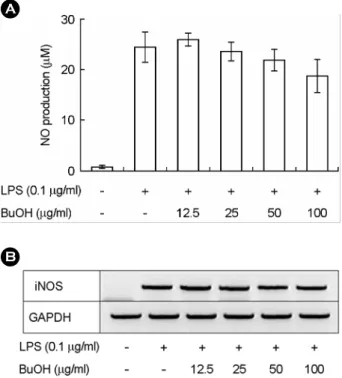
In order to test anti-inflammatory effect of S. herbacea in vitro, we intended to determine whether ethyl acetate fraction and n-butanol fraction of this plant modulated the LPS-induced NO production and iNOS expression in RAW- 264.7 cells. We first determined the cytotoxicity of ethyl acetate fraction and n-butanol fraction of S. herbacea to RAW264.7 cells. The pretreatment of unstimulated cell lines RAW264.7, with both fractions prepared from S. herbacea for 18 h, did not significantly affect cell viability (data not shown). Fig. 4A shows that ethyl acetate fraction of S.
herbacea dose-dependently suppressed NO production in RAW264.7 cells, which was stimulated by 0.1 µg/ml of
LPS. Next, we examined whether ethyl acetate extract of S.
herbacea modulated the expression of iNOS, which is an inducible enzyme that produces NO in response to a LPS.
As shown in Fig. 4B, ethyl acetate fraction of the plant suppressed the LPS-induced iNOS expression in a dose- dependent manner. Since n-butanol fraction of this plant showed the anti-oxidative activity even less than ethyl acetate fraction, we determined whether n-butanol extract modulated NO production and iNOS expression in RAW- 264.7 cells, stimulated by LPS (0.1 µg/ml). The n-butanol fraction of this plant did not suppress the LPS-induced NO production and iNOS expression. These results suggest that the inhibitory of ethyl acetate fraction on NO production and iNOS expression may not be simply due to its radical scavenging activity, but may be due to the modulation of signaling pathway, induced by LPS stimulation, for NO production.
3. S. herbacea extracts modulate the expression of pro-inflammatory cytokines, which are stimulated by LPS
IL-1β, GM-CSF, and IL-6 are known to be pro- inflammatory cytokines, with a multitude of biological activities that are linked to the immunopathology of acute or chronic inflammatory diseases such as septic shock and rheumatoid arthritis, and autoimmune diseases (Eigler et al.,
Fig. 6. The effects of ethyl acetate (EA) extract of S. herbacea on the mRNA expression of IL-1β, GM-CSF, and IL-6 in LPS- activated RAW264.7 cells. RAW264.7 cells (1×10⁶ cells/ml) were stimulated by a LPS (0.1 µg/ml) and incubated with the indicated concentration of the plant's ethyl acetate (EA) fraction. The pre- paration of total RNA and RT-PCR were performed as described in Materials & Methods. The figures represent the results of three independent experiments.
Fig. 5. The effects of n-butanol (BuOH) fraction of S. herbacea on the production of NO and on the mRNA expression of iNOS in LPS-activated RAW264.7 cells (1×10⁶ cells/ml). RAW264.7 cells (1×10⁶ cells/ml) were stimulated by a LPS (0.1 µg/ml) and incubated with n-butanol (BuOH) fraction of S. herbacea. A.
Supernatants were collected after 18 h and nitrite formation was determined using Griess' reagent. Means ± SEM was calculated from three independent experiments that were performed in tripli- cate. B. The preparation of total RNA and RT-PCR were performed as described in Materials & Methods. The figures represent the results of three independent experiments. RAW264.7 cells (1×
106 cells/ml) were stimulated by a LPS (0.1 µg/ml) and incubated with the indicated concentration of the plant's n-butanol (BuOH) fraction.
A
B
1997; Charo and Taubman, 2004; Hamilton and Anderson, 2004). Therefore, we addressed whether ethyl acetate and n-butanol extracts of S. herbacea modulated the expression of IL-1β, GM-CSF, and IL-6 mRNA. Fig. 6 shows that solvent extracts do not have any effect on the expression of GM-CSF and IL-6, which is stimulated by 0.1 µg/ml LPS.
The ethyl acetate extract of the plant significantly suppressed the expression of IL-1β mRNA. Macrophages are known to be a kind of inflammatory and immune cells. They play an important role in the both innate and acquired immunity.
That is, they directly eat the foreign materials, called pha- gocytosis, and act as antigen presenting cell to help helper T cell or cytotoxic T cell. The activated-macrophages by bacterial endotoxin such as lipopolysaccharide (LPS) release inflammatory mediator (i.e., NO and various cytokines).
Therefore, the inhibitory effect of testing materials in the LPS-induced NO production of macrophage (i.e., RAW- 264.7 cells) is good indicator of anti-inflammaroy charac- teristic in vitro assay. In this regard, ethyl acetate fraction of S. herbacea suppressed the NO production, iNOS expression, and IL-1β expression in the LPS-activated RAW264.7 cells (Fig. 4 and Fig 6). Although n-butanol fraction showed anti-oxidative activity, it did not modulate the NO production, iNOS expression, and inflammatory cytokines' expression (Fig. 5 and data not shown). The prescription in traditional medicines for the treatment of intestinal ailments, nephropathy, and hepatitis in Oriental countries seems to be, at least, due to the some components of ethyl acetate fraction showing potential anti-oxidative and anti-inflammatory characteristics rather than those of other fractions. Therefore, these results suggest the possi- bility that solvent-extracted fraction of S. herbacea could be developed as functional food or Oriental medicine with anti-oxidative and anti-inflammatory properties.
Acknowledgements
This work was supported in part by the Korea Science and Engineering Foundation (KOSEF, RO1-2004-000- 10764-0 to M. H. R., H. J. P. and J. Y. C.) and BK21 (to W.
M. L.).
REFERENCES
Aruoma OI. Methodological considerations for characterizing potential antioxidant actions of bioactive components in plant foods. Mutat Res. 2003. 523-524: 9-20.
Blois MS. Antioxidant determinations by the use of a stable free radical. Nature 1958. 181: 1199-1200.
Charo IF, Taubman MB. Chemokines in the pathogenesis of vascular disease. Circ Res. 2004. 95: 858-866.
Cho JY, Park SC, Kim TW, Kim KS, Song JC, Kim SK, Lee HM, Sung HJ, Park HJ, Song YB, Yoo ES, Lee CH, Rhee MH.
Radical scavenging and anti-inflammatory activity of extracts from Opuntia humifusa Raf. J Pharm Pharmacol. 2006. 58:
113-119.
Chung YC, Chun HK, Yang JY, Kim JY, Han EH, Kho YH, Jeong HG. Tungtungmadic acid, a novel antioxidant, from Salicornia herbacea. Arch Pharm Res. 2005. 28: 1122-1126.
Eigler A, Sinha B, Hartmann G, Endres S. Taming TNF: strategies to restrain this proinflammatory cytokine. Immunol Today 1997. 18: 487-492.
Gallucci S, Provenzano C, Mazzarelli P, Scuderi F, Bartoccioni E.
Myoblasts produce IL-6 in response to inflammatory stimuli.
Int Immunol. 1998. 10: 267-273.
Halliwell B, Aeschbach R, Loliger J, Aruoma OI. The characteri- zation of antioxidants. Food Chem Toxicol. 1995. 33: 601 -617.
Hamilton JA, Anderson GP. GM-CSF Biology. Growth Factors 2004. 22: 225-231.
Hogg N. Free radicals in disease. Semin Reprod Endocrinol. 1998.
16: 241-248.
Hong S, Kim SH, Rhee MH, Kim AR, Jung JH, Chun T, Yoo ES, Cho JY. In vitro anti-inflammatory and pro-aggregative effects of a lipid compound, petrocortyne A, from marine sponges.
Naunyn Schmiedebergs Arch Pharmacol. 2003. 368: 448-456.
Ihm BS, Lee JS. The strategies of Salicornia herbacea and Suaeda Japonica for coping with environmental fluctuation of salt marsh. Korean J Environ Biol. 1986. 4: 15-25.
Im SA, Kim K, Lee CK. Immunomodulatory activity of polysac- charides isolated from Salicornia herbacea. Int Immunophar- macol. 2006. 6: 1451-1458.
Im SA, Lee YR, Lee YH, Oh ST, Gerelchuluun T, Kim BH, Kim Y, Yun YP, Song S, Lee CK. Synergistic activation of mono- cytes by polysaccharides isolated from Salicornia herbacea and interferon-gamma. J Ethnopharmacol. 2007. 111: 365-370.
Kim Y, Min HY, Park HJ, Lee EJ, Park EJ, Hwang HJ, Jin C, Lee YS, Lee SK. Suppressive effects of nitric oxide production and inducible nitric oxide synthase (iNOS) gene expression by Calystegia soldanella methanol extract on lipopolysaccharide- activated RAW 264.7 cells. Eur J Cancer Prev. 2004. 13:
419-424.
Lee KY, Lee MH, Chang IY, Yoon SP, Lim DY, Jeon YJ. Macro- phage activation by polysaccharide fraction isolated from Salicornia herbacea. J Ethnopharmacol. 2006. 103: 372-378.
Lee YS, Lee HS, Shin KH, Kim BK, Lee S. Constituents of the halophyte Salicornia herbacea. Arch Pharm Res. 2004. 27:
1034-1036.
Lee YS, Lee S, Lee HS, Kim BK, Ohuchi K, Shin KH. Inhibitory effects of isorhamnetin-3-O-beta-D-glucoside from Salicornia herbacea on rat lens aldose reductase and sorbitol accumu- lation in streptozotocin-induced diabetic rat tissues Consti- tuents of the halophyte Salicornia herbacea. Biol Pharm Bull.
2005. 28: 916-918.
Lundberg IE. Clinical symptoms in patients with myositis-an acquired metabolic myopathy? Idiopathic inflammatory myo- pathies: why do the muscles become weak? Curr Opin Rheu-
matol. 2003. 15: 675-678.
Mates JM, Perez-Gomez C, Nunez de Castro I. Antioxidant enzymes and human diseases. Clin Biochem. 1999. 32: 595 -603.
Min BM. Seed distribution and burial properties of Suaeda Japonica in tidal-flat. Korean J Ecol. 2005. 28: 141-147.
Noro T, Oda Y, Miyase T, Ueno A, Fukushima S. Inhibitors of xanthine oxidase from the flowers and buds of Daphne genkwa. Chem Pharm Bull. 1983. 31: 3984-3987.
Shankar H, Garcia A, Prabhakar J, Kim S, Kunapuli SP. P2Y12 receptor-mediated potentiation of thrombin-induced throm- boxane A2 generation in platelets occurs through regulation of Erk1/2 activation. J Thromb Haemost. 2006. 4: 638-647.
Tori M, Ohara Y, Nakashima K, Sono M. Caffeic and coumaric acid esters from Calystegia soldanella. Fitoterapia 2000. 71:
353-359.
Walsh LJ. Mast cells and oral inflammation. Crit Rev Oral Biol Med. 2003. 14: 188-198.
Wiseman SA, Balentine DA, Frei B. Antioxidants in tea. Crit Rev Food Sci Nutr. 1997. 37: 705-718.