Copyright © 2012 Korean Neurological Association 151
Print ISSN 1738-6586 / On-line ISSN 2005-5013 http://dx.doi.org/10.3988/jcn.2012.8.2.151 CASE REPORT
J Clin Neurol 2012;8:151-154
Bilateral Optic Disc Drusen Mimicking Papilledema
Alparslan Şahin,a Abdullah Kürşat Cingü,a Şeyhmus Ari,a Yasin Çinar,b İhsan Çaçaa
aDepartment of Ophthalmology, Dicle University Faculty of Medicine, Diyarbakır, Turkey
bDepartment of Ophthalmology, Ergani State Hospital, Diyarbakır, Turkey
Received March 11, 2011 Revised April 21, 2011 Accepted April 21, 2011 Correspondence Alparslan Şahı̇n, MD Department of Ophthalmology, Dicle University Faculty of Medicine, Diyarbakır 21280, Turkey
Tel +90 412 2488001-4748 Fax +90 412 2488440
E-mail [email protected]
BackgroundzzOptic disc drusen, which are calcified deposits that form anterior to the lamina cribrosa in the optic nerve, may mimic papilledema.
Case ReportzzWe report herein three cases referred to us with suspicion of disc swelling and papilledema. Following ophthalmologic evaluation with B-scan ultrasound, red-free fundus photography, and computed tomography, the diagnosis of papilledema was excluded in all cas- es and optic disc drusen was diagnosed.
ConclusionszzClinical suspicion of optic disc drusen in cases presenting with swelling of the optic nerve head is important in order to avoid unnecessary interventions and anxiety. The re- ported cases highlight the commonly encountered clinical presentations and the practical as- pects of diagnosis and management of optic disc drusen. J Clin Neurol 2012;8:151-154 Key Wordszz optic disc drusen, papilledema, differential diagnosis.
Open Access
cc This is an Open Access article distributed under the terms of the Cre- ative Commons Attribution Non-Commercial License (http://creative- commons.org/licenses/by-nc/3.0) which permits unrestricted non-com- mercial use, distribution, and reproduction in any medium, provided the ori- ginal work is properly cited.
Introduction
Optic disc drusen (ODD), which are congenital and develop- mental anomalies of the optic nerve head, are hyaline-con- taining bodies that, over time, appear as elevated, lumpy ir- regularities on the anterior portion of the optic nerve. Buried ODD cannot be detected, but superficial ODD may be diag- nosed by fundus examination. ODD primarily result from ax- onal degeneration, or may form secondarily from compres- sion of the preliminary nerve fibers and blood vessels by these extracellular preliminary deposits.1,2 Patients with ODD are often asymptomatic, with the condition being found inci- dentally during fundus examination. Visual acuity is well pre- served but the visual fields of these patients can be abnormal and may deteriorate over time.1
ODD may mimic papilledema. Thus, clinical suspicion of ODD is important in cases with optic disc swelling in order to avoid unnecessary interventions, such as lumbar puncture.
Several tests can aid the diagnosis of ODD, such as red-free fundus photography, ophthalmic ultrasonography (USG),
computed tomography (CT), optical coherence tomography (OCT), and scanning laser ophthalmoscopy.
It is important to consider ODD in the differential diagnosis of optic nerve swelling, and particularly in cases of papillede- ma. We presented herein three cases of ODD that had previ- ously been misdiagnosed as papilledema. Informed consent was obtained from all patients.
Case Report
Case 1
A 20-year-old female patient was referred from another clinic with a diagnosis of bilateral papilledema. On ocular examina- tion, a posterior capsular cataract was the only remarkable finding of both anterior segments. Her best corrected visual acuity was 8/10 in the right eye and 10/10 in the left eye. Fun- doscopy revealed swelling and a fluffy appearance of both optic discs. Red-free fundus photography revealed autofluo- rescence of the optic discs (Fig. 1). B-scan USG and orbital CT revealed calcification on the optic nerve head (Figs. 2 and 3). The patient was found to have no general or neurological signs. She was diagnosed with bilateral ODD. The diagnosis was explained and the patient discharged.
Bilateral Optic Disc Drusen Mimicking Papilledema
152 J Clin Neurol 2012;8:151-154
Case 2
A 14-year-old female was admitted to a pediatric clinic with a complaint of headache. She was considered to have bilateral papilledema and consulted with our department for lumbar puncture evaluation. Her best corrected visual acuity was 10/10 in both eyes. Anterior segment evaluation was also found to be normal. Examination of the fundus revealed mild swelling and ill-defined boundaries of the optic disc. Visual field anal- ysis did not reveal any defect, including enlargement of the blind spot. The diagnosis of ODD was confirmed by CT and B-scan USG, which yielded a calcified image of the optic nerve head.
Case 3
A 22-year-old male was referred to our neurology clinic with a diagnosis of papilledema, and then to our department with bi-
lateral disc swelling. His visual acuity and anterior segment examination were unremarkable. The fundus examination re- vealed bilateral disc swelling. The visual field test and Ichihara color test findings were normal. The CT and B-scan USG were unremarkable except for bilateral calcification of the op- tic nerve heads. The diagnosis was changed accordingly, pre- vious treatment was stopped, and the patient was discharged.
Discussion
ODD are yellow, opalescent, hyaline excrescences derived from calcified axonal debris present on the surface of the op- tic disc or buried within it. They reportedly occur in 0.3-2%
of the general population, are bilateral in 70% of cases, and have no sex predilection.1,3 ODD are inherited in an irregular dominant fashion.4 Thus, it may be useful to examine relatives of the patients.
The ophthalmoscopic appearance of ODD depends upon their location within the optic nerve head. They may be bur- ied (usually in young patients)1 or superficial. The diagnosis of ODD is very easy in cases with superficial ODD, but most young children with ODD present with elevated optic discs Fig. 1. The red-free fundus photographs of case 1, revealing autofluorescence of the optic discs.
Fig. 2. Axial CT of the brain and orbits of case 1. Note the hyper- dense area on both optic nerve heads due to calcification.
Fig. 3. B-scan ultrasonography of case 1 showing a hyperechoic region over the optic nerve head.
Şahin A et al.
www.thejcn.com 153 due to buried drusen.5 Therefore, children and young adults
are particularly vulnerable to misdiagnosis, such as papill- edema.
Papilledema due to increased intracranial pressure is asso- ciated with optic disc hyperemia and a swollen peripapillary retina, which obscures the retinal vessels at the disc margin.
The detection of soft exudates and peripapillary hemorrhage is more likely with papilledema. Furthermore, a spontaneous ve- nous pulsation is absent in papilledema. There is often a se- vere frontal headache and it is occasionally associated with nausea and vomiting. Apart from the difficulties associated with examining young children, accurate imaging and lumbar puncture assessment may require separate anesthetics.
Impairment of visual acuity is rare in ODD, but insignificant visual field defects may occur in up to half of cases.6 The fre- quency of associated visual field defects is significantly lower for buried ODD identified by USG than for visible ODD.7 Lee and Zimmerman8 reported that the most common visual field defects in ODD are inferior nerve fiber bundle defects, en- largement of the blind spot, and generalized constriction.
USG, CT, fluorescein angiography, and OCT are required for a correct diagnosis of ODD.1,9 However, ODD are gener- ally not calcified in young patients and USG cannot detect them in such cases. Orbital and cranial CT are suitable for ex- cluding the diagnosis of an intracranial mass lesion and possi- bly to simultaneously detect buried drusen of the optic nerve head at the same time.10 A very recent study has shown that OCT can differentiate papilledema from ODD. Although we performed B-scan USG in all patients with ODD, they still harbored anxiety regarding the possibility of having intracra- nial pathology. Thus, CT was performed in all patients at the neurology clinic to exclude any intracranial pathology. ODD may be diagnosed incidentally during ophthalmological ex- aminations. In patients without any neurological signs, no further examination or radiologic imaging is required after confirming the diagnosis of ODD with autofluorescence of
the optic discs on red-free fundus photography and a hypere- choic region over the optic nerve head on orbital B-scan USG.
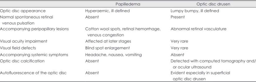
The clinical differences between papilledema and ODD are listed in Table 1.
While there is no effective treatment for ODD, regular ex- aminations should be carried out in order to rule out accompa- nying disorders such as elevated intraocular pressure and sub- retinal neovascularization.1
All three of our patients expressed anxiety regarding the possibility of intracranial pathology. We diagnosed ODD via a fundus examination, autofluorescence of the optic discs, and B-scan USG. We then further confirmed the diagnosis of ODD with CT to assure the patients they did not have any intracra- nial pathology.
Swelling of the optic discs does not always mean elevated intracranial pressure, and therefore other causes should be kept in mind before coming to a final conclusion based on all relevant clinical findings. Importantly, the early diagnosis of ODD spares the patient unnecessary anxiety about intracrani- al disease and prevents unnecessary interventions. Misdiagno- sis may increase the incidence of somatization of these pa- tients, particularly in the young. The ophthalmologist, neuro- logist, and the reporting radiologist must therefore consider ODD when dealing with atypical cases of disc swelling.
Conflicts of Interest
The authors have no financial conflicts of interest.
Acknowledgements
This study was presented as a poster at the 44th National Congress of Turkish Ophthalmology Society, in Antalya, TURKEY (29 Septem- ber-03 October 2010).
REFERENCES
1. Auw-Haedrich C, Staubach F, Witschel H. Optic disk drusen. Surv Ophthalmol 2002;47:515-532.
2. Tso MO. Pathology and pathogenesis of drusen of the optic nervehead.
Ophthalmology 1981;88:1066-1080.
Table 1. Clinical differences between papilledema and optic disc drusen
Papilledema Optic disc drusen
Optic disc appearance Hyperaemic, ill defined Lumpy bumpy, ill defined
Normal spontaneous retinal venous pulsation
Absent Present
Accompanying peripapillary lesions Cotton wool spots, retinal hemorrhage, venous congestion
Abnormal retinal vasculature
Visual acuity impairment Affected at later stages Very rare
Visual field defects Blind spot enlargement Very rare
Accompanying systemic symptoms Headache, nausea, vomiting Absent
Optic disc calcification Absent Detected with computed tomography and/
or ocular ultrasound
Autofluorescence of the optic disc Absent Evident especially in superficial optic disc drusen
Bilateral Optic Disc Drusen Mimicking Papilledema
154 J Clin Neurol 2012;8:151-154
3. Davis PL, Jay WM. Optic nerve head drusen. Semin Ophthalmol 2003;
18:222-242.
4. Lorenizen SE. Drusen of the optic disk, an irregularly dominant hered- itary affection. Acta Ophthalmol (Copenh) 1961;39:626-643.
5. Boldt HC, Byrne SF, DiBernardo C. Echographic evaluation of optic disc drusen. J Clin Neuroophthalmol 1991;11:85-91.
6. Antcliff RJ, Spalton DJ. Are optic disc drusen inherited? Ophthalmol- ogy 1999;106:1278-1281.
7. Wilkins JM, Pomeranz HD. Visual manifestations of visible and buried
optic disc drusen. J Neuroophthalmol 2004;24:125-129.
8. Lee AG, Zimmerman MB. The rate of visual field loss in optic nerve head drusen. Am J Ophthalmol 2005;139:1062-1066.
9. Patel NN, Shulman JP, Chin KJ, Finger PT. Optical coherence tomog- raphy/scanning laser ophthalmoscopy imaging of optic nerve head drusen. Ophthalmic Surg Lasers Imaging 2010;41:614-621.
10. Kurz-Levin MM, Landau K. A comparison of imaging techniques for diagnosing drusen of the optic nerve head. Arch Ophthalmol 1999;117:
1045-1049.