ABSTRACT
Background: In this article, we estimated the combined effect of radiotherapy (RT) with ultrasound (US) wave and the ability of gold nanoparticles (GNPs) to improve their combined therapeutic effects.
Methods: At first, HeLa cells received the various treatment modalities: RT (6 MV; 0.5, 1, and 2 Gy), US irradiation (1 MHz; 0.5, 1, and 1.5 W/cm2, 1 minute), and RT+US. Afterwards, the enhanced effect of US on RT was evaluated. Then, the effect of the synthesized GNPs at different concentrations (0.2, 1, and 5 µg/mL, 24 hours) was evaluated to assess the effect on HeLa cells combined with RT+US. Cell survival rates in the different treatment groups at 24, 48, and 72 hours post-treatment were evaluated using 3-(4,5-dimethylthiazol-2-yl)-2,5- diphenyltetrazolium bromide and trypan blue assays.
Results: Our results show US irradiation could enhance the effect of RT at the same radiation dose and could be utilized as a sensitizer agent for RT. Moreover, our findings indicate RT+US in combination with different nanoparticle concentrations could enhance the effect of RT+US so that they can improve the treatment results up to 9.93 times and act as sonodynamic- radiosensitivity. These results also indicate that the combination of RT with US along with GNPs has synergistic effects compared to RT or US alone. Cell survival results show that combining the low US waves (1.5 W/cm2), GNPs (5 μg/mL), and X-rays (2 Gy) increase the cytotoxicity on HeLa cell up to 95.8%.
Conclusion: We concluded that GNPs could act as a good sensitizing agent in RT+US irradiation and could result in the synergistic effects.
Keywords: Cancer; Radiotherapy; Sonodynamic Therapy; Gold Nanoparticles; MTT Assay;
Trypan Blue Exclusion Assay
INTRODUCTION
Radiotherapy (RT) is a non-invasive method and is considered as one of the most widely used therapeutic methods for cancer treatment.1,2 More than half of all cancer patients receive radiation during their treatments.3 In the RT, the high energy radiation beam emitted from an external source is used to destroy the cancer cells.4 However, in contrast, normal tissue tolerance around cancer cells restrains the treatment efficacy of cancer cells.5 In addition,
Original Article
Received: Apr 24, 2019 Accepted: Jul 24, 2019 Address for Correspondence:
Hadi Akbari-Zadeh, MSc
Department of Medical Physics, School of Medicine, Isfahan University of Medical Sciences, Azadi Square, Hezar-Jarib Street, Isfahan 81746-73461, Iran.
E-mail: hadi.akbari.zadeh@gmail.com
© 2019 The Korean Academy of Medical Sciences.
This is an Open Access article distributed under the terms of the Creative Commons Attribution Non-Commercial License (https://
creativecommons.org/licenses/by-nc/4.0/) which permits unrestricted non-commercial use, distribution, and reproduction in any medium, provided the original work is properly cited.
ORCID iDs Ahmad Shanei
https://orcid.org/0000-0001-5999-0932 Hadi Akbari-Zadeh
https://orcid.org/0000-0002-9424-027X Disclosure
The authors have no potential conflicts of interest to disclose.
Author Contributions
Conceptualization: Shanei A. Data curation:
Akbari-Zadeh H. Investigation: Akbari-Zadeh H. Project administration: Shanei A. Writing - original draft: Akbari-Zadeh H. Writing - review
& editing: Shanei A.
Ahmad Shanei and Hadi Akbari-Zadeh
Department of Medical Physics, School of Medicine, Isfahan University of Medical Sciences, Isfahan, Iran
Investigating the Sonodynamic- Radiosensitivity Effect of Gold Nanoparticles on HeLa Cervical Cancer Cells
Oncology & Hematology
conventional RT methods can result in serious side effects because there is no discrimination between cancer cells and surrounding normal tissue. Therefore, some undesirable outcomes may occur due to healthy tissue irradiation. It has always been the researchers' efforts to find a way to increase therapeutic efficacy and, at the same time, to reduce side effects.6 In this regard, the combined therapeutic methods have been reported to be more effective in achieving these goals.7-9
Another treatment regime with an undeniable role in medicine is the therapeutic ultrasound (US). US, which is based on non-ionizing radiation,10 is easy and cheap to produce and has greatly improved the field of diagnostic imaging.11 US also has an important role in therapeutic regimens because it increases the permeability of substances within cancerous cells, activating sensitizers12 and imposing heat damage to cancer cells by raising their temperature, the so-called hyperthermia effect.13 The use of US irradiation can also promote the RT for the treatment of cancer tissue and reduce the side effects.8
One of the ways to increase the treatment efficacy and to reduce side effects is using sensitizer during irradiation.14 Sensitizers are substances that generate more anticancer effects when combined with radiation and due to the localization of energy cause less unwanted effects. They increase the therapeutic effects in several ways, including increasing the probability of radiation interaction,15 producing more reactive oxygen species (ROS)16 and arresting cells at the most sensitive cycle of cell proliferation (G2/M phase).17
In recent decades, nanotechnology has made great progress and has opened new horizons for other subspecialties in different fields.18 As sensitizer, nanoparticles can improve clinical application problems of some sensitizers, such as hydrophobic. Removing this problem prevents them from aggregation and supports easier injection, better bio-distribution and increased efficacy of sensitizers. On the other hand, the large surface area of nanoparticles has the capability of becoming better sensitizer through modification and functionalization.19 Also, the enhanced permeation and retention theory (EPR effect) predicted that nanoparticles accumulate more in cancerous tissue compared to normal tissue. This particular property can lead to localize the penetration of nanoparticles into cancerous cells as the therapeutic agent and decrease the unwanted effects due to various treatment modalities.2,20 Based on these characteristics, nanoparticles can be a good candidate as new sensitizers.
Between all the studied nanoparticles, gold nanoparticles (GNPs) have been shown to provide some unique advantages which make them ideal for applications in medicine. Easy synthesis, low toxicity, ability to control size and shape during production, biocompatibilities,21 special optical properties, high uptake in mammalian cells, higher atomic number relative to human tissues,22 high ability to absorb light relative to organic dyes (millions of times),23 and anti-angiogenesis property24 are among the reasons that are of great interest for such nanoparticles. These properties have led to the use of these nanoparticles as a sensitizer in combination with different modalities.24-26
Up to now, many studies have proven the combination of sensitizers with various radiation modalities can increase treatment efficiency together with reduced side effects.24,26 Addition of sensitizer to RT may increase radiosensitivity of the cancer cells,5 and also applying US wave also causes synergetic effects.9 In addition, the combination of sensitizers with US radiation is known as a roughly new approach for cancer treatment, called sonodynamic therapy.27
Very few studies have evaluated the combined effect of RT with US wave along with nano- sensitizers, as far as we know.9 To the best of our knowledge, no studies have investigated the sonodynamic-radiosensitivity (SRS) effects of GNPs when combined with low-intensity US and X-rays as combined effects of sonodynamic therapy with RT.
Consequently, the purpose of this study was to investigate the effect of US irradiation alone on RT as well as to measure the SRS factor due to the different concentrations of GNPs (0.2, 1, and 5 μg/mL) and US irradiation with the various intensities (1 MHz; 0.5, 1 and 1.5 intensities, 60 seconds) when combined with 6 MV X-ray radiation (0.5, 1, and 2 Gy) on the HeLa cell line.
METHODS
Synthesis of GNPs
All the chemical compounds used in this research were purchased from Merck (Darmstadt, Germany) and Fluka (Buchs, Switzerland). Hydrogen tetrachloroaurate (III) trihydrate (HAuCl4.3H2O, 99.5% purity) and sodium citrate dihydrate (C6H5Na3O7.2H2O) were purchased from Merck.
GNPs were prepared by the chemical reduction of HAuCl4 in the presence of citrate.28 The preparation method was as follows: glassware used in this experimental procedure was cleaned in aqua regia, washed with deionized water, and then dried in oven. The 5 mL of 0.2% (w/w) HAuCl4 aqueous solution and 90 mL of deionized H2O were heated under reflux and stirring. Then, 5 mL of 1% (w/w) sodium citrate dihydrate solution was added in one- step. The pale-yellow colour of the solution was changed to deep red. The solution was refluxed for 15 minutes after the colour change. Next, the solution temperature was allowed to reach room temperature. Finally, the GNPs were purified by filtration, centrifugation and double re-precipitation from distilled H2O.
Characterization techniques
UV-visible (UV-vis) absorption spectroscopic measurements were performed using UV-vis spectrometer (Agilent Cary 100; Agilent Technologies, Inc., Santa Clara, CA, USA), by using quartz cells of 1 cm path length and water as the reference solvent at room temperature.
Morphology and size of GNPs were determined using transmission electron microscopy (TEM, Zeiss EM 900; Carl Zeiss AG, Oberkochen, Germany).
Cell culture
We used a cervical carcinoma cells line (HeLa cells) in this study. This cell line was obtained from the Pasteur Institute of Iran. The cells were cultured in High-Glucose Dulbecco's Modified Eagle's Medium (DMEM) supplemented with fetal bovine serum (FBS, 10%) and pen-strep (1%) inside T-25 cell culture flasks. They were kept inside the incubator containing 5% CO2 and adequate humidity at 37°C. The cells were checked every day and, if necessary, their culture medium was changed. This was done approximately every three days. HeLa cells were seeded in 96-well and 24-well plates (Zhejiang Sorfa Life Science Research Co., Ltd., Huzhou, China) and interventions were begun after the incubation of the cells for 24 hours in these wells.
Cells doubling time calculation
One of the most important features of the cell is the time it takes to duplicate (Doubling Time). To measure this time, 20,000 cells/well were first seeded in a 24-well plate, and then every day, the number of cells for three wells were counted two times. This process was continued for up to 10 days. The information was plotted in a semi-logarithmic diagram, showing the gradient of the graph representing the doubling time.
US and RT irradiation procedure
In this study, US waves were used at a frequency of 1 MHz and intensities of 0.5, 1, and 1.5 W/cm2. These waves were produced using a therapeutic US generator (Novin, 215X, joint product of Iran and England) with 4 cm2 transducer area. The cells were irradiated vertically for 60 seconds.
RT was performed with a superficial X-ray irradiator (Philips Medical Systems, Amsterdam, The Netherlands) operating at 6 MV and 80 mA. The cells were irradiated at doses of 0.5, 1, and 2 Gy at a dose rate of 100 rad/min. Dosimetry was performed with a 2570/1 Farmer Dosimeter electrometer (NE Technology Ltd., Cambridge, UK).
Cell survival assay
The 3-(4,5-dimethylthiazol-2-yl)-2,5-diphenyltetrazolium bromide (MTT) assay Cell viability was measured by MTT assay. This test evaluates the metabolic activity of mitochondria in live cells and is one of the most common tests for measuring cell survival.
First, we completely removed the culture medium, and then 90 μL medium without FBS and 10 μL MTT (50 mg/mL in phosphate-buffered saline [PBS]) were added to each well. Then, the plate was incubated again for 3 hours. After this time, the live cells change the colourless tetrazolium compound to an insoluble blue formazan crystal. The formation of these crystals indicated the mitochondria were healthy and their amount determined the number of living cells. To determine the amount of formed crystals, we added 100 μL of DMSO to each well and then read the optical absorptions at 570 nm through the ELISA reader (Bio-Rad, Hercules, CA, USA).
Cell survival was represented as:
Cell Survival = Optical Density of Treated Samples
Optical Density of Control Samples × 100 (1)
Trypan blue exclusion assay
Trypan blue staining is another assay of cell survival assessment. We can use this test to dye dead cells. In the live cell, the cell membrane controls the entry of substances into the membrane. But due to lack of this control in the dead cells, trypan blue can penetrate through the membrane and turn them to blue.
In this study, trypan blue assay was done as follows: Initially, the cells were detached from the surface of the 24-well plate and after centrifugation; trypan blue was added to them.
Finally, the counting process of the number of the dead and live cells was performed under a microscope using a hemocytometer.
Viability is also indicated by:
Viability = Number of Counted Live Cells
Total Number of Cells Counted × 100 (2) Experimental method
HeLa cells were seeded in 96-well and 24-well Sorfa plates with 4,000 and 50,000 cells in each well, respectively. After planting, they were allowed 24 hours to adhere to the bottom of the plates' surface. Then, cells were treated with different concentrations of nanoparticles for another overnight.5,29 After completion of this time, the cell culture medium was completely removed, and the cells were washed three times with PBS to ensure that any nanoparticle was in the well. The fresh culture medium was then added to the well and exposed to X-ray irradiation. Then cells were exposed to US waves. Finally, cell survival was investigated using MTT and trypan blue assay 24, 48, and 72 hours after the intervention. We used 96-well plates for the MTT assay and 24-well plates for the trypan blue assay. Table 1 shows the brief processes of the current experiment.
SRS ratio of GNPs
The SRS effect of GNPs at various concentrations in combination with different radiation conditions can be derived from cell survival.
SRS was determined using Equation 3:
SRS = Cell Survival US + RT
Cell Survival US + RT + NPs (3)
Statistics method
The data were analyzed using SPSS version 22 by one-way analysis of variance test and also Tukey's post hoc multiple comparisons for all groups (P < 0.05). Data exhibited on the curves were stated as mean ± standard error of mean and other data were stated as mean ± standard deviation.
Ethical statements
This article does not contain any studies on human or animal subjects performed by the any of the authors.
Table 1. The types of intervention in different treatment groups; the interval between each intervention was 24 hours
Groups Cell seeding Adding GNPs RT/US irradiation Cell survival assay
(24 hr) Cell survival assay
(48 hr) Cell survival assay (72 hr)
Control
* * * *
GNPs only
* * * * *
RT only
*
RT* * *
US only
*
US* * *
Radiation group
*
RT and US* * *
GNPs + radiation group
* *
RT and US* * *
GNPs = gold nanoparticles, RT = radiotherapy, US = ultrasound.
RESULTS
The results of the MTT assay (24 and 48 hours) and the trypan blue assay for the various treatment modalities combined with different concentration nanoparticles as well as the synthesis and characterization of nanoparticles and the cell characteristic are expressed completely in the Supporting File (Supplementary Data 1). The purpose of the current experiment was to assess the role of US wave in the enhancement of RT along with GNPs. The Fig. 1 explains the idea of the present study.
The combination effects of X-rays beam with US waves on cell survival The combined effects of RT with US waves on cell survival were evaluated using the MTT and trypan blue assays. We evaluated the effects of changes in the intensity and the dose at three different incubation times (24, 48, and 72 hours) following the intervention. Information from both cell survival tests indicates because of the destructive effects of both different modalities; the combination groups (RT+US) were significantly different from RT groups or US groups alone in the same radiation (RT, US) condition (P < 0.05) (Supplementary Figs. 1 and 2). In other words, US irradiation could enhance the effect of RT and that could act alone as sensitizer agent for RT.
Also, the effect of changes due to the US intensity on cell survival depends on RT doses and incubation time. Our results show the radiation treatment (RT+US) at 0.5 Gy and various US intensity (0.5, 1, 1.5 W/cm2), 24 hours after incubation did not effect the cell survival between all intensities (P < 0.05) (Supplementary Fig. 1A). However, 48 and 72 hours after incubation, only the 1.5 W/cm2 intensity in combination with X-ray beam (0.5 Gy) caused a significant difference in comparison with the two other intensities (P < 0.05) (Supplementary Fig. 1B and Fig. 2). Also, our results indicated that, among all the intensities (0.5, 1, and 1.5 W/cm2) added to 1 Gy, only 1.5 W/cm2 was significantly different from the rest at 24 and 48 hours (P < 0.05). Nevertheless, with an increase the incubation time to 72 hours in the same dose, the change in any intensity makes a significant difference in cell survival (P < 0.05) (Fig. 2 and Supplementary Fig. 1).
The highest effect was observed at 2 Gy in combination with 1.5 W/cm2 intensity in which the cell survival was decreased up to 41.41% (Fig. 2). A significant difference was observed between all intensities in this dose (P < 0.05).
Trypan blue results for all the different incubation times (24, 48, and 72 hours) approved the results of MTT assay. The cell survival outcome of this assay was approximately similar to the results of the MTT assay (Supplementary Fig. 2).
Administration of GNPs into cervical cancer cells
GNPs X-ray radiation
US wave Interaction between GNPs and
radiation into cervical cancer cells Increased efficacy of radiation therapy due to GNPs in combination with US
as post-treatment Fig. 1. The schematic illustration of the current experiment.
GNPs = gold nanoparticles, US = ultrasound.
The combination effects of RT with US waves on cell survival in the presence of GNPs
At first, in order to investigate the combined effects of RT and US waves in the presence of GNPs, it was necessary to evaluate and measure the cytotoxicity level of nanoparticles on HeLa cells in order to select the appropriate concentrations. For this purpose, we conducted an initial study to evaluate the cytotoxicity of various nanoparticles concentrations (0.2, 1, 5, 25, and 50 μg/mL) on the HeLa cells. We found in the concentrations ≤ 5 μg/mL had no toxicity.
Consequently, these concentrations (0.2, 1, and 5 μg/mL) were used in this study. Moreover, the GNPs sizes were about 13 nm and their shapes were spherical (Supplementary Fig. 3).
The results indicate that the presence of GNPs in the combination of RT with US wave (GNPs+RT+US) can improve the efficiency of radiation treatment (RT+US). These results show a significant difference between combined groups (GNPs+RT+US) and radiation groups (RT+US) at the same radiation condition (P < 0.05) (Supplementary Figs. 4-6).
The concentration effect of nanoparticles on the combined group's cell survival
(GNPs+RT+US) varies in different conditions. After 24 hours of treatment, it is most observed that, the adding 5 μg/mL of GNPs to the radiation treatment groups (RT+US) produces the considerable differences compared to the two other concentrations (0.2 and 1 μg/mL) in the similar radiation group (P < 0.05) (Supplementary Fig. 4).
The MTT's results showed the change effect of concentration on cell survival in the combined groups (GNPs+RT+US) has improved with reaching time to 48 hours after the treatment.
These results indicated that in 0.5 Gy, only 5 μg/mL of GNPs in the combined group (GNPs+RT [0.5 Gy]+US [0.5, 1, and 1.5 W/cm2]) can cause a significant difference compared to other concentrations (GNPs [0.2 and 1 μg/mL]+RT [0.5 Gy]+US [0.5, 1, and 1.5 W/cm2]) in the same radiation condition (P < 0.05) (Supplementary Fig. 5A). But comparing the data of combined group (GNPs+RT+US) under the same radiation conditions at doses of 1 and 2 Gy indicates that the change in the nanoparticles' concentration causes a significant difference in the cell survival (P < 0.05) (Supplementary Fig. 5B and C).
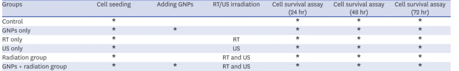
Finally, the lowest survival was obtained in all combined groups (GNPs+RT+US) after 72 hours post-treatment. The concentration change effect was more visible at this time because of the reduction in the cell survival of the combined group (GNPs+RT+US) (Fig. 3).
0 0 Gy (only US) 0.5 Gy 1 Gy 2 Gy
X-ray doses 20
40 60 80 100
Cell survival, % RT
RT + 0.5 W/cm2 RT + 1 W/cm2 RT + 1.5 W/cm2
Fig. 2. Effect obtained using MTT assay in a change of US intensity in combination with different doses of RT after 72 hours incubation times.
MTT = 3-(4,5-dimethylthiazol-2-yl)-2,5-diphenyltetrazolium bromide, US = ultrasound, RT = radiotherapy.
Comparison of different concentrations at this combined group (GNPs+RT [2 Gy]+US [1.5 W/
cm2]) indicated a significant difference in the different concentrations after 24 and 48 hours (P < 0.05). However, due to the high reduction in the cell survival in this combination (72 hours), it is no longer possible to differentiate between concentrations of 1 and 5 μg/mL (P > 0.05). The combined group results for 72 hours are shown in Fig. 3.
The effect of change in any used experimental factors (nanoparticle concentration, US intensity, RT dose, and incubation time) on the cell survival of the combined groups (GNPs+RT+US) indicate these effects are dependent on each other, and when each of these factors increases, the change effects of the rest become more evident.
The presence of nanoparticle in radiation treatment regime (RT+US) resulted in the reduced significance in cell survival and increased the cytotoxicity (P < 0.05). Cell survival decreases
A
0 0.5 W/cm2 1 W/cm2 1.5 W/cm2 US intensity
20 40 60 80
Cell survival, % RT + US
0.2 µg + RT + US 1 µg + RT + US 5 µg + RT + US
B
0 0.5 W/cm2 1 W/cm2 1.5 W/cm2 US intensity
20 40 60 80
Cell survival, %
C
0 0.5 W/cm2 1 W/cm2 1.5 W/cm2 US intensity
20 40 60 80
Cell survival, %
US + 1 Gy US + 1 Gy + 0.2 µg US + 1 Gy + 1 µg US + 1 Gy + 5 µg
US + 2 Gy US + 2 Gy + 0.2 µg US + 2 Gy + 1 µg US + 2 Gy + 5 µg
Fig. 3. The dependence of cellular survival changes on US intensity and nanoparticle concentrations in (A) 0.5 Gy, (B) 1 Gy, and (C) 2 Gy dose of X-rays 72 hours after the intervention with MTT assay.
US = ultrasound, MTT = 3-(4,5-dimethylthiazol-2-yl)-2,5-diphenyltetrazolium bromide, RT = radiotherapy.
with increasing RT doses, US intensity, concentration of nanoparticles and incubation time.
Overall, the results shown the highest and the lowest reduction in cell survival (95.8%, 41.2%) were obtained in these combined groups, (GNPs [5 μg/mL]+RT [2 Gy]+US [1.5 W/
cm2], 72 hours) and (GNPs [0.2 μg/mL]+RT [0.5 Gy]+US [0.5 W/cm2], 24 hours), respectively.
(Fig. 3A and Supplementary Fig. 4A).
We used another test to evaluate the cell survival to ensure MTT assay results. Trypan blue assay was performed for all groups after interventions at three different times (24, 48, and 72 hours).
Their results were approximately consistent with MTT assay results (Supplementary Figs. 7-9).
The results of this study using the trypan blue assay also showed the addition of nanoparticles to the combination of RT and US wave produces a significant difference in cell viability (P <
0.05). Trypan blue data also indicated the concentration change effect appears more visible with an increased incubation time. Also, the highest reduction in cell survival was 94.9% in this group (GNPs [5 μg/mL]+RT [2 Gy]+US [1.5 W/cm2], 72 hours) (Supplementary Fig. 9C).
As stated above, the significant differences in the reduction of survival fraction can be seen in the radiation treatment group (RT+US) compared to the treated combined groups (GNPs+RT+US) at different nanoparticle concentrations and considerable differences existed between each of these component radiation groups alone (RT or US) and with radiation (RT+US) at the same radiation condition, suggesting a synergistic effect.
SRS effect of GNPs
The SRS effect of GNPs at various concentrations in combination with different radiation conditions calculated according to equation 3. We calculated this parameter for each cell survival assay, including both MTT and the trypan blue assays (Supplementary Tables 1 and 2). These calculations were done in different combinations of RT dose (0.5, 1, and 2 Gy), US radiation (0.5, 1, and 1.5 W/cm2) and the concentrations of nanoparticles (0.2, 1, and 5 μg/
mL) at three different incubation times (24, 48, and 72 hours). These results showed that, by increasing any of these 3 factors, the SRS factor of nanoparticles increased (Table 2). The lowest SRS factor was observed at nanoparticles concentration of 0.2 μg/mL in combination with 0.5 W/cm2 and 0.5 Gy. By increasing any of these parameters, the SRS factor of
nanoparticles was increased. As expected, the maximum value of SRS factor occurred when the maximum value of each parameter was used (Table 2). Comparison of SRS value with incubation time indicated that there was an increase by increasing the incubation time from 24 to 72 hours.
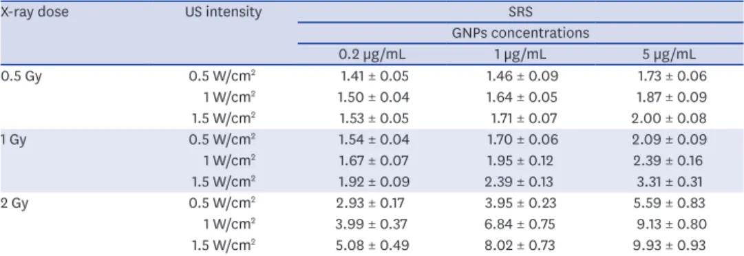
Table 2. The value of calculated SRS ratio in 0.5, 1, and 2 Gy with MTT assay 72 hours after the intervention
X-ray dose US intensity SRS
GNPs concentrations
0.2 µg/mL 1 µg/mL 5 µg/mL
0.5 Gy 0.5 W/cm2 1.41 ± 0.05 1.46 ± 0.09 1.73 ± 0.06
1 W/cm2 1.50 ± 0.04 1.64 ± 0.05 1.87 ± 0.09
1.5 W/cm2 1.53 ± 0.05 1.71 ± 0.07 2.00 ± 0.08
1 Gy 0.5 W/cm2 1.54 ± 0.04 1.70 ± 0.06 2.09 ± 0.09
1 W/cm2 1.67 ± 0.07 1.95 ± 0.12 2.39 ± 0.16
1.5 W/cm2 1.92 ± 0.09 2.39 ± 0.13 3.31 ± 0.31
2 Gy 0.5 W/cm2 2.93 ± 0.17 3.95 ± 0.23 5.59 ± 0.83
1 W/cm2 3.99 ± 0.37 6.84 ± 0.75 9.13 ± 0.80
1.5 W/cm2 5.08 ± 0.49 8.02 ± 0.73 9.93 ± 0.93
SRS = sonodynamic-radiosensitivity, MTT = 3-(4,5-dimethylthiazol-2-yl)-2,5-diphenyltetrazolium bromide, US = ultrasound, GNPs = gold nanoparticles.
DISCUSSION
One of the most important goals of any therapeutic modality is to increase the efficacy of treatment and reduce the side effects to improve the quality of the patient's life. This study was conducted to evaluate the efficacy of using combined treatment modalities such as RT in combination with sonodynamic therapy and to measure and evaluate the SRS effect of GNPs in this combination.
For this purpose, this study can be assessed in two ways: i) the combined effect of X-ray+US wave and ii) the SRS role of GNPs.
The results of cell survival at the different incubation times (24, 48, and 72 hours) indicated the combination of RT with US waves have a significant difference compared to any other treatment modality. It was observed with increasing RT dose or US wave intensity, the cell survival was significantly decreased (P < 0.05). The maximum decrease of these combinations (RT+US) was approximately 59% and occurred 72 hours after the treatments in this condition at (RT [2 Gy]+US [1.5 W/cm2]) (Fig. 2).
Jun-Qun et al.8 conducted a study on 89 patients with rectal cancer who received high- intensity US radiation (3.5 MHz and 1,300–1,700 W/cm2) during their treatments. Their patients were exposed to US for 60–90 minutes (firing time 0.13 to 0.20 seconds, interval time between shots 0.2 to 0.3 seconds) in each treatment session after receiving 2–3 Gy of X-ray. They found 22.5% of the patients were fully recovered and the size of the tumor in 64%
of the patients had a reduction of more than 50%, without showing any complication.
Also, the results of the histological analysis of Czarnota et al.30 during the combination of US wave and RT on mice showed that 44% of cells could be killed with 2 Gy dose in combination with 10 W/cm2 intensity (US exposed for 750-ms in 5 minutes).
The results of the above studies confirm our results on the combination of X-rays and US. The differences in the rate of treatment efficiency can vary in different experimental conditions such as the type of study (in vitro or in vivo), type of cell, the US wave's characteristic (frequency, intensity, and exposure time), and the characteristic of X-ray (beam energy and radiation dose).
Ionizing radiation affects the cells in two ways, directly and indirectly. In the direct way, the radiation interacts with the cells and affects them.31 Many studies have described that DNA, as the most important target, can be damaged due to direct X-rays interactions.32 While, for the indirect effects, the radiation interacts with the environment around the cells and leads to the production of ROS. ROS can cause a decrease in cell survival by damaging other components such as DNA, proteins, lipids, and mitochondria.31
US waves cause the biological effects through mechanisms such as cavitation, increase in temperature and mechanical stress. Nevertheless, the most important effect is the cavitation phenomenon.33 It should be noted that this phenomenon causes the production of bubbles.
Some bubbles rise to a certain size and start to oscillate about their equilibrium radius for many acoustic cycles (non-inertial cavitation). This oscillation causes stress and dysfunction in the cells.27 But the most important effect happens when these bubbles collapse (inertial cavitation). Collapse of these bubbles causes high temperature and pressure (5,000 K and
800 Pa)34 and also produces a light that is called sonoluminescence. Some pyrolysis reaction can occur in the medium for such an energy release and this results in ROS production. Also, the producing sonoluminescence can be absorbed by other materials.33
The results of the addition of GNPs in different concentrations to X-ray and US wave showed the SRS effect of nanoparticle at all concentrations (Table 2). The value of SRS was increased with increasing nanoparticle concentration, US intensity and, X-ray dose. The SRS value of nanoparticles showed that they can improve treatment efficacy up to 9.93 times (Table 2). In addition, the results indicate that the combination of these factors can reduce cell survival up to 95.8% (Fig. 3C).
Kuo et al.9 have studied the sensitizing effect of protoporphyrin IX conjugated to SO2
nanoparticles and decorated with gold nanoparticle on cervical carcinoma cells (HeLa). They found that, adding this sensitizer (0.5 μg/mL) to US wave (1 MHz, 0.4 W/cm2, 10 minutes with 20% duty ratio) and RT (6 MV, 2 Gy) indicated the combination therapy was more effective than single treatment. In this study, this combination can reduce cell survival (up to 50%) 24 hours after the intervention. This difference in the results of our studies with the mentioned studies can be due to different experimental conditions, as previously stated.
The different mechanisms have been proposed for the sensitizing role of GNPs when combined with RT or US irradiation. For the radiosensitivity effect of GNPs, it can be mentioned that the atomic number of the GNPs was approximately 11-times of the tissue atomic number. This causes considerable increases in the number of x-ray interactions in the tissue which mainly depends on the energy of the beam.5,15 Also, Roa et al.17 showed that GNPs can arrest the cells in their G2/M stage of cell proliferation. This is the most sensitive stage in the cell proliferation to radiation.
There are some reasons for the sonosensitizing effect of GNPs when combined with US wave.
The existence of a particle in medium, reduces the cavitation threshold.35 GNPs have high absorption coefficients and can easily absorb sonoluminescence. Brazzale et al.23 stated that the absorption of these lights will quickly increase the temperature of the nanoparticles and causes more ROS. This might be considered as another reason for the sonosensitizing effect of GNPs in combination with US wave.
These results showed the combination of US and X-rays with and without GNPs as a sensitizer agent is far more potent than of these modalities alone, which is in line with our findings. Cell survival assays have been performed using MTT assays that consider the mitochondrial activity in the cell and this result was confirmed with the trypan blue assay. We believed that one of the key basic mechanisms to reduce the cell survival in this study can be due to the increased damage to DNA by X-rays and the role of amplified ROS in mitochondria which increase the damaging DNA and prevent its repair as well as the probability increased temperature into cancer cells due to gold nanoparticle in interaction with US wave.
In conclusion, there is a significant effect on HeLa cells when X-ray radiation is combined with US compared to RT alone. Moreover, the results of this study indicated that, the combination of X-rays with US could have a profound effect on cell survival when the cells were conjugated with GNPs as the nano-sensitizer which they can highly increase the treatment efficacy. Therefore, this combination could result in the synergistic effect.
ACKNOWLEDGMENTS
This study is part of an MSc thesis in the Medical Physics and was performed in the Department of Medical Physics, School of Medicine, Isfahan University of Medical Sciences, Isfahan, Iran.
SUPPLEMENTARY MATERIALS
Supplementary Data 1
Investigating the sonodynamic-radiosensitivity effect of gold nanoparticles on HeLa cervical cancer cells
Click here to view
Supplementary Table 1
The value of calculated SRS ratio in different conditions of intervention with MTT assay 24, 48, and 72 hours after the intervention
Click here to view
Supplementary Table 2
The value of calculated SRS ratio in different conditions of intervention with trypan blue assay 24, 48, and 72 hours after the intervention
Click here to view Supplementary Fig. 1
Effect obtained using MTT assay in a change of US intensity in a combination with different doses of RT after (A) 24, (B) 48, and (C) 72 hours incubation times.
Click here to view Supplementary Fig. 2
Effect obtained using the trypan blue assay in change of US intensity in a combination with different doses of X-rays (A) 24, (B) 48, and (C) 72 hours incubation times.
Click here to view Supplementary Fig. 3
(A) UV-Vis absorption spectra of GNPs, (B) size distribution histogram of GNPs, and (C) TEM image of GNPs.
Click here to view Supplementary Fig. 4
The dependence of cellular survival changes on US intensity and nanoparticle concentrations in (A) 0.5, (B) 1, and (C) 2 Gy dose of X-rays 24 hours after the intervention with MTT assay.
Click here to view
Supplementary Fig. 5
The dependence of cellular survival changes on US intensity and nanoparticle concentrations in (A) 0.5, (B) 1, and (C) 2 Gy dose of X-rays 48 hours after the intervention with MTT assay.
Click here to view Supplementary Fig. 6
The dependence of cellular survival changes on US intensity and nanoparticle concentrations in (A) 0.5, (B) 1, and (C) 2 Gy dose of X-rays 72 hours after the intervention with MTT assay.
Click here to view Supplementary Fig. 7
The dependence of cellular survival changes on US intensity and nanoparticle concentrations in (A) 0.5, (B) 1, and (C) 2 Gy dose of X-rays 24 hours after the intervention with trypan blue assay.
Click here to view Supplementary Fig. 8
The dependence of cellular survival changes on US intensity and nanoparticle concentrations in (A) 0.5, (B) 1, and (C) 2 Gy dose of X-rays 48 hours after the intervention with trypan blue assay.
Click here to view Supplementary Fig. 9
The dependence of cellular survival changes on US intensity and nanoparticle concentrations in (A) 0.5, (B) 1, and (C) 2 Gy dose of X-rays 72 hours after the intervention with trypan blue assay.
Click here to view
REFERENCES
1. Khoei S, Mahdavi SR, Fakhimikabir H, Shakeri-Zadeh A, Hashemian A. The role of iron oxide nanoparticles in the radiosensitization of human prostate carcinoma cell line DU145 at megavoltage radiation energies. Int J Radiat Biol 2014;90(5):351-6.
PUBMED | CROSSREF
2. Fakhimikabir H, Tavakoli MB, Zarrabi A, Amouheidari A, Rahgozar S. The role of folic acid-conjugated polyglycerol coated iron oxide nanoparticles on radiosensitivity with clinical electron beam (6 MeV) on human cervical carcinoma cell line: in vitro study. J Photochem Photobiol B 2018;182:71-6.
PUBMED | CROSSREF
3. Brun E, Sanche L, Sicard-Roselli C. Parameters governing gold nanoparticle X-ray radiosensitization of DNA in solution. Colloids Surf B Biointerfaces 2009;72(1):128-34.
PUBMED | CROSSREF
4. Fakhimikabir H, Tavakoli MB, Zarrabi A, Amouheidari A, Rahgozar S. Could FA-PG-SPIONs act as a hyperthermia sensitizing agent? An in vitro study. J Therm Biol 2018;78:73-83.
PUBMED | CROSSREF
5. Khoshgard K, Hashemi B, Arbabi A, Rasaee MJ, Soleimani M. Radiosensitization effect of folate- conjugated gold nanoparticles on HeLa cancer cells under orthovoltage superficial radiotherapy techniques. Phys Med Biol 2014;59(9):2249-63.
PUBMED | CROSSREF
6. Her S, Jaffray DA, Allen C. Gold nanoparticles for applications in cancer radiotherapy: mechanisms and recent advancements. Adv Drug Deliv Rev 2017;109:84-101.
PUBMED | CROSSREF
7. Liu Y, Liu X, Jin X, He P, Zheng X, Dai Z, et al. The dependence of radiation enhancement effect on the concentration of gold nanoparticles exposed to low- and high-LET radiations. Phys Med 2015;31(3):210-8.
PUBMED | CROSSREF
8. Jun-Qun Z, Guo-Min W, Bo Y, Gong-Xian W, Shen-Xu H. Short-term results of 89 cases of rectal carcinoma treated with high-intensity focused ultrasound and low-dose radiotherapy. Ultrasound Med Biol 2004;30(1):57-60.
PUBMED | CROSSREF
9. Kuo WJ, Wang YC, Chen MH, Tung FI, Liu TY. Study of a novel vehicle developed for enhancing the efficacy of radiation therapy. Ceram Int 2017;43:S789-96.
CROSSREF
10. Kwee RM, Kwee TC. Ultrasonography in diagnosing clinically occult groin hernia: systematic review and meta-analysis. Eur Radiol 2018;28(11):4550-60.
PUBMED | CROSSREF
11. Xu Y, Wang Y, Yuan J, Cheng Q, Wang X, Carson PL. Medical breast ultrasound image segmentation by machine learning. Ultrasonics 2019;91:1-9.
PUBMED | CROSSREF
12. Wood AK, Sehgal CM. A review of low-intensity ultrasound for cancer therapy. Ultrasound Med Biol 2015;41(4):905-28.
PUBMED | CROSSREF
13. Al-Bataineh O, Jenne J, Huber P. Clinical and future applications of high intensity focused ultrasound in cancer. Cancer Treat Rev 2012;38(5):346-53.
PUBMED | CROSSREF
14. Ebrahimi Fard A, Tavakoli MB, Salehi H, Emami H. Synergetic effects of docetaxel and ionizing radiation reduced cell viability on MCF-7 breast cancer cell. Appl Cancer Res 2017;37(1):29.
CROSSREF
15. Rahman WN, Bishara N, Ackerly T, He CF, Jackson P, Wong C, et al. Enhancement of radiation effects by gold nanoparticles for superficial radiation therapy. Nanomedicine (Lond) 2009;5(2):136-42.
PUBMED | CROSSREF
16. Zhang Y, Xu Z, Ding J, Tan C, Hu W, Li Y, et al. HZ08 suppresses RelB-activated MnSOD expression and enhances radiosensitivity of prostate cancer cells. J Exp Clin Cancer Res 2018;37(1):174.
PUBMED | CROSSREF
17. Roa W, Zhang X, Guo L, Shaw A, Hu X, Xiong Y, et al. Gold nanoparticle sensitize radiotherapy of prostate cancer cells by regulation of the cell cycle. Nanotechnology 2009;20(37):375101.
PUBMED | CROSSREF
18. Ebrahimi Fard A, Zarepour A, Zarrabi A, Shanei A, Salehi H. Synergistic effect of the combination of triethylene-glycol modified Fe3O4 nanoparticles and ultrasound wave on MCF-7 cells. J Magn Magn Mater 2015;394:44-9.
CROSSREF
19. Sazgarnia A, Shanei A, Meibodi NT, Eshghi H, Nassirli H. A novel nanosonosensitizer for sonodynamic therapy: in vivo study on a colon tumor model. J Ultrasound Med 2011;30(10):1321-9.
PUBMED | CROSSREF
20. Maeda H. The enhanced permeability and retention (EPR) effect in tumor vasculature: the key role of tumor-selective macromolecular drug targeting. Adv Enzyme Regul 2001;41(1):189-207.
PUBMED | CROSSREF
21. Shah M, Badwaik VD, Dakshinamurthy R. Biological applications of gold nanoparticles. J Nanosci Nanotechnol 2014;14(1):344-62.
PUBMED | CROSSREF
22. Shanei A, Shanei MM. Effect of gold nanoparticle size on acoustic cavitation using chemical dosimetry method. Ultrason Sonochem 2017;34:45-50.
PUBMED | CROSSREF
23. Brazzale C, Canaparo R, Racca L, Foglietta F, Durando G, Fantozzi R, et al. Enhanced selective sonosensitizing efficacy of ultrasound-based anticancer treatment by targeted gold nanoparticles.
Nanomedicine (Lond) 2016;11(23):3053-70.
PUBMED | CROSSREF
24. Shanei A, Sazgarnia A, Tayyebi Meibodi N, Eshghi H, Hassanzadeh-Khayyat M, Esmaily H, et al.
Sonodynamic therapy using protoporphyrin IX conjugated to gold nanoparticles: an in vivo study on a colon tumor model. Iran J Basic Med Sci 2012;15(2):759-67.
PUBMED
25. Eshghi H, Sazgarnia A, Rahimizadeh M, Attaran N, Bakavoli M, Soudmand S. Protoporphyrin IX-gold nanoparticle conjugates as an efficient photosensitizer in cervical cancer therapy. Photodiagnosis Photodyn Ther 2013;10(3):304-12.
PUBMED | CROSSREF
26. Zhang XD, Wu D, Shen X, Chen J, Sun YM, Liu PX, et al. Size-dependent radiosensitization of PEG-coated gold nanoparticles for cancer radiation therapy. Biomaterials 2012;33(27):6408-19.
PUBMED | CROSSREF
27. Rosenthal I, Sostaric JZ, Riesz P. Sonodynamic therapy--a review of the synergistic effects of drugs and ultrasound. Ultrason Sonochem 2004;11(6):349-63.
PUBMED | CROSSREF
28. Grabar KC, Freeman RG, Hommer MB, Natan MJ. Preparation and characterization of Au colloid monolayers. Anal Chem 1995;67(4):735-43.
CROSSREF
29. Geng F, Song K, Xing JZ, Yuan C, Yan S, Yang Q, et al. Thio-glucose bound gold nanoparticles enhance radio-cytotoxic targeting of ovarian cancer. Nanotechnology 2011;22(28):285101.
PUBMED | CROSSREF
30. Czarnota GJ, Karshafian R, Burns PN, Wong S, Al Mahrouki A, Lee JW, et al. Tumor radiation response enhancement by acoustical stimulation of the vasculature. Proc Natl Acad Sci U S A 2012;109(30):E2033-41.
PUBMED | CROSSREF
31. Pouget JP, Mather SJ. General aspects of the cellular response to low- and high-LET radiation. Eur J Nucl Med 2001;28(4):541-61.
PUBMED | CROSSREF
32. Iyer R, Lehnert BE. Effects of ionizing radiation in targeted and nontargeted cells. Arch Biochem Biophys 2000;376(1):14-25.
PUBMED | CROSSREF
33. Canavese G, Ancona A, Racca L, Canta M, Dumontel B, Barbaresco F, et al. Nanoparticle-assisted ultrasound: a special focus on sonodynamic therapy against cancer. Chem Eng J 2018;340:155-72.
PUBMED | CROSSREF
34. Trendowski M. The promise of sonodynamic therapy. Cancer Metastasis Rev 2014;33(1):143-60.
PUBMED | CROSSREF
35. Sazgarnia A, Shanei A, Taheri AR, Meibodi NT, Eshghi H, Attaran N, et al. Therapeutic effects of acoustic cavitation in the presence of gold nanoparticles on a colon tumor model. J Ultrasound Med 2013;32(3):475-83.
PUBMED | CROSSREF