ORIGINAL ARTICLE
DOI: 10.4174/jkss.2011.80.6.420
JKSS
Journal of the Korean Surgical Society pISSN 2233-7903ㆍeISSN 2093-0488
Received August 7, 2010, Accepted February 14, 2011 Correspondence to: Chan Yong Park
Department of Surgery, Chonnam National University Hospital, Chonnam National University Medical School, 671 Jebongno, Dong-gu, Gwangju 501-757, Korea
Tel: +82-62-220-6456, Fax: +82-62-227-1635, E-mail: [email protected]
cc Journal of the Korean Surgical Society is an Open Access Journal. All articles are distributed under the terms of the Creative Commons Attribution Non-Commercial License (http://creativecommons.org/licenses/by-nc/3.0/) which permits unrestricted non-commercial use, distribution, and reproduction in any medium, provided the original work is properly cited.
Inguinal hernia repair in patients with liver cirrhosis accompanied by ascites
Young Hoe Hur, Jung Chul Kim, Dong Yi Kim, Shin Kon Kim, Chan Yong Park
Department of Surgery, Chonnam National University Hospital, Chonnam National University Medical School, Gwangju, Korea
Purpose: We describe the clinical characteristics and assess the outcomes and stability of inguinal hernia repair under local anesthesia for patients with liver cirrhosis accompanied by ascites. Methods: We retrospectively reviewed the medical re- cords of 22 patients with cirrhosis and ascites who underwent mesh plug hernia repair performed by a single surgeon from January 2002 to August 2009, and the clinical characteristics and outcomes of the patients were analyzed. Results:
Twenty-two patients were included in the study. Fifteen (68.2%) were Child’s class B and seven (31.8%) were Child’s class C.
Hernia repairs were successful without major complications or recurrence in all patients. Minor complications occurred in only three patients, consisting of two hematomas and one case of scrotal swelling. Complications were resolved sponta- neously without the need for blood transfusion or reintervention. Thirteen patients died during follow-up (59.1%); eight of these patients died within 1 year after hernia repair. However, there was no 30-day postoperative mortality. Five of the eight patients who died were Child’s class B and the remaining three patients were Child’s class C. Deaths were all related to cir- rhotic complications, and there was no operation-related mortality. Conclusion: Inguinal hernia repairs under local anes- thesia in patients with cirrhosis accompanied by ascites were performed safely and effectively. Therefore, surgical repair is recommended even in patients with refractory ascites and poor hepatic function to prevent life-threatening complications or severe pain and improve quality of life.
Key Words: Inguinal hernia repair, Local anesthesia, Liver cirrhosis, Ascites
INTRODUCTION
Patients with a cirrhotic liver accompanied by ascites show an increased incidence rate of inguinal hernias in comparison to the general population. However, the opti- mal management of inguinal hernia in patients with cir- rhosis is still undefined, because patients with liver cir- rhosis have a limited hepatic reserve and are vulnerable to physiological stress.
There is increasing interest in the use of local anesthesia for inguinal hernia repair, as it seems to be a preferable technique for inguinal hernia repair in patients with cir- rhosis accompanied by ascites. The present study was per- formed to assess the outcome and stability of inguinal her- nia repair under local anesthesia in patients with cirrhosis accompanied by ascites.
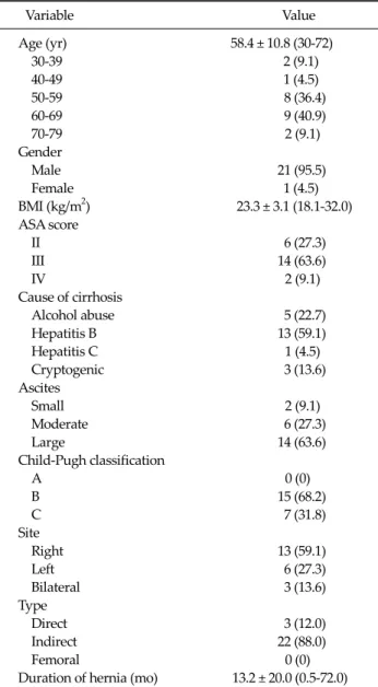
Table 1. Demographics of patients with cirrhosis accompanied by ascites and inguinal hernia
Variable Value
Age (yr) 58.4 ± 10.8 (30-72)
30-39 2 (9.1)
40-49 1 (4.5)
50-59 8 (36.4)
60-69 9 (40.9)
70-79 2 (9.1)
Gender
Male 21 (95.5)
Female 1 (4.5)
BMI (kg/m2) 23.3 ± 3.1 (18.1-32.0) ASA score
II 6 (27.3)
III 14 (63.6)
IV 2 (9.1)
Cause of cirrhosis
Alcohol abuse 5 (22.7)
Hepatitis B 13 (59.1)
Hepatitis C 1 (4.5)
Cryptogenic 3 (13.6)
Ascites
Small 2 (9.1)
Moderate 6 (27.3)
Large 14 (63.6)
Child-Pugh classification
A 0 (0)
B 15 (68.2)
C 7 (31.8)
Site
Right 13 (59.1)
Left 6 (27.3)
Bilateral 3 (13.6)
Type
Direct 3 (12.0)
Indirect 22 (88.0)
Femoral 0 (0)
Duration of hernia (mo) 13.2 ± 20.0 (0.5-72.0) Values are presented as mean ± SD (range) or number (%).
BMI, body mass index; ASA, American Society of Anesthe- siologists.
METHODS
A retrospective analysis of the medical records of 22 adult patients who complained of inguinal pain or dis- comfort and treated surgically by a single surgeon for in- guinal hernia at our medical institution from January 2002 to August 2009 was performed to evaluate age, gender, cause of liver cirrhosis, severity of liver cirrhosis, clinical laboratory findings, underlying diseases, anesthetic meth- ods, surgery, and complications.
Our choice for local anesthesia is a 50:50 mixture of 2%
lidocaine and 0.5% bupivacaine with 1/100,000 epine- phrine. An average of 30 mL of this mixture is usually suf- ficient for a unilateral inguinal hernia repair. Subdermal infiltration, intradermal injection, ilioinguinal/iliohypo- gastric nerves block (injection at the point 2.5 cm medial to the anterior superior iliac spine [ASIS] and 1 cm cephalad toward a reference line connectiong umbilicus and ASIS), deep subcutaneous injection, subaponeurotic injection, Infiltration at the level of the pubic tubercle, and around the neck of a hernial sac were done sequentially.
In patients with concurrent presence of ascites, the amount of ascites was determined based on preoperative abdominal ultrasound or abdominal computed tomog- raphy scans. The amount of ascites is categorized into three groups (small - few collections of fluid, largest di- mension generally less than 3 cm; moderate - multiple col- lections of fluid, smallest dimension more than 3 cm; large - generalized fluid, floating bowel). In this study, there was no patient with refractory ascites which is defined as as- cites that cannot be mobilized or the early recurrence of which cannot be satisfactorily prevented by medical therapy. The severity of cirrhosis is classified with the Child-Pugh score.
RESULTS
Patient characteristics (Table 1)
The patients consisted of 21 men and one woman. Their mean age was 58.4 years; there were nine patients (40.9%) in their sixties and eight patients (36.4%) in their fifties.
The mean body mass index was 23.3 kg/m2.
The patients were assessed based on the American Society of Anesthesiologists (ASA) score; there were six cases of ASA II (27.3%), 14 of ASA III (63.6%), and two (9.1%) of ASA IV (9.1%). Causes of liver cirrhosis included hepatitis B (n = 13, 59.1%), alcohol abuse (n = 5, 22.7%), hep- atitis C (n = 1, 4.5%), and idiopathic causes (n = 3, 13.6%).
The severity of liver cirrhosis was assessed based on the Child-Pugh classification; there were 15 cases (68.2%) of grade B and seven cases (31.8%) of grade C. The study pop-
Table 2. Laboratory findings of patients with cirrhosis accompanied by ascites and inguinal hernia
Variable Value
Albumin (g/dL) 3.1 ± 0.6 (1.9-4.1) Total bilirubin (mg/dL) 1.5 ± 1.2 (0.23-5.26) Platelet count (×103/μL) 107.9 ± 72.9 (36-300) Hemoglobin (g/dL) 11.6 ± 1.9 (7.5-15.0)
INR 1.28 ± 0.16 (1.01-1.57)
Values are presented as mean ± SD (range).
INR, international normalized ratio.
Table 3. Underlying disease other than liver cirrhosis and viral hepatitis in patients with cirrhosis accompanied by ascites and inguinal hernia (n = 22)
Variable Value
Underlying disease 17 (77.3)
Cardiac disease 4 (18.2)
Diabetes mellitus 7 (31.8)
Cerebrovascular accident 2 (9.1)
Lung disease 2 (9.1)
Chronic renal failure 1 (4.5)
Benigh prostatic hyperplasia 0 (0)
History of laparotomy 6 (27.3)
Hepatocellular carcinoma 11 (50.0)
TACE only 10 (45.5)
TACE + RFA 1 (4.5)
Values are presented as number (%).
TACE, transarterial chemoembolization; RFA, radiofrequency thermal ablation.
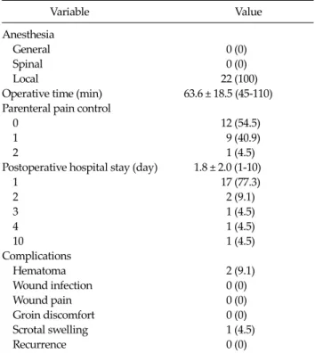
Table 4.Operation and outcomes in patients with cirrhotic accom- panied by ascites and inguinal hernia (n = 22)
Variable Value
Anesthesia
General 0 (0)
Spinal 0 (0)
Local 22 (100)
Operative time (min) 63.6 ± 18.5 (45-110) Parenteral pain control
0 12 (54.5)
1 9 (40.9)
2 1 (4.5)
Postoperative hospital stay (day) 1.8 ± 2.0 (1-10)
1 17 (77.3)
2 2 (9.1)
3 1 (4.5)
4 1 (4.5)
10 1 (4.5)
Complications
Hematoma 2 (9.1)
Wound infection 0 (0)
Wound pain 0 (0)
Groin discomfort 0 (0)
Scrotal swelling 1 (4.5)
Recurrence 0 (0)
Values are presented as number (%) or mean ± SD (range).
ulation consisted of 14 (63.6%), 6 (27.3%), and 2 (9.1%) cas- es of severe, moderate, and mild ascites, respectively. The patients included 13 cases of right-sided hernia (59.1%), six of left-sided hernia (27.3%), and three of bilateral her- nia (13.6%). All three bilateral cases were of the indirect type. There were 22 cases (88.0%) of indirect type and 3 cases (12.0%) of direct type. The mean period of symptoms was 13.2 months.
Laboratory findings (Table 2)
The mean albumin level was 3.1 g/dL (range, 1.9 to 4.1 g/dL), and mean total bilirubin was 1.5 mg/dL (range, 0.23 to 5.26 mg/dL). The mean platelet count was 107.9 × 10³/μL (range, 36 to 300 × 103/μL); mean hemoglobin level was 11.6 g/dL (range, 7.5 to 15.0 g/dL); and mean international normalized ratio was 1.28 (range, 1.01 to 1.57).
Underlying diseases (Table 3)
Underlying diseases other than liver cirrhosis were present in 77.3% of cases (17/22), the most common of which was hepatocellular carcinoma (HCC), which was present in 50.0% of cases (11/22). This was followed by dia- betes mellitus in seven cases (31.8%) and a history of lapa- rotomy in six cases (27.3%).
Of 11 patients with HCC, ten underwent transarterial chemoembolization (TACE) and the remaining patient re- ceived concomitant medication for TACE with radio- frequency thermal ablation (RFA) prior to inguinal hernia repair.
Anesthetic methods and surgical treatment out- comes (Table 4)
Surgery was performed under local anesthesia in all pa- tients and the mean surgical time was 63.6 minutes. Drain was not used in all patients. Parenteral analgesics for post- operative pain control were administered at a mean fre- quency of 0.50 times (range, 0 to 2 times); they were ad- ministered once in nine patients (40.9%) and twice in one
patient (4.5%). No further parenteral analgesics were needed following the surgery in the remaining 12 patients (54.5%). The mean postoperative length of hospital stay was 1.8 days. Seventeen patients (77.3%) were discharged the day following surgery. Two patients (9.1%) were dis- charged on day 2, and one patient was discharged on day 10. In patient who was discharged on day 10, the length of hospital stay was prolonged for ascites control.
There were 3 cases (13.6%) of postoperative complica- tions, including 2 cases of hematoma and one case of scro- tal swelling. However, these cases improved following conservative treatment. There was no case of serious com- plications, such as wound infection or recurrence.
There were 13 (59.1%) postoperative deaths, and 8 of these patients died within 1 year after hernia repair.
However, there was no 30-day postoperative mortality.
Five of the eight patients who died were Child’s class B and the remaining three were Child’s class C. Deaths were all related to cirrhotic complications, but there were no cases of operation-related mortality.
DISCUSSION
Adult cases of indirect inguinal hernia originate from an unclosed processus vaginalis and an enlarged internal inguinal ring. An increase in abdominal pressure plays a role in promoting the occurrence of this disease entity.
Direct inguinal hernia originates from weakening of the posterior wall of the inguinal canal composed of the trans- versalis fascia and an increase in size of the abdominal ring. An increase in abdominal pressure has been reported to occur due to obesity, intestinal obstruction, chronic bronchitis, voiding difficulty caused by prostate hyper- plasia, constipation, and liver cirrhosis accompanied by ascites. Indications and the natural course have not been established in detail for patients with liver cirrhosis who have poor systemic status, and particularly those accom- panied by ascites. Related reports are very limited in scope [1]. Both hernia and liver cirrhosis have detrimental effects on the quality of life (QoL). Surgical treatment in patients with liver cirrhosis increases the risk of developing anes- thetic or postsurgical complications. It is possible that liver
cirrhosis could lead to a serious condition in patients with liver cirrhosis who concurrently have an inguinal hernia or those who develop intestinal obstruction due to in- carceration or strangulation; these cases require special attention.
In principle, all cases of inguinal hernia should be treat- ed by hernia repair. However, there is controversy regard- ing the feasibility of hernia repair in patients with poor systemic status. Particularly in patients with inguinal her- nia who concurrently have liver cirrhosis accompanied by ascites, there is controversy regarding whether hernia re- pair should be performed because of the risk of mortality due to anesthetic complications, postsurgical complica- tions, recurrence, and liver cirrhosis itself [1]. Horn et al.
[2] reported that hernia repair in patients with advanced portal hypertension and ascites should be approached with caution and treated conservatively whenever possi- ble, because cirrhosis increases the risk of significant peri- operative complications, such as infection, recurrence, and ascites leakage. However, Hurst et al. [3] reported that life-threatening complications from inguinal hernia repair in patients with cirrhosis and ascites are uncommon.
Morbidity and long-term mortality rates in these patients are due to progression of the underlying liver disease.
Hurst et al. [3] reviewed 18 patients with cirrhosis accom- panied by ascites and groin hernia (20 inguinal and one femoral), and 11 underwent repair of their groin hernia (a total of 13 repairs). Ten hernia repairs were performed electively, two were performed urgently because of recent difficult reduction, and one was performed emergently for incarceration without strangulation. No major and four minor postoperative complications occurred, and there were no cases of perioperative death or ascites leakage.
Hurst et al. [3] reported that of 18 patients with cirrhosis and an inguinal hernia, the cause of cirrhosis was alcohol abuse in 14 cases, viral hepatitis in one case, and crypto- genic cirrhosis in three cases. Among 15 patients with cir- rhosis and an inguinal hernia, Kim et al. [1] reported that the causes of cirrhosis were ethanol abuse in five cases and viral hepatitis (hepatitis B) in 10 cases. In the present study population of 22 patients, the causes of cirrhosis were ethanol abuse in 5 cases, viral hepatitis in 14 cases (hepatitis B, n = 13; hepatitis C, n = 1), and idiopathic in 3
cases.
The evaluation and management of ascites in patients with known cirrhosis are very important, as this manifes- tation of portal hypertension has a detrimental effect on prognosis. Patients with cirrhosis have a significant risk of adverse outcome after abdominal wall hernia repair com- pared to non-cirrhotic patients, particularly those who un- dergo emergent surgery. Ideally, patients with cirrhosis should undergo elective hernia repair after medical opti- mization [4,5]. In the present study, patients had all re- sponsive ascites and had been treated continuously at de- partment of hepatology for control of ascites with appro- priate sodium restriction and diuretic therapy and im- provement of general condition before elective hernia repairs. There was no outstanding change in Child-Pugh score just before surgery.
Rarely, massively dilated veins (1.5 to 2.0 cm in diame- ter) mimicking inguinal hernias have been noted entering the spermatic cord at the internal inguinal ring. Given the unusual presentation of these dilated veins, the use of pre- operative Doppler ultrasound is advocated in patients with cirrhosis and suspected inguinal hernias [2]. There- fore, it is mandatory to postulate the possibility of dilated veins in patients with liver cirrhosis suspected to have an inguinal hernia.
Refractory ascites is associated with poor prognosis in patients with cirrhosis and is an indication for liver transplantation. In patients who concurrently have an in- guinal hernia, elective surgery can also be performed prior to liver transplantation or hernia repair can also be per- formed along with liver transplantation [2,6].
Ennaifer et al. [7] reported that the Child’s score is a good index for assessing prognosis in patients with cirrhosis. Their overall survival rate was 47% at 5 years.
Hurst et al. [3] reported that the survival rate was approx- imately 75% at 2 years, and only 28% survived to 5 years.
In the present study, the survival rate was approximately 68% at 2 years, and 28% survived to 4 years.
Patti et al. [8] used the Short Form-36 (SF-36) ques- tionnaire to evaluate QoL in patients with cirrhosis under- going inguinal hernia repair to identify optimal manage- ment of symptomatic inguinal hernia in patients with cirrhosis. All eight SF-36 domains and the mental compo-
nent summary and physical component summary scores improved remarkably after hernia repair, especially in pa- tients in Child’s class C and/or those with refractory ascites. The authors reported that inguinal hernia repair is a safe procedure for treatment of symptomatic inguinal hernia in patients with cirrhosis. The improvement in QoL represents a clear-cut indication for elective hernia repair.
Patients with cirrhosis frequently have comorbidities resulting in gastrointestinal, lung, or cervical cancer, among others. A significant proportion of patients with cirrhosis develop HCC during the course of the disease [9].
In this study, HCC developed in 11 (50.0%) patients; ten were treated with TACE and one was treated with both TACE and RFA.
Franzetta et al. [10] reported that patients with cirrhosis undergoing elective or emergent surgery can incur sig- nificant preoperative risks and postoperative complica- tions, thus increasing their mortality rate. The presence of tensive ascites, low albumin level, prothrombin time, or activated partial thromboplastin time, together with an emergent operation showed significant associations with mortality rates of 7.1% in Child’s class A, 23% in class B, and 84% in class C.
Kim et al. [1] performed hernia repairs under local anes- thesia in three of 15 patients with cirrhosis, spinal anes- thesia in 7 cases, general anesthesia in 3 cases, and epi- dural anesthesia in 2 cases. In the present study, we per- formed hernia repairs under local anesthesia without an- esthesia-related complications in all 22 patients with cir- rhosis, suggesting that other types of anesthesia increase anesthesia-related complications. If needed, a low dose of demerol and valium was administered for sedation and pain control.
Dissection of the hernia sac can lead to pain during her- nia repair. At this time, pain is relieved with infiltration of local anesthetics into the neck of the hernia sac. When se- vere ascites is associated with hydrocele, the distal part of indirect hernial sac is not dissected excessively to the end and the mid portion of the hernia sac is cut. The proximal portion of hernial sac is inverted by using mesh plug after ligation. In addition, the distal part of hernial sac is cut ver- tically after aspiration of ascites and is fixed after eversion.
Hernial sac is very thin and easily torn in patients with cir-
rhosis accompanied by ascites. Therefore, careful dis- section of the hernial sac is needed to prevent leakage of ascites. Although the tear of the hernial sac is developed, it is no severe problem and only meticulous repair of the tearing site and compression dressings are enough.
Furthermore, these methods are helpful to prevent post- operative seroma and any problem related with wound healing. There are four sizes (small, medium, large, and extra large) in plug. Extra large (4.1 × 5.0 cm) plug is large enough to cover the large internal ring and very large di- rect hernia.
When severe ascites is associated with hydrocele, the distal part of indirect hernial sac is not dissected ex- cessively to the end and the mid portion of the hernia sac is cut. The proximal portion of hernial sac is inverted by using mesh plug after ligation. In addition, the distal part of hernial sac is cut vertically after aspiration of ascites and is fixed after eversion.
Patti et al. [8] reported no major complications after her- nia repair for symptomatic inguinal hernias in 32 patients with cirrhosis. In this study, 15 (68.2%) patients were in Child’s class B and seven (31.8%) were in Child’s class C.
All hernia repairs were performed under local anesthesia and were successful with no major complications or recurrence. Only three minor complications occurred, consisting of two hematomas and one case of scrotal swelling. Both of the two patients showing hematoma af- ter hernia repair underwent hematoma removal under lo- cal anesthesia within 2 to 3 days and then were recovered without any specific problem. It seems very difficult to de- velop massive bleeding in mesh plug hernia repair since it does not involve entering the abdominal cavity. Meticu- lous bleeding control is very important and compression dressing may also be helpful. In the present study, platelet replacements were performed in only two patients whose platelet counts were less than 50 × 103/μL.
Thirteen patients died during the follow-up period (59.1%); eight of these patients died within 1 year after her- nia repair. However, there were no cases of 30-day post- operative mortality. Five of the eight patients who died were Child’s class B, while the other three patients were Child’s class C. Deaths were all related to cirrhotic compli-
cations, and there was no operation-related mortality.
In conclusion, elective hernia repair in patients with cir- rhosis accompanied by ascites and an inguinal hernia can be performed safely without severe complications, even in patients in Child’s class C or in those with refractory ascites. Furthermore, hernia repair under local anesthesia in these patients is safer than that under general or spinal anesthesia.
CONFLICTS OF INTEREST
No potential conflict of interest relevant to this article was reported.
REFERENCES
1. Kim JW, Keum JH, Kim CS, Moon HY, Koo BW. Inguinal herniorrhaphy in patient with cirrhosis of liver and ascites.
J Korean Surg Soc 1997;53:275-9.
2. Horn TW, Harris JA, Martindale R, Gadacz T. When a her- nia is not a hernia: the evaluation of inguinal hernias in the cirrhotic patient. Am Surg 2001;67:1093-5.
3. Hurst RD, Butler BN, Soybel DI, Wright HK. Management of groin hernias in patients with ascites. Ann Surg 1992;
216:696-700.
4. Herrera JL. Current medical management of cirrhotic ascites. Am J Med Sci 1991;302:31-7.
5. Carbonell AM, Wolfe LG, DeMaria EJ. Poor outcomes in cirrhosis-associated hernia repair: a nationwide cohort study of 32,033 patients. Hernia 2005;9:353-7.
6. Marsman HA, Heisterkamp J, Halm JA, Tilanus HW, Metselaar HJ, Kazemier G. Management in patients with liver cirrhosis and an umbilical hernia. Surgery 2007;142:
372-5.
7. Ennaifer R, Ouakaa-Kchaou A, BelHadj N, Elloumi H, Gargouri D, Kochlef A, et al. Prognostic factors for surviv- al in cirrhosis. Tunis Med 2007;85:1039-43.
8. Patti R, Almasio PL, Buscemi S, Famà F, Craxì A, Di Vita G.
Inguinal hernioplasty improves the quality of life in pa- tients with cirrhosis. Am J Surg 2008;196:373-8.
9. Francoz C, Durand F. The risk of surgery in patients with cirrhosis. Acta Gastroenterol Belg 2008;71:42-6.
10. Franzetta M, Raimondo D, Giammanco M, Di Trapani B, Passariello P, Sammartano A, et al. Prognostic factors of cir- rhotic patients in extra-hepatic surgery. Minerva Chir 2003;58:541-4.