Ⅰ. Introduction
Prosthetic treatment is required when teeth are lost due to caries, periodontal disease, trauma. This may be crown and bridge, removable partial denture, complete denture or implant prosthesis.
Recently, implant prosthesis has come to play an important role in prosthodontic treatment. It is now possible to almost completely restore patient chewing ability, esthetics, speech and psy- chologic comfort.
Mandibular movement have been investigated to evaluate and analyze chewing pattern, mal- occlusion, Temporomandibular disorder, mandibu- lar surgery, speech. Methods for measuring mandibular movement have been developed con- tinuously throughout the years. In 1889, Luce mea- sured mandibular movement with still photography.
Cinephotography by Hildebrand, gnathic replicator by Cannon, stereophotography by Erdman, pho- toelectric device by Gillings, radionuclide track- ing device by Salomon and various other method had been used9,12,13,37,41).
Kinesiograph designed by Jankelson et al is used for jaw tracking16), however this device is limited to incisor point checking. It is impossible to check condylar and molar point with this device,
but it is easy to place and also possible to check jaw tracking with EMG data by Hannam2,10,11,17,33,42)
. Although complete denture rehabilitation can restore a patient’s appearance and perceived so- cial role, other aspects of impaired oral function associated with being edentulous can be com- pensated for only to a limited extent3). Implant pros- thesis is reported to improve oral function to a de- gree approaching that of dentate persons with re- spect to chewing efficiency and muscle activity19). The purpose of this study was to explore the mandibular movement of complete implant sup- ported fixed prosthesis wearing patients and compare that movement with that of natural den- tition and complete denture wearing patients.
Ⅱ. Materials and Method 1. Control and experimental group
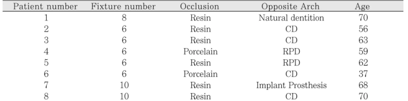
The experimental group is comprized of 8 pa- tients(Table 1). Implant subjects were chosen ac- cording to the following criteria:
(1) full arch fixed implant prosthesis in mandible (2) 6-10 fixtures
(3) occlusal surface : resin or porcelain occlu- sion
대한치과보철학회지:Vol. 37, No. 4, 1999
Jaw movement analysis in subjects with implant-supported prosthesis
Yang-Soo Kim, D.D.S., Yung-Soo Kim, D.D.S., M.S.D., Ph.D., M.Sc.<O.S.U.>, Chang-Whe Kim, D.D.S., M.S.D., Ph.D., Yong-Ho Kim, D.D.S., M.S.D., Ph.D.
Department of Prosthodontics, College of Dentistry, Seoul National University
(4) opposite arch: natural dentition, complete denture, removable partial denture (5) Angle Class I molar relationship
(6) No evidence of temporomandibular joint prob- lem or masticatory dysfunction
Implant prosthesis wearers consisted of 8 women (Average age : 60.6 years).
Two different control group were selected.
The first group had normal dentition and the sec- ond one was patients wearing complete den- tures.
Normal dentition group was chosen according to the following criteria
(1) complete number of natural teeth retained except the third molar
(2) Angle class I occlusal relationship (3) no masticatory function problem
(4) no history of orthodontic treatment and pros- thetic treatment.
Five female students of The Seoul National University were selected to be subjects for the nat- ural dentition control (Average age : 25.5 years).
Complete denture wearers
(1) Angle class I occlusal relationship (2) no masticatory functional problem (3) minimum 3 months denture use (4) full arch complete dentures
The complete denture wearers consisted of 8 women ( Average age : 64.3 years).
2. Recording method
Mandibular movement was recorded with a Sirognathograph (Siemens, Benshiem, Germany) and analyzed with the BioPAK program (Bioresearch, Milwaukee, U.S.A.).
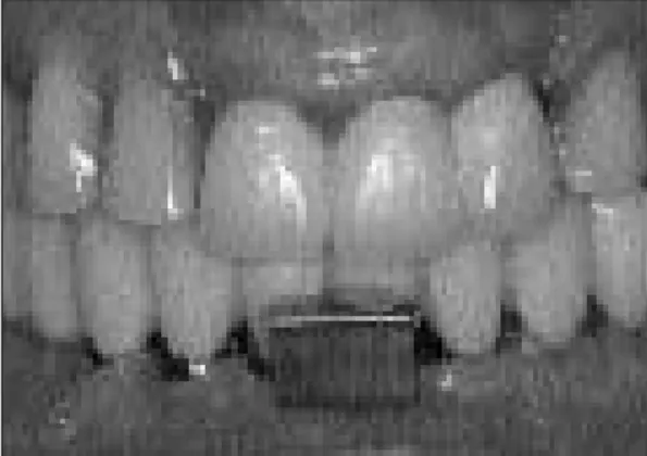
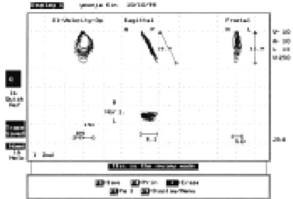
Patients were seated with the Frankfort plane parallel to the floor. A magnet was placed hori- zontally with the center of the magnet at the mandibular labial frenum and on the gingival third of central incisor. The magnet was positioned par- allel to the interpupillary line. Stomahesive (BioResearch, Milwaukee, U.S.A.) was used for attachment of the magnet45)(Fig. 2).
Sirognathograph sensor array comes with one elastic band used for its placement on the patient’
s head, but it is not possible to stably place it. The system was adjusted with a head band system of Saphon Visitrainer.
Upper crossbar was placed parallel to inter- pupillary line and the side bar was parallel to the Frankfort plane. The sensor array was aligned with the magnet using a magnet position bar (Fig. 1).
3. Border and functional movement
Unilateral chewing and border movement were analyzed. Yurkstas and Manly recommended the use of peanuts, ham, and carrots as test food of choice23). Raw carrots were used for fuctional uni- lateral chewing.
Table 1. Information of fully implant supported fixed prosthesis
1 8 Resin Natural dentition 70
2 6 Resin CD 56
3 6 Resin CD 63
4 6 Porcelain RPD 59
5 6 Resin RPD 62
6 6 Porcelain CD 37
7 10 Resin Implant Prosthesis 68
8 10 Resin CD 70
(CD : complete denture, RPD : removable partial denture)
Patient number Fixture number Occlusion Opposite Arch Age
Carrots were chopped to the standardized size of 1.0cm×1.0cm×0.5cm. The sample weighed 0.5g. 6 pieces were used, so the total weight was 3g. Yukstas, Manly recommended 3 - 5g for sta- bility testing of complete dentures. The patient was advised to chew the sample food ten times uni- laterally on the patient’s preferred side. This test was repeated two times.
The executed movements were recorded with a Sirognathograph and a BioPAK system. To record border movement in frontal and sagittal plane, the subject was induced to carry out the exercise three times. And border movement in frontal and sagittal plane was recorded three times repeatedly. To measure the border movement in sagittal plane, the patient was told to close in cen-
tric occlusion first and then protrude to the maximum with sliding contact of teeth and open the mouth to the maximum and close the mouth in maximal retrusion (Fig. 5).
For measuring border movements in frontal plane, the patient was induced to close in centric occlusion and slide the mandible to the left and right to the maximum (Fig. 6).
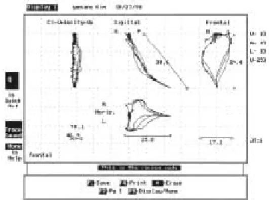
After the measurement, the movement of chewing cycle was analyzed. Movements were di- vided into 10 segments. Horizontal and vertical range of movement in frontal plane, anterior and posterior range of movement in sagittal plane, max- imum moving distance of incisor point, maximum opening and closing velocity of movement were mea- sured (Fig. 3, Fig. 4).
Fig. 1. Sirognathograph in place on a subject. Fig. 2. Position of magnet on the prosthesis.
Fig. 3. Unilateral chewing pattern. Fig. 4. Sweep display of chewing pattern.
4. Statistical Analysis
For statistical analysis, Microsoft Excel (ver- sion : Office 98, Microsoft) and SPSS program (Ver 7.5). Oneway ANOVA test and Scheffe’s multi- ple range analysis were run.
Ⅲ. Results
1) Height of the chewing cycle in frontal plane
a) raw data analysis
There were no significant differences among groups. The increasing order in mean was CD group, implant prosthesis group and natural dentition group(Fig. 8, Table 2).
b) ratio analysis
To know the range of motion within border move- ment, we measured the ratio of functional move- ment data and border movement data.
Fig. 5. Border movement in sagittal plane.
Fig. 7. The analysis criteria of chewing cycle.
Fig. 6. Border movement in frontal plane.
1. Height of chewing cycle 2. Width of chewing cycle 3. Opening angle
4. Closing angle
5. Chewing angle = |Closing angle - Opening angle|
1. Anterior and posterior range of chewing cycle 2. Maximum moving distance of chewing cycle
Analyzed data=(functional movements/ border movements) * 100
There were no significant differences among groups. Age, patient’s attitude to the test, and unfavorable prosthesis may affect border and func- tional movement. In complete denture wearing group and implant prosthesis group, border movement was restricted. Relatively the ratio of complete denture wearing patients and implant prosthesis was increased(Fig. 9, Table 3).
2) Width of the chewing cycle in frontal plane a) raw data analysis
The value in the implant group was significantly
lower than that of the natural dentition group and the CD group. Intragroup difference was higher in natural dentition and complete denture wear- ing group than implant group(Fig. 10, Table 4).
b) ratio analysis
Two different chewing pattern could be observed in frontal plane. One was a grinding pattern, the other is a chopping pattern, Consistency and tough- ness of food, type of prosthesis, shape of the oc- clusal surface, chewing habit, occlusion type and other factors may influence shape of chew- ing pattern(Fig. 11, Table 5).
It was impossible to discriminate the differences Fig. 8. Raw datas of the height of the chewing
cycle in frontal plane
Table 2. Result of Scheffe’s analysis in the height of the chewing cycle in frontal plane.
CD 22 13.1605
Scheffe Implant 24 13.5171
Natural 14 14.0821
p .359
* : Subset for alpha = .05
Fig. 9. Ratio data of the height of the chewing cycle in fontal plane.
Table 3. Results of Scheffe’s analysis in the height of the chewing cycle in frontal plane
CD 14 43.1657
Scheffe Implant 24 43.2650
Natural 22 44.1741
p .831
* : Subset for alpha = .05
Group N 1*
Group N 1*
among the ratio data of the test group in the width of the chewing cycle.
3) Chewing angle in frontal plane
The value in the complete denture group was significantly lower than that of the natural den- tition group(Fig. 16, Table 10).
4) Maximum moving distance incisor point in sagittal plane
a) raw data analysis
There were no significant differences between the natural dentate person and implant group. The
maximum moving distance in the complete den- ture group were significantly lower than that of the natural dentition group(Fig. 12, Table 6).
b) ratio analysis
The values seen in the implant group were sig- nificantly different from the CD group and the nat- ural dentition group. The ratio seen in implant group was lower than that of other groups.
Border movement of the CD group was limited ex- tensively, so the ratio of the CD group was higher than raw data results(Fig. 13, Table 7).
Table 4. Result of Scheffe’s analysis in the width of the chewing cycle in frontal plane
CD 24 3.9492
Scheffe Implant 14 4.9613
Natural 22 5.3043
p 1.000 .689
1 < 2 at P<0.05
Group N 1 2
Fig. 10. Raw data of the width of the chewing cycle in frontal plane.
Fig. 11. Ratio data of the width of the chewing cycle in frontal plane.
Table 5. Result of Scheffe’s analysis in the width of the chewing cycle in frontal plane
CD 14 25.9029
Scheffe Implant 24 27.5688
Natural 22 29.0559
p .488
* : Subset for alpha = .05
Group N 1*
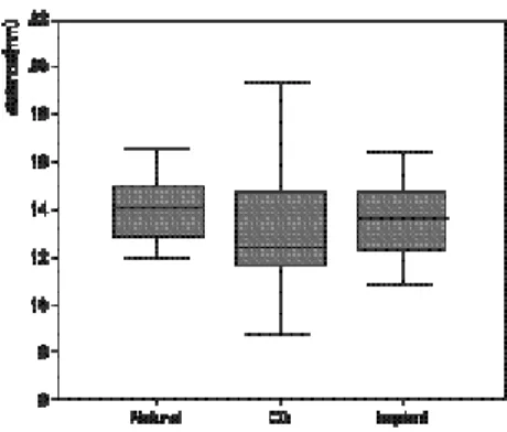
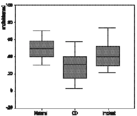
5) Maximum anterior and posterior range of movement in sagittal plane
a) raw data analysis
The values in the complete denture group and the implant group were significantly lower than that of the natural dentition group(Fig. 14, Table 8).
b) ratio data analysis
The value seen in the implant group was sig- nificantly lower than the natural dentition group(Fig. 15, Table 9). Chewing pattern in sagittal plane was analyzed on two criteria.
One is the maximum moving distance of in-
cisor point and the other is the anterior and pos- terior range of movement. Both values were lower in the implant group than the natural den- tition group.
It can be assumed that chewing pattern in the implant group was more confined to the frontal plane than other group because the two values mea- sured were lower.
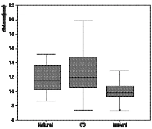
6) Maximum opening velocity
The value of the test groups increased in following order, the natural dentition group, the CD group, and the implant prosthesis group. The val- ue in the natural dentition group was significantly Fig. 16. Raw data of chewing angle.
Table 10. Results of Scheffe’s analysis
CD 22 30.0859
Scheffe Implant 24 40.8379 40.8379
Natural 14 50.1986
p .124 .202
1 < 2 at P<0.05
Table 6. Results of Scheffe’s analysis
CD 22 14.9082
Scheffe Implant 24 15.5221 15.5221
Natural 14 16.7721
p .548 .090
1 < 2 at P<0.05 Fig. 12. Raw data of the maximum moving distance
in sagittal plane.
Group N 1 2
Group N 1 2
Fig. 13. Ratio data of the maximum moving distance in sagittal plane.
Table 7. Results of Scheffe’s analysis
CD 24 35.0408
Scheffe Implant 22 39.2769
Natural 14 40.0257
p 1.000 .874
1 < 2 at P<0.05
Table 8. Results of Scheffe’s analysis
CD 22 5.7386
Scheffe Implant 24 6.3592
Natural 14 8.0443
p .468 1.000
1 < 2 at P<0.05 Fig. 14. Raw data of the maximum anterior and-
posterior range of movement in sagittal plane.
Group N 1 2
Group N 1 2
Fig. 15. Ratio data of the maximum anterior and- posterior range of movement in sagittal plane.
Table 9. Results of Scheffe’s analysis
CD 24 20.2013
Scheffe Implant 22 24.2614 24.2614
Natural 14 27.8714
p .073 .123
1 < 2 at P<0.05
Group N 1 2
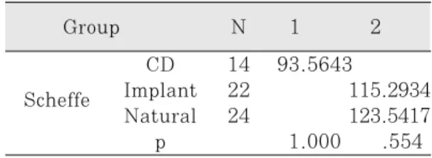
lower than that of the CD and the implant prosthesis group(Fig. 17, Table 11).
7) Maximum closing velocity
The value in the test group increased in following order, the CD group, the natural dentition group, and the implant prosthesis group. But there were no significant differences among the test groups(Fig. 18, Table 12).
IV. Summary of the results 1. Natural dentition
Border movement guiding by subject oneself was
rather smooth and natural in comparison to complete denture wearer and implant supported prosthesis wearer. Maximum moving range of in- cisor point and anterior and posterior range of move- ment was higher than the other group. Chewing angle was also higher than the other group.
But maximum opening velocity was the lowest among the test group(Fig. 19).
2. Complete denture
Some patient showed restricted border and functional movement. They were hard to chew car- rot. The other patient showed fluent border and functional movement. It might be due to ridge con- Table 11. Results of Scheffe’s analysis
CD 14 93.5643
Scheffe Implant 22 115.2934
Natural 24 123.5417
p 1.000 .554
1 < 2 at P<0.05 Fig. 17. Raw data of maximum opening velocity.
Fig. 18. Raw data of maximum closing velocity.
Table 12. Results of Scheffe’s analysis
Group N 1*
CD 22 64.9341
Scheffe Implant 14 69.0221
Natural 24 71.7267
p .402
* : Subset for alpha = .05
Group N 1 2
Group N 1*
dition, retention and stability of denture. Intragroup difference was higher than the other group.
Width of the chewing cycle was higher than the other groups, but standard deviation was too high to be reliable. Maximum moving distance of in- cisor point and anterior and posterior range of move- ment was lower than the others. Chewing angle was also lower that the others(Fig. 20, 21).
3. Implant prosthesis
Chewing pattern was slightly different as op-
posing arch types. Patients wearing maxillary com- plete denture had unnatural and rather limited border movement than patients with maxillary com- plete implant supported prosthesis and patients with upper natural dentition. Chewing pattern at frontal plane was rhythmic but the movement was hard to find core of movement. Patients did easily crush the carrot. Height of the chewing cy- cle was not significantly different from the others.
Width of the chewing cycle was significantly lower than the others. Maximum moving range of incisor point were not significantly different from Fig. 19. Left side unilateral chewing of one test den-
tate control.
Fig. 20. Left side unilateral chewing of complete den- ture wearer who had poor residual alveo- lar ridge.
Fig. 21. Left side unilateral chewing of complete den- ture wearer who had good residual alveolar ridge.
Fig. 22. Left side unilateral chewing of patient who had complete implant supported pros- thesis in maxilla and mandible.
the others. Maximum anterior and posterior range of movement in sagittal plane was signif- icantly lower than natural dentition group.
Chewing angle was not significantly different from the others. Maximum opening and closing velocity was the highest(Fig. 22).
Ⅴ. Discussion
Mastication is considered a rhythmic event with timing that is generated in the reticular substance of the brain stem and can be modified by inputs from the central and peripheral nervous system26). Habitual mastication is a rhythmic event26-29). Natural chewing pattern is characteristic for each individual1). But determining a mean chew- ing movement can be difficult28). All recordings showed a large variety of individual movement pat- terns. Because of the variety of individual move- ments the measurement revealed relatively high standard deviation4).
In order for jaw tracking to be acceptable, it must be carried out in a standardized manner, and spe- cific movements should be measured which have been shown in controlled clinical recordings to be reproducible. As with all human movement there is a broad range of intra and interindivid- ual values15). Type of food, emotional and psy- chological status, sex difference may take part in chewing movement.
At first, the test food was standardized into same size and weight. Many studies prove that chew- ing pattern is changed in relation to the resistance of food. Width of the chewing stroke increased when chewing hard foods8).When soft food is chewed the angulated patterns appear much less frequent- ly and the slim and drop shaped forms are fa- vored35). Chewing hard food is characterized by shorter times for the chewing cycles and greater vertical and sagittal amplitudes of the movements.
Longer vertical dimensions of the pattern and small- er frontal widths obviously lead to smaller that is
steeper angles of approach to the intercuspal po- sition. Ahlgren found that a wide grinding chew- ing cycle was used with gum, whereas carrots and peanuts called for a more vertical chopping stroke1). To compare the data, the standardized sample food must be used. Yurkstas and Manly recommended the use of peanuts, ham or carrots for test food. Kapur recommended the use of raw carrot23). Raw carrot recommended for the stability test of complete denture. Because raw carrot have different resistance nature according to the area, we used the outerside portion of carrot away from the center.
The patient usually chewed unilaterally at a pre- ferred side. Preferred side is not associated with handedness43). No association was found be- tween chewing side preference and handedness, footedness, eyedeness or earedness14). If several chewing cycles are computed to assess a mean chewing movement, cycles toward opposite sides tend to abate reciprocally. Therefore statistical stud- ies usually have been performed on unilateral vol- untary masticatory movements26).
Although chewing on only one side of the jaw may be necessary to analyze the pattern of jaw movements during enforced unilateral chewing to classify chewing strokes, any method of investi- gating masticatory function that uses an apparatus that interferes with subconscious chewing or imposes voluntary control over the chewing cycle, introduces a degree of bias and may therefore in- validate the observation30). In research on mas- tication, there is a need to distinguish between ha- bitual and deliberate unilateral chewing. Deliberate unilateral chewing is the accepted model for study of the influence of a specific factor upon mas- tication38).
Patient’s cooperation to the test have an effect on the results. Sirognathograph can measure mandibular movement easily and quickly. But this device presses the nasion area forcefully. Patients feel pain, when the test have lasted more than ten
minutes. Patient who were cooperative to the test were selected. Chewing movement may vary according to the emotional and physical status of the patient31,32). A slight alteration in the masti- catory pattern in repeated registrations, might mean an adaptation to the test situation. If this is the case, initial training of the test situation as was done in the present study would reduce the risk of registered changes to be affected by adaptation18).
Sex difference also might have an effect on the result. Because patients were almost female, only female patients were selected. Differences were found between men and women with respect to cycle time, velocity of movement, dimensions of the chewing envelope, and duration of the pause in the intercuspal position. The sex of the subject have been shown to effect mandibular displace- ment30).
Accuracy of device can have an effect on the re- sults. Mandibular kinesiograph provides an accuracy of 0.1mm for resolution of madibular po- sitions in the vicinity of occlusion. The accuracy and linearity of the electrognathographic method have been tested, with an accuracy of 1%
achieved in the spatial domain to which chewing movements are confined35)Sirognathograph tend- ed to underestimate the true jaw opening by about 13% and that this relationship was linear for open- ings of less that 45mm. Beyond 45mm the rela- tionship was no longer linear40). Functional movement may be less than 45mm, so we can have the linear data and compare the data among group.
Sirognathograph yields an error in presentation on the borders of the prism-shaped area of up to 10%. Correct positioning of the magnet obtains a spatial region of nearly 15×15mm horizontal- ly and 25 mm vertically. Inside this region non- linearities are less than 1%. Error of the readout of the graphic tracing was 0.5mm31). Michler25)and Maruyama24) tested the Sirognathograph and found that a 10 degree rotation of magnet caused
a 1mm displacement. Additional errors due to head movement during the recording are always pos- sible. To avoid error due to head movement, we alter the device with head cap system of Saphon Visitrainer.
There were no significant differences among the test groups in the height of the chewing cycle in the frontal plane. The mean in increasing order is CD group, implant prosthesis group and nat- ural dentition group. But border movement of CD group and implant prosthesis group was re- stricted, so the ratio of functional movement and border movement in increasing order was nat- ural dentition group, implant prosthesis group and CD group.
Intragroup differences was too high to standardize the mean data in the width of the chewing cycle in frontal plane. But the data of implant prosthesis group was significantly lower than those of oth- er groups. Majority of implant prosthesis group have upper complete denture and worn resin teeth occlusion, so the chewing pattern was different from natural dentition group. Opening and closing path of chewing cycle was more vertical than other groups. The width of the habitual chewing move- ments at the first segment on the frontal projection was greater in denture wearerrs. Unilateral chewing cycles had greater dimensions with wide and curved opening strokes in denture wearers. Incisor point movements during later- al contact movements in patients whose dentures had nonanatomically formed teeth differed es- sentially from the same movements in dentate sub- jects4).
The mean increasing order of chewing angle was CD, implant and natural dentition group. The chew- ing angle of CD group was significantly lower than that of natural dentition group. CD group had the lowest maximum moving distance values of the incisor point. The values of CD group were sig- nificantly lower than those of natural dentition group. There were no significant differences be-
tween natural dentition group and implant pros- thesis group in the maximum moving distance val- ues of the incisor point. In anterior and posteri- or range of chewing cycles in sagittal plane, the natural dentition group have the highest value.
The values of natural dentition group were sig- nificantly higher than that of CD and implant group.
There is no significant differences among the test groups in the maximum closing velocity. But the closing velocity of complete denture group was low- er than that of implant and natural dentition group.
Mandibular movement of complete denture wearer have remarkable contrast according to the condition of residual alveolar ridge, retention and stability of denture, age, occlusion, and shape of denture teeth. Patient with good resid- ual alveolar ridge and well fabricated denture had not remarkable difference against natural dentition.
They have rhythmic chewing pattern and natural border movement. Chewing movement of denture wearers with good alveolar ridge occurred with- in the scope of approximately the same area as that of dentate subjects31). There was no difference of chewing angle size and velocity of movement22). But patient with poor residual alveolar ridge have unnatural chewing pattern and restricted border movement. Chewing patterns of the complete den- ture wearers with poor alveolar ridge were char- acterized by less vertical opening and less lateral excursions than those of the subjects with natural teeth39).These differences suggested that complete denture wearers avoid extreme chewing movements to avoid dislocating the dentures. The poor fit which leads to reduced chewing efficiency and relapsing denture sore spots was the main reason for edentulous patient seeking implantation in the mandible4). Denture wearers had a shallow angle of disocclusion in the frontal view compared with the natural dentition subjects5). The test sub- jects showed a wide intraindividual variation in chewing patterns in the complete denture situation19).Smaller chewing cycles was associated
with slower mandibular velocity in the group of complete denture wearers compared with dentate persons19).
Denture wearers do not prefer anatomic over nonanatomic posterior teeth to any significant de- gree. Denture teeth of both 0 degree and 30 de- gree designs produced a rounded, poorly de- fined intercuspal position in contrast to a sharp well defined intercuspal position in the natural den- tition subjects5).
The characteristic findings of the more anterior chewing movements of the edentulous subjects, particularly the females may possibly be ex- plained by several factors. 1) there is an anterior migration of the edentulous mandible. 2) the ver- tical dimensions of occlusion is reduced during the establishment of interarch relationships 3) the re- duced cusp height of the artificial teeth could cause difficulties in localizing the bolus and require a stronger force to crush the carrots. 4) consider- ing the direction of muscle fibers that are primarily engaged in the closing movement and the fact in denture wearers maximum intercuspation is achieved in centric relation position, it appears that the mandible is placed more anteriorly in the at- tempt to find optimum muscle length for gener- ating the strongest contractions. However. the pos- terior borders of the chewing movement en- velopes for denture wearers generally are placed more posteriorly in comparison to those of den- tate subjects, particularly in male subjects4).
Implant prosthesis have been reported to im- prove oral function to a degree approaching that of dentate persons with respect to chewing effi- ciency and muscle activity19).
Patients with implant supported fixed prosthesis chew carrot easily and naturally irrespective of op- posite arch status. Patients with resin occlusal sur- face had chopping type chewing pattern accord- ing to wear of resin teeth and altered occlusal pat- tern. Patient with porcelain superstructure had natural grinding type chewing pattern.
General chewing pattern underwent only mi- nor changes. However some chewing cycle pa- rameters showed obvious alterations. Increased velocity and displacement especially in the open- ing phase. The cycle duration showed a tenden- cy to decrease due to significant shortening of the occlusion phase. This can be attributed to the sta- bilization of the occlusion provided by the fixed den- ture in the mandible19).
5 patients of implant prosthesis group wore com- plete maxillary dentures, it was not expected that the effect of the implant prosthesis treatment would result in an improvement that would approach den- tate individuals19). Consequently, analyzed pa- rameters in implant prosthesis patient improved but did not fully coincide with dentate persons.
2 patients have opposite upper RPD, and 1 pa- tient have opposing full arch implant supported prosthesis. Patient with opposite RPD or full arch implant supported prosthesis have unrestricted border movement and grinding type chewing pattern. Implant prosthesis treatment actually in- fluences mandibular border movements as well as chewing patterns.
Female dentate subjects recorded a greater chew- ing width on the frontal projection30). Female den- tate subjects have the lowest opening velocity.
Female subjects perform the chewing move- ments in a different manner as a result of culture.
Perhaps female subjects are taught to perform chew- ing movements more slowly and well measured, while the male subjects do so more strongly and with less conscious control31).
Ⅵ. Conclusions
On the basis of the analysis of chewing move- ment of complete implant supported fixed pros- thesis wearers, complete denture wearers and nat- ural dentate subjects recorded using a Sirogn- athograph, the following conclusions are drawn : 1. No difference was found in the height of
chewing cycle in frontal plane among the test groups(p>0.05).
2. Differences were found in the width of chew- ing cycles in frontal plane between implant and natural dentition group and between implant and complete denture group(p<0.05).
3. Differences were found in the anterior and pos- terior range of chewing cycles in sagittal plane between implant and natural denti- tion group and between implant and complete denture group(p<0.05).
4. No difference was found in the chewing angle in frontal plane between implant and natur- al dentition group and between implant and com- plete denture group(p>0.05).
5. No difference was found in the maximum moving distance of incisor point in frontal plane between implant and natural denti- tion group and between implant and complete denture group(p>0.05).
6. The maximum opening velocity of natural dentition group was significantly lower than that of complete denture wearer and implant pros- thesis wearer(p<0.05).
7. No difference was found in the maximum closing velocity among the test groups(p>0.05).
8. There is great individuality in the mastication.
References
1. Ahlgren J, Mechanism of mastication, Acta odont scand, 1966;24(Suppl. 44):1-109.
2. Lewin A, Lemmer J, The measurement of jaw movement. Part II, J Prosthet Dent 1976;36(3):312-318.
3. Bates JF, Stafford GD, Harrison A, Masticatory function-a review of the literature. I The form of the masticatory cycle, J Oral Rehabil, 1975;2:281-301.
4. Benzing U, Weber H, Simonis A, Engel E, Changes in chewing patterns after implantation in the edentulous mandible, Int J Oral
Maxillofac Implants, 1994;9(2):207-213.
5. Colaizzi FA, Michael CG, Javid NS, Gibbs CH, Condylar and incisal border movements: A comparative study of complete denture wear- ers and natural dentition subjects, J Prosthet Dent, 1988;59(4):453-459.
6. Feine JS, Hutchins, MO, Lund JP, An eval- uation of the criteria used to diagnose mandibular dysfunction with the mandibu- lar kinesiograph, J Prosthet Dent, 1988;
60(3):374-379.
7. Gibbs CH, Messerman T, Reswick JB, Derda HJ, Functional movements of the mandible, J Prosthet Dent, 1971;26(6):604-620.
8. Gibbs CH, Lundeen HC, Advances in occlu- sion, Joun Wright. PSG Inc., 1982; 2-32.
9. Gillings BRD, Photoelectric mandibulography:
A technique for studying jaw movements, J Prosthet Dent, 1967;17(2):109-121.
10. Hannam AG, De Cou RE, Scott JD, Wood WW, The kinesiographic measurement of jaw movement, J Prosthet Dent, 1980;44:
88-93 .
11. Hannam AG, Scott JD, De Cou RE, A com- puter based system for the simultaneous measurement of muscle activity and jaw movement during mastication in man, Archs oral Biol, 1977;22:17-23.
12. Hickey JC, Allison ML, Woelfel JB, Boucher CO, Stacy RW, Mandibular movements in three dimensions, J Prosthet Dent, 1963;13(1):72-91.
13. Hildebrand GY, Studies in the masticatory movements of the human lower jaw, Skand- inavisches Archiv fur Physiologie, 1931;61:1- 190.
14. Hoogmartens MJ, Caubergh MAA, Chewing side preference in man correlated with hand- edness, footedness, eyedness, and eardness, Electromyogr Clin Neurophysiol, 1987;27:
293-300.
15. Howell PGT, Ellis S, Johnson CWL, Watson
IB, The recording and analysis of EMG and jaw tracking. II. reproducibility of jaw track- ing, J Oral Rehabil, 1993;20:33-43.
16. Jankelson B, Swain CW, Crane PF, Radke JC, Kinesiometric instrumenatation: a new technology, JADA, 1975;90;834-840.
17. Jemt T, Olsson K, Computer-based analysis of the single chewing cycle during mastication in repeated registrations, J Prosthet Dent, 1984;52(3):437-443.
18. Jemt T, Lindquist L, Hedegard B, Changes in chewing patterns of patients with complete dentures after placement of osseointegrated implants in the mandible, J Prosthet Dent, 1985;53(4):578-583.
19. Jemt T, Hedegard B, Reproducibility of chewing rhythm and of mandibular dis- placement during chewing, J Oral Rehabil, 1982;9:531-537.
20. 정재균, 김창회, 자연치군과 총의치군의 저작운 동에 관한 연구, 대한치과보철학회지, 1987;
25(1):181-193.
21. Karlsson S, Recording of mandibular move- ments by intra-orally placed light-emitting diodes, Acta Odontol Scand, 1977;35:111.
22. Kapur K, Yurkstas A, Test foods for measuring masticatory performance of denture wearers, J Prosthet Dent 1964;14(3):483-491.
23. Lemmer J,Lewin A, The measurement of jaw movement. Part I, J Prosthet Dent, 1976;
36(2):211-218.
24. Maruyama T, Kuwabara T, Nakamura Y, Akanishi M, Miyauchi S, Kuroda T, A new mandibular movement recording and analyzing system composed of Sirognathograph and a personal computer and its clinical application, J Osaka Univ Dent Sch, 1984;24:97.
25. Michler L, Bakke M, Moller E, Graphic assessment of natural mandibular move- ments, J Craniomandibular Disorders, 1987;1:97-114.
26. Mongini F, Tempia-Valenta G, Benvegnu,
Computer-based assessment of habitual mastication, J Prosthet Dent, 1986;55(5):638- 649.
27. Mongini F, Tempis-Valenta G, A graphic and statistical analysis of the chewing move- ments in function and dysfunction, J Cranio Prac, 1984;2(2):125-134.
28. Mongini F, Fabiris E, Tempia-Valenta G., A computerized system to study masticatory function, J Craniomand Prac, 1984;2(3):226- 231.
29. Mongini F, Tempia-Valenta G, Habitual mastication in dysfunction: A computer based analysis, J Prosthet Dent, 1989;
61(4):484-493.
30. Neill DJ, Howell PGT, Computerized kine- siography in the study of mastication in dentate subjects, J Prosthet Dent, 1986;
55(5):629-637.
31. Postic SD, Krstic MS,Teodosijevic MV, A com- parative study of the chewing cycles of den- tate and denture wearing subjects, Int J Prosthodont 1992;5(3):244-256.
32. Postic SD, Teodosijevic MV, Krstic MS, Graphic assessment of interincisal point movements during chewing of hard and soft foods, Quintessence Int, 1991;22(8):623-630.
33. PGT Howell, The recording and analysis of EMG and jaw tracking. I the recording pro- cedure, J Oral Rehabil 1992;19:595-605.
34. Proschel P, An extensive classification of chewing patterns in the frontal plane, J Craniomandibular Pract, 1987;5:56-63.
35. Proschel P, Hofmann M, Frontal chewing pat- terns of the incisor point and their dependence on resistance of food and type of occlusion, J Prosthet Dent 1988;59(5):617-624.
36. Rugh JD, A telemetry system for measuring chewing behaviors in humans, Behav Res Meth
Instr, 1971;3:73-77.
37. Salomon JA, Waysenson BD, Computer monitored radionuclide tracking of three dimensional mandibular movements. Part I : Theoretical approach, J Prosthet Dent, 1979;41(3):340-344.
38. Stohler CS, A comparative electromyographic and kinesiographic study of deliberate and habitual mastication in man, Archs oral Biol, 1986;31(10):669-678.
39. Tallgren A, Mizutani H, Tryde G, A two year kinesiographic study of mandibular movement patterns in denture wearers, J Prosthet Dent, 1989;62(5):594-600.
40. Throckmorton GS, Teenier TJ, Ellis III E, Reproducibility of mandibular motion and muscle activity levels using a commercial com- puter recording system, J Prosthet Dent, 1992;
68(2):348-354.
41. Waysenson BD, Salomon JA, Three dimen- sional recordings of envelopes of motion related to mandibular movements, J Prosthet Dent, 1977;38(1):52-601991;36(1):65-75.
42. Willding RJC, Lewin A., A computer analy- sis of normal human masticatory move- ments recorded with a sirognathograph, Archs Oral Biol,
43. Willding RJC, Lewin A. A model for optimum functional human jaw movements based on values associated with preferred chewing patterns, Archs oral Biol, 1991;36(7):519- 523.
44. Yaffe A, Hochman N, Ehrlich J, A functional aspect of anterior attrition or flaring and mode of treatment, Int J Prosthodont, 1992;
5(3):437-443.
45. BioPAK Operator’s Manual. Bioresearch Inc., 1992.
임플랜트 보철 환자의 하악 운동에 관한 연구를 위해 완전 고정성 보철 치료를 한 8명의 환자와 상하악 총의치를 장착한 8명의 환자, 그리고 5명의 자연치아 피검자를 대상으로 Sirognathograph(Siemens, Benshiem, Germany)와 BioPAK program(BioResearch, Milwaukee, U.S.A.)으로 한계운동과 당근 시편에 대한 편측 저작운동에 대한 검사를 시행하였다.
편측 저작 운동은 전두면에서의 저작운동의 높이와 폭, 전두면에서의 저작각도, 시상면에서의 절치기 준점의 최대 이동거리, 시상면에서의 저작운동의 전후방 이동거리, 최대개구속도, 최대폐구속도의 기준 점으로 각 저작주기의 성적을 평균처리 하여 다음과 같은 결론을 얻었다.
1. 전두면에서의 저작운동의 높이는 자연치, 총의치, 임플랜트 군간에 유의성있는 차이가 없었다 (p>0.05).
2. 전두면에서의 저작운동의 폭은 임플랜트 군이 자연치, 총의치 군에 비해 유의성있게 작은 수치를 보 였다(p<0.05).
3. 전두면에서의 저작각도 분석시 임플랜트 군과 자연치 군, 임플랜트 군과 총의치 군간에 유의성있는 차이가 없었다(p>0.05).
4. 시상면에서의 절치기준점의 최대 이동거리 분석시 임플랜트 군과 자연치 군, 임플랜트 군과 총의치 군간에 유의성 있는 차이가 없었다(p>0.05).
5. 시상면에서의 전후방이동거리는 자연치 군이 임플랜트 군, 총의치 군에 비해 유의성있게 크게 나타났 다(p<0.05).
6. 최대개구속도는 자연치 군이 총의치 군과 임플랜트 군에 비해 유의성있게 작은 수치를 보였다 (p<0.05).
7. 최대폐구속도는 각 군간에 유의성 있는 차이가 없었다(p>0.05).
8. 저작운동은 개인간의 차이가 커서 명확한 결론을 유추하기가 어려웠다.
임플랜트 보철 환자의 하악 운동에 관한 연구
서울대학교 치과대학 치과보철학교실 김양수∙김영수∙김창회∙김영호 ABSTRACT
주요어 : 완전 고정성 임플랜트 보철물, 하악 운동, Sirognathograph