피질골의 내측 비후에 따른 전자하 비전형 대퇴골 골절에서의 골수강내 금속정 수술의 실패
노영호ㆍ강기문ㆍ김희중*ㆍ남광우
제주대학교병원 정형외과, 서울대학교병원 정형외과*
Failure of Intramedullary Nailing for Subtrochanteric Atypical Femoral Fractures Caused by Endosteal Cortical Thickening
Young Ho Roh, M.D., Kimoon Kang, M.D., Hee Joong Kim, M.D., Ph.D.*, Kwang Woo Nam, M.D., Ph.D.
Department of Orthopaedic Surgery, Jeju National University Hospital, Jeju, Department of Orthopaedic Surgery, Seoul National University Hospital*, Seoul, Korea
Received September 25, 2019 Revised October 4, 2019 Accepted October 13, 2019 Correspondence to:
Kwang Woo Nam, M.D., Ph.D.
Department of Orthopaedic Surgery, Jeju National University Hospital, 15 Aran 13-gil, Jeju 63241, Korea Tel: +82-64-717-1690 Fax: +82-64-717-2714 E-mail: [email protected] Financial support: This work was supported by the Academic Research Foundation of Jeju National University Institute of Medical Science in 2014.
Conflict of interests: None.
Purpose: Recent literature has noted incidences of subtrochanteric atypical femoral fractures (AFFs) in patients who have taken long-term bisphosphonates (BPs). Most cases of subtrochanteric AFFs have been treated with intramedullary nailing and cases of delayed union have been reported. On the other hand, there is no data available on the complications associated with endosteal thickening or cortical thickening. This study evaluated the results of surgical treatment according to the endosteal thickening of the lateral cortex in subtrochanteric AFFs.
Materials and Methods: Investigation was performed at the Department of Orthopaedic Surgery, Jeju National University Hospital. The study consisted of patients with subtrochanteric AFFs, defined by the American Society for Bone and Mineral Research (ASBMR) major criteria, who underwent intramedul- lary nailing from March 2012 to October 2014. The cases were categorized into two groups based on the presence of endosteal thickening. The evaluation included the demographic data, radiographic data of initial reduction state, and duration of BPs.
Results: The demographic data and duration of BPs were similar in the two groups. On the other hand, varus reduction (Group I: 12.5% vs. Group II: 78.9%; p=0.001), delayed union (Group I: 0% vs. Group II: 70.0%; p=0.003), nonunion (Group I: 0% vs. Group II: 47.4%; p=0.017), and union time (Group I:
5.5 months vs. Group II: 8.3 months; p<0.001) were significantly different in the two groups.
Conclusion: Endosteal thickening of the lateral cortex in subtrochanteric AFFs was identified as an in- dependent factor that decides the reduction of the fracture and nonunion. The endosteal thickening should be removed to obtain anatomical alignment for successful surgical results.
Key Words: Femur, Femoral fractures, Subtrochanteric, Bone nails, Ununited, Insufficiency fracture
Copyright © 2019 The Korean Fracture Society. All rights reserved.
This is an Open Access article distributed under the terms of the Creative Commons Attribution Non-Commercial License (http://creativecommons.org/licenses/by-nc/4.0) which permits unrestricted non-commercial use, distribution, and reproduction in any medium, provided the original work is properly cited.
Introduction
During the last two decades, bisphosphonates (BPs) have
been the most commonly prescribed anti-resorptive agents for the prevention of hip and vertebral fractures in osteopo- rotic patients. However, prolonged BPs therapy was report-
ed to be associated with atypical femoral fractures (AFFs) in subtrochanteric and diaphyseal area.1-3)
The task force of the American Society for Bone and Mineral Research (ASBMR) reported that a causal rela- tionship between BPs and AFFs had not been established.4) However, recent reports have suggested that prolonged BPs therapy may severely suppress bone turnover and impair bone remodeling, eventually leading to accumulation of micro-damage, which results in AFFs.5-8) The updated case definition of AFFs is a fracture located along the femoral diaphysis from just distal to the lesser trochanter to just proximal to the supracondylar flare. These insufficiency fractures can be seen on plain radiographs as simple trans- verse patterns with unicortical beaking or flaring and hy- pertrophy of the lateral cortex.4,9)
There is no established management protocol with strong evidence for the AFFs.10) However, a number of reports suggest a multidisciplinary approach, including discontinu- ation of BPs, adequate consumption of calcium and vitamin D, and consideration of teriparatide, a recombinant form of parathyroid hormone.11) Banffy et al.12) and Egol et al.13)sug- gested that non-operative management of AFFs has a high likelihood of failure with the majority of impending frac- tures progressing to complete fractures. However, surgical treatment of AFFs is associated with a high rate of intra- operative and post-operative complications. Compared to the traditional femoral fractures, many literatures report that AFFs are more frequently associated with delayed union, nonunion, implant failure and reoperations.
We hypothesized that endosteal thickening of the lat- eral cortex, can interfere with intramedullary reaming and
nailing. It consequently results in the varus reduction of the fracture, resulting in postoperative complications such as delayed union and nonunion. Therefore, we assessed whether endosteal thickening of the lateral cortex affects fracture reduction and surgical outcomes in subtrochanteric AFFs treated with intramedullary devices.
Materials and Methods
1. Target patients and study design
Our study consisted of patients with AFFs, defined by the ASBMR major criteria (Table 1) who underwent intra- medullary nailing from March 2012 to October 2014 in Jeju National University Hospital. Inclusion criteria were patients with AFFs resulting from no or low-energy trauma, ages of 50 years and older, and community ambulation. Injury mechanism of fall was defined as a drop down from stand- ing height or less. Patients were excluded if they had a clear trauma history, pathologic fracture related to malignancy, or metabolic bone disease other than osteoporosis.
A total 27 cases (22 patients) were included in this study.
The mean age was 66.1±6.8 years (range, 57-87 years).
The mean body mass index (BMI) was 24.3±3.3 kg/m2 (range, 19.8-32.1 kg/m2). The lowest bone mineral density (BMD) of the contralateral hip was measured using dual energy X-ray absorptiometry, and T-scores were used as a reference. The lowest T-score of the contralateral hip as –2.2±1.1 (range, –4.7 to 0.4). According to the American Society of Anesthesiologist (ASA) classification, there were 15, 8, and 4 patients in ASA class I, II, and II, respectively.14)
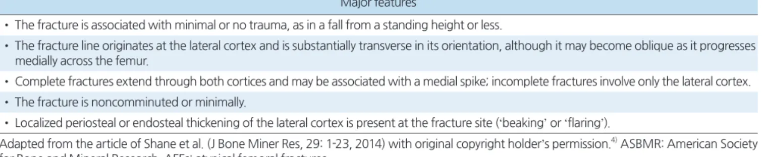
Table 1. ASBMR Task Force 2013 Case Definition of AFFs Baseline4)
Major features
• The fracture is associated with minimal or no trauma, as in a fall from a standing height or less.
• The fracture line originates at the lateral cortex and is substantially transverse in its orientation, although it may become oblique as it progresses medially across the femur.
• Complete fractures extend through both cortices and may be associated with a medial spike; incomplete fractures involve only the lateral cortex.
• The fracture is noncomminuted or minimally.
• Localized periosteal or endosteal thickening of the lateral cortex is present at the fracture site (‘beaking’ or ‘flaring’).
Adapted from the article of Shane et al. (J Bone Miner Res, 29: 1-23, 2014) with original copyright holder’s permission.4) ASBMR: American Society for Bone and Mineral Research, AFFs: atypical femoral fractures.
BPs have been used in all registered patients, with an aver- age period of 4.7±2.4 years (range, 0.3-10 years). Alendro- nate, was prescribed to 14 patients (63.6%), risedronate to 3 patients (13.6%), and ibandronate to 5 patients (22.7%).
Five patients took drug holidays from BPs for at least 1 year due to a prolonged use. Prodromal symptoms developed in 16 of the 27 fractures (59.3%). The mean follow-up period was 26.4±15.8 months (range, 12-66 months). There were 11 cases with fractures on the right side and 16 cases with fractures on the left side.
This is a retrospective case-control study in Jeju National University Hospital. We obtained approval from institu- tional review board of Jeju National University Hospital (IRB No. 2019-09-001) and the informed consent was waived.
2. Surgical technique and implant
Every operation was performed under aseptic condi- tions in accordance with the established procedure speci- fied in the appropriate surgical technique manual. Closed reduction was carried out on the fracture table for traction, and quality of reduction was confirmed by fluoroscopy. If adequate reduction could not be achieved by such method, bone hooks or long forceps were used additionally to aid anatomic reduction. The entry point of the nail on the
antero-posterior (AP) and lateral (LAT) views were the tip and midpoint of the greater trochanter, respectively. While continually checking the aforementioned fluoroscopic views, lag screws and blades were inserted into the center of the femoral head. Every single procedure was performed by a single experienced surgeon (K.W.N.).
The implants used in the surgery were Proximal Femoral Nail Antirotation II, Expert Asian Femoral Nail (PFNA- II, A2FN; DePuy Synthes, Oberdorf, Switzerland) and long ITST nail (Intertrochanteric/Subtrochanteric Fixation System; Zimmer, Warsaw, IN, USA). In this study, the sizes of implant were classified as long and short on the basis of 240 mm. Sitting was allowed starting first postoperative day while wheelchair and partial weight bearing was begun be- tween the third and the seventh postoperative days depend- ing on the degree of reduction, systemic condition and pain.
Weight bearing was gradually increased according to the extent of fracture union on radiography.
3. Outcomes measurements
Cortical thickening, femoral bowing angle, fracture re- duction status and bone union were evaluated for all par- ticipating patients on plain radiographs at various points:
immediately after the surgery, postoperative 6th week,
A B C D
Fig. 1. (A) Atypical subtrochanteric frac- ture in the right femur, antero-posterior (AP) view. Endosteal thickening of the lateral cortex at the fracture site is de- picted by a white arrow. (B) Atypical sub- trochanteric fracture in the left femur, AP view. Endosteal thickening of the lateral cortex at the fracture site is depicted by a white arrow. (C) Atypical subtrochanteric fracture in the right femur, AP view. The lateral cortex thickness at the fracture site does not differ from the distal part (white arrowhead). (D) Atypical subtrochanteric fracture in the left femur AP view. Peri- osteal thickening of the lateral cortex is observed, but endosteal cortical thicken- ing is not observed (white arrowhead).
postoperative 3rd, 6th and 12th months, and annually henceforward. Endosteal cortical thickening is defined as the increased cortical thickness of the fracture line just distal to the fracture site and formation of endosteal callus, seen as
“beaking” or “flaring”4) in the medial margin of the lateral cortex (Fig. 1).15) In this study, patients without endosteal cortical thickening were classified as group I, and patients observed to have endosteal cortical thickening in the frac- ture site were classified as group II.
The method described by Yau et al.16) was used to mea- sure femoral bowing angles. According to this method, femoral diaphysis was divided into 4 equal parts at the coronal and sagittal planes. A line that best described the midpoint of the endosteal canal was drawn in each quarter.
The overall coronal and sagittal femoral bowing were mea- sured the angle between the proximal and distal quarters of the femoral diaphysis in the AP and LAT views, respectively (Fig. 2).
Radiologic union is defined as bridging of the fracture site by callus or bone at a minimum of three cortices. Cortical healing is assessed in four anatomic femur regions (medial, lateral anterior and posterior) using AP and LAT views of plain radiography (Fig. 3).17,18) Delayed union was defined as lack of evidence of fracture site union even 6 months post- operatively.19) The reduction status was evaluated by com- paring the plain radiographs immediately after surgery with contralateral side. A varus reduction was defined as angula- tion of more than 10° at the fracture site in the subtrochan- teric area (Fig. 4).20,21) Angular deformity less than 10° at the site of fracture was defined as acceptable reduction.
Radiological assessment was carried out by two observ- ers (Y.H.R. and K.W.N.), and the mean values were calcu- lated. Evaluation and measurement of the plain radiographs were done using a picture archiving and communication
A B
Fig. 2. (A) Coronal plane femoral bowing was measured as the angula- tion between the proximal and distal quarters of the femoral diaphysis in the femur antero-posterior view. (B) Sagittal plane femoral bowing was measured as the angulation between the proximal and distal quarters of the femoral diaphysis in the femur lateral view.
A B C D
Fig. 3. (A, B) Atypical subtrochan- teric fracture in the left femur, antero- posterior (AP) and lateral (LAT) views.
Shortening, varus deformation, and total displacement of fracture were observed.
Endosteal thickening of the lateral cortex at the fracture site is depicted by a white arrowhead. (C, D) These are left femur AP and LAT views taken nine months af- ter surgery. The atypical subtrochanteric fracture was fixed with a long cephalo- medullary nail and cerclage cable. Callus formation was observed at four cortical regions (medial, lateral, anterior and pos- terior cortex) in the fracture site marked with white arrows.
system (PACS) (INFINITT; INFINITT Healthcare, Seoul, Korea).
4. Statistical analysis
The continuous variables between the group I and the group II were analyzed using the Student t-test and Mann–
Whitney test. In contrast, the chi-square test and Fisher’s exact test were used for categorical variable analysis. Fur- thermore, Fisher’s exact test and linear by linear association with 95% confidence intervals were used in the univariate analysis to assess the individual effects of nonunion. A mul- tiple logistic regression analysis was performed to identify the independent nonunion predictors. Cox proportional hazard model was used as a survival analysis for the union differences between the two groups with an end point of nonunion. Statistical analysis was performed using the IBM SPSS Statistics (ver. 20.0; IBM Co., Armonk, NY, USA).
Statistical significance was defined as a p-value <0.05.
Results
There were 8 cases in group I and 19 cases in group II.
Baseline characteristics such as age, height, weight, BMI,
BMD, duration of BPs, presence of BPs holiday, prodromal symptoms and follow-up period did not differ significantly between the two groups (Table 2).
A B C D
Fig. 4. (A) Atypical subtrochanteric frac- ture in the right femur, antero-posterior (AP) view. (B) This is right femur AP view taken immediately after surgery. The atypical subtrochanteric fracture was fixed with a long cephalo-medullary nail.
Com pared to the contralateral side, 13 varus reduction was observed (asterisk).
(C) This is the right femur AP view taken 12 months after surgery. Breakage of the distal interlocking screw (white ar- row) and nonunion of the fracture are ob served. (D) This is the right femur AP view after performing re-operation using a plate.
Table 2. Baseline Characteristics Included in This Study
Variable Group I (n=8) Group II (n=19) p-value
Age (yr) 68.0±9.2 65.4±5.6 0.367
Height (cm) 157.3±4.8 153.1±5.6 0.098
Weight (kg) 58.6±5.3 55.7±8.1 0.368
BMI (kg/m2) 23.9±3.2 24.6±3.4 0.621
BMD (T-score) –2.3±0.9 –2.2±1.2 0.952
ASA classification 0.506
1 4 (50.0) 11 (57.9)
2 2 (25.0) 6 (31.6)
3 2 (25.0) 2 (10.5)
Duration of BPs (yr) 5.4±3.1 4.5±2.0 0.375
Presence of drug holiday 0.558
Yes 1 (12.5) 4 (21.1)
No 7 (87.5) 15 (78.9)
Prodromal symptoms 0.675
Yes 4 (50.0) 12 (63.2)
No 4 (50.0) 7 (36.8)
Follow-up (yr) 2.0±1.2 2.3±1.4 0.616
Side (right:left) 2:6 (25:75) 9:10 (47:53) 0.280 Values are presented as mean±standard deviation or number (%).
BMI: body mass index, BMD: bone mineral density, ASA: American Society of Anesthesiologists, BPs: bisphosphonates.
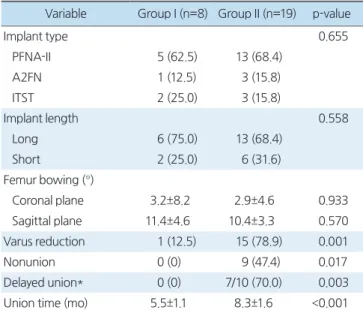
Implant type (p=0.655) and length (p=0.558) did not differ significantly between the two groups. The coronal plane femoral bowing angle was 3.2°±8.2° in group I and 2.9°±4.6° in group II (p=0.933). The sagittal plane femoral bowing angle was 11.4°±4.6° in group I and 10.4°±3.3°
in group II (p=0.570). Varus reduction occurred in total 16 cases, with 1 patient in group I and 15 cases in group II. Varus reduction occurred significantly more in group II (Group I: 12.5% vs. Group II: 78.9%; p<0.001), and they all showed more than 10° at the fracture site. Nonunion occurred in total 9 cases, all of them were group II and this result statistically significant (Group I: 0% vs. Group II:
47.4%; p=0.017). All patients with nonunion were con- tinuously observed in postoperative radiographs. Revision surgery was performed on all the patients. Delayed union occurred in total of 7 group II patients, and this result also statistically significant (Group I: 0% vs. Group II: 70.0%;
p=0.003). The mean union time was observed to be 5.5±
1.1 months in group I and 8.3±1.6 months in group II. The bone healing period was 2.8 months longer on average in group II and it was statistically significant (p<0.001) (Table 3).
Univariate analyses were used to assess the individual effects of nonunion in all patients. A total of nine non- unions occurred (33.3%). Age (p=0.231), BMI (p=0.153), BMD (p=0.143), ASA classification (p=0.532), duration of BPs (p=0.854), presence of BPs holiday (p=0.675), pro- dromal symptoms (p=0.551), implant type (p=0.865), im- plant length (p=0.550), and femur bowing (coronal plane;
p=0.626, sagittal plane; p=0.925) did not have statistically significant associations. However, statistically significant differences were observed for endosteal cortical thickening (p=0.026) (Table 4).
Table 3. Intra- and Postoperative Variables
Variable Group I (n=8) Group II (n=19) p-value
Implant type 0.655
PFNA-II 5 (62.5) 13 (68.4)
A2FN 1 (12.5) 3 (15.8)
ITST 2 (25.0) 3 (15.8)
Implant length 0.558
Long 6 (75.0) 13 (68.4)
Short 2 (25.0) 6 (31.6)
Femur bowing (°)
Coronal plane 3.2±8.2 2.9±4.6 0.933
Sagittal plane 11.4±4.6 10.4±3.3 0.570
Varus reduction 1 (12.5) 15 (78.9) 0.001
Nonunion 0 (0) 9 (47.4) 0.017
Delayed union* 0 (0) 7/10 (70.0) 0.003
Union time (mo) 5.5±1.1 8.3±1.6 <0.001
Values are presented as mean±standard deviation or number (%).
*This subject was presented as a percentage of patients who were finally bone healing. PFNA-II: Proximal Femoral Nail Antirotation II, A2FN: Expert Asian Femoral Nail, ITST: Intertrochanteric/Subtrochan- teric.
Table 4. Univariate Comparison between the Union and Nonunion Groups
Variable Union (n=18) Nonunion (n=9) p-value
Age (yr) 63.7±7.0 69.1±6.6 0.231
BMI (kg/m2) 22.9±3.4 25.2±3.1 0.153
BMD (T-score) –2.0±0.8 –2.7±1.5 0.143
ASA classification 0.532
1 10 (55.6) 5 (55.6)
2 5 (27.8) 3 (33.3)
3 3 (16.7) 1 (11.1)
Duration of BPs (yr) 4.7±2.6 4.9±2.0 0.854
Presence of drug holiday 0.675
Yes 3 (16.7) 2 (22.2)
No 15 (83.3) 7 (77.8)
Prodromal symptoms 0.551
Yes 11 (61.1) 5 (55.6)
No 7 (38.9) 4 (44.4)
Cortical thickening 0.026
Yes 10 (55.6) 9 (100)
No 8 (44.4) 0 (0)
Implant type 0.865
PFNA-II 12 (66.7) 6 (66.7)
A2FN 3 (16.7) 1 (11.1)
ITST 3 (16.7) 2 (22.2)
Implant length 0.550
Long 13 (72.2) 6 (66.7)
Short 5 (27.8) 3 (33.3)
Femur bowing (°)
Coronal plane 3.4±6.3 2.1±4.7 0.626
Sagittal plane 10.7±4.7 10.8±2.9 0.925
Values are presented as mean±standard deviation or number (%).
BMI: body mass index, BMD: bone mineral density, ASA: American Society of Anesthesiologists, BPs: bisphosphonates, PFNA-II: Proximal Femoral Nail Antirotation II, A2FN: Expert Asian Femoral Nail, ITST:
Intertrochanteric/Subtrochanteric.
Multiple logistic regression analyses performed to iden- tify independent factors associated with nonunion with variables found that endosteal cortical thickening was the independent predictor of nonunion (odds ratio: 7.198; 95%
confidence interval: 1.718-30.154; p=0.007) (Table 5).
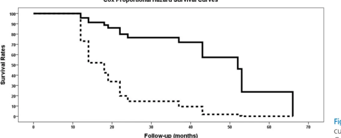
Cox proportional hazard model shows the difference in survival curves between two groups for the nonunion (p=0.025) (Fig. 5).
Discussion
Increased bone turnover and insufficient bone heal- ing due to long-term BP use are the keys to understanding the pathogenesis of AFFs.3) Repeated micro-damage to the lateral aspect of the femur, where withstands the maximal tensile stress, also plays an important role. In a physiologic femur model, Koch demonstrated how the highest tensile stress falls upon on the lateral cortex in the subtrochanteric region of femur.22) These features of the subtrochanteric AFFs cause poor union rates and numerous postoperative complications.
The average bone union time of subtrochanteric AFFs is reported to be more than 8 months,4,23,24) longer than that of the typical femoral fractures at an average of 3 to 6 months.25,26) Prasarn et al.27) has analyzed factors affect- ing healing time in subtrochanteric AFFs and demonstrated a 44% fracture healing complication rates in his series of subtrochanteric AFFs, but most of those complications were treated with plate fixation. In addition, a systematic review by Koh et al.2) has demonstrated that there was a significantly greater need of revision surgery in plate fixa- tion group over intramedullary nailing group (31.3% vs.
12.9%). The current surgical intervention of choice that al- lows endochondral ossification for AFFs is intramedullary nailing.10,11) However, no specific intramedullary device was advocated in current literature for the treatment of sub- trochanteric AFFs. In our study, we used three models of intramedullary nails, which fixation the femoral head with single blade, single lag screw, and two lag screws. Spinelli et al.28) has demonstrated that in patients with osteoporosis and a high risk of fracture in femur neck and intertro- chanteric area, it may be advantageous to use a cephalo- medullary nail for prevention.
Cephalo-medullary nails allow for static and dynamic locking options on distal screws. Dynamically locked screws have the advantage of providing rotational stability while allowing for axial compression of the fracture. The dynamic screw position can also be used to enhance the formation of the callus if the fracture gap remains after nail insertion.
However, it has been proved that because the process of
Table 5. Logistic Regression Analysis of Nonunion
Variable B value 95% CI p-value
Age 0.956 0.885-1.033 0.257
BMI 0.789 0.611-1.018 0.068
BMD 0.813 0.753-1.125 0.192
Cortical thickening 7.198 1.718-30.154 0.007 CI: confidence interval, BMI: body mass index, BMD: bone mineral density.
Fig. 5. Cox proportional hazard survival curve (solid line: Group I, dotted line:
Group II).
callus formation takes longer for AFFs, it may be safer to select a static screw position. No large-scale studies have been conducted to demonstrate superiority between distal locking options, and there is no univocal consensus.29,30)
Yang et al.31) have analyzed other factors that can po- tentially affect healing time in AFFs. Subtrochanteric AFFs commonly have disproportionate medullary space and intramedullary device size when compared to AFFs that occur in the femoral diaphysis. This can aggravate rota- tional instability, and subtrochanteric AFFs are at a higher risk of delayed union and nonunion. In our study, the total nonunion rate was 33.3%, similar to the reported rate of revision surgery due to nonunion or implants failure in vari- ous studies.32-34) Subtrochanteric AFFs tend to show much higher rates of nonunion compared to two percent reported in typical femoral fractures.35)
Another possible variable for predicting a fixation failure of the subtrochanteric AFFs is the reduction status. Egol et al.13) reported that varus reduction of the fracture site re- quired an average of 3.7 months or more for bone union compared to that of acceptable reduction. In our study, the radiological union time for group I was average of 5.5 months, and for group II was 8.3 months, indicating simi- larity to that of the previous study. In addition, consider- ing that the varus reduction rate was significantly higher in group II, it is logical to assume that the long union period in group II was directly related to varus reduction. Lim et al.36) also reported the importance of acceptable reduction in the healing of AFFs. The reduction status on the coronal plane and the residual gap in the fracture area were im- portant factors causing delayed union and nonunion. They concluded that the remaining gap of more than 20% of the cortical thickness on the anterior and lateral sides of the fracture could cause problems with the bone union.
As demonstrated in this study, endosteal thickening of the lateral cortex in subtrochanteric AFFs is a factor that interferes with acceptable reduction and negatively affects surgical outcomes. However, there are not many studies that try to identify the factors affecting cortical thickness.32,33) It has been postulated that cortical thickening is due to long- term BPs use, but the study of Lenart et al.37) reported that
cortical thickening was observed in AFF patients who had never used BPs. In addition, Giusti et al.38) demonstrated that cortical thickness is not different in AFF patients with or without exposure to BP therapy and that cortical thick- ness does not increase with prolonged use of BPs.
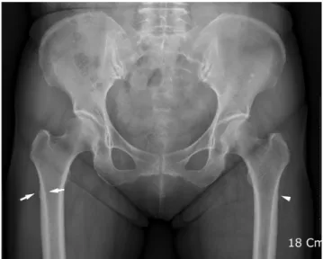
There was no mention in the literature about the direc- tion of thickening the cortex. There have also been no pre- vious studies on factors affecting endosteal cortical thicken- ing. However, our study results show that the direction of cortical thickness is closely related to the fracture reduction and surgical results. The interesting fact is that one of the patients in this study had a different direction in cortex thickening between the two sides (Fig. 6).
There are several studies that have investigated on fac- tors that promote fracture union and improve the results of surgery in subtrochanteric AFFs. The ASBMR recom- mended that the medullary canal should be over-reamed to a minimum diameter of at least 2.5 mm larger than the in- tramedullary nail diameter in order to prevent varus reduc- tion and compensate for the narrow medullary diameter.4) A retrospective study of Lovy et al.39) has reported that percu- taneous injections of bone marrow aspirate concentrate and demineralized bone matrix on fracture sites can be used to avoid delayed union and nonunion. Another study by Yeh
Fig. 6. Thickening of the lateral cortex in subtrochanteric regions is ob- served on both sides. On the right side, both endosteal and periosteal cortical thickening are observed (white arrows), whereas only perios- teal cortical thickening is observed on the left side (white arrowhead).
et al.40) demonstrated that teriparatide treatment in patients with subtrochanteric AFFs may help in fracture healing, hip function recovery, and pain relief.
The advantage of our research is that it is a single cen- ter research. All operations were performed by one skilled orthopedic surgeon. This will produce consistent results in surgery compared to studies involving multiple orthopedic surgeons in multi center research. It can also reduce the number of variables that can occur during the treatment period, thereby increasing the reliability of this study. An- other advantage of our research is use of inferential statisti- cal analysis to enhance the validity of the conclusions made.
However, there are some limitations that need to be acknowledged and addressed regarding this study. First, the main limitation of the current study is its retrospective design. There would be observer bias, including no stan- dardized quality and duration of follow-up, missing data, and inability to control confounding variables. Second, the follow-up period is relatively short, leading to an inability to represent long-term outcomes of subtrochanteric AFFs.
Third, due to the uncommon nature of these fractures, there is a relatively small study population. Due to such limita- tions, it was difficult to retain sufficient power for each variable used for analysis in this study; consequently, future study needs to compensate for such short comings to pro- vide more concrete correlations.
Conclusion
This study suggests that femoral endosteal thicken- ing of the lateral cortex in the fracture site is an obstacle in intramedullary nail fixation for subtrochanteric AFFs.
Endosteal callus acts as a ‘malpositioned poller screw’, in- terrupts reduction and contributes to varus deformation during nail advancement. As a result, it is necessary to make every effort to correct this as it is an independent factor in increasing the delayed union and nonunion. The only strict acceptable reduction is the key to successful management of subtrochanteric AFFs. As such, intentionally excessive over- reaming during operation is essential.
Acknowledgements
We thank Dr. Seung Jin Yoo for English-editing of the manuscript.
요 약
목적: 대부분의 전자하 비전형 대퇴골 골절은 골수강내 금속
정을 사용하여 치료하며 합병증으로 지연유합, 불유합 등이 보고되었다. 본 연구의 목적은 전자하 비전형 대퇴골 골절에 서 골절 부위의 외측 피질골의 내측 비후 여부에 따른 수술적 치료의 결과를 알아보고자 함이다.대상 및 방법: 2012년 3월부터 2014년 10월까지 비전형 전자
하 골절을 진단받은 환자 중 골수강내 금속정 고정술을 받은 환자를 대상으로 하였다. 평가항목으로는 인구학적 정보, 방 사선검사상 내반정복 및 유합, 비스포스포네이트 제제 사용 기간이 포함되었다.결과: 전체 27예에서 내측 비후가 없는 그룹 I과 있는 그룹 II
로 구분하였다. 두 군 간의 인구학적 정보의 차이는 없었으며 내반정복(그룹 I: 12.5% vs. 그룹 II: 78.9%; p=0.001), 지연 유합(그룹 I: 0% vs. 그룹 II: 70.0%; p=0.003), 불유합(그룹 I: 0% vs. 그룹 II: 47.4%; p=0.017), 유합시간(그룹 I: 5.5개월 vs. 그룹 II: 8.3개월; p<0.001)은 통계적으로 유의하게 차이 가 있었다.결론: 비전형 전자하 골절에서 외측 피질골의 내측 비후는 골
절의 정복을 방해하고 불유합을 유발하는 독립인자로 확인 되었다. 성공적인 수술결과를 얻기 위해 내측 비후를 제거하 고 해부학적 정복을 얻는 것이 중요하다.색인 단어: 대퇴골, 대퇴골 골절, 전자하부, 골수정, 불유합,
부전골절ORCID
노영호, https://orcid.org/0000-0002-0703-4970 강기문, https://orcid.org/0000-0003-0687-9637 김희중, https://orcid.org/0000-0002-3994-5672 남광우, https://orcid.org/0000-0003-1096-149X
References
1. Lloyd AA, Gludovatz B, Riedel C, et al: Atypical fracture with
long-term bisphosphonate therapy is associated with altered cortical composition and reduced fracture resistance. Proc Natl Acad Sci U S A, 114: 8722-8727, 2017.
2. Koh A, Guerado E, Giannoudis PV: Atypical femoral fractures related to bisphosphonate treatment: issues and controversies related to their surgical management. Bone Joint J, 99: 295-302, 2017.
3. Gedmintas L, Solomon DH, Kim SC: Bisphosphonates and risk of subtrochanteric, femoral shaft, and atypical femur fracture:
a systematic review and meta-analysis. J Bone Miner Res, 28:
1729-1737, 2013.
4. Shane E, Burr D, Abrahamsen B, et al: Atypical subtrochanteric and diaphyseal femoral fractures: second report of a task force of the American Society for Bone and Mineral Research. J Bone Miner Res, 29: 1-23, 2014.
5. Schilcher J, Sandberg O, Isaksson H, Aspenberg P: Histology of 8 atypical femoral fractures: remodeling but no healing. Acta Orthop, 85: 280-286, 2014.
6. Schilcher J: Epidemiology, radiology and histology of atypical femoral fractures: development of understanding. Acta Orthop, 84: 1-26, 2013.
7. Goh SK, Yang KY, Koh JS, et al: Subtrochanteric insufficiency fractures in patients on alendronate therapy: a caution. J Bone Joint Surg Br, 89: 349-353, 2007.
8. Koh JS, Goh SK, Png MA, Kwek EB, Howe TS: Femoral corti- cal stress lesions in long-term bisphosphonate therapy: a herald of impending fracture? J Orthop Trauma, 24: 75-81, 2010.
9. Neviaser AS, Lane JM, Lenart BA, Edobor-Osula F, Lorich DG: Low-energy femoral shaft fractures associated with alen- dronate use. J Orthop Trauma, 22: 346-350, 2008.
10. Balach T, Baldwin PC, Intravia J: Atypical femur fractures as- sociated with diphosphonate use. J Am Acad Orthop Surg, 23:
550-557, 2015.
11. Zheng N, Tang N, Qin L: Atypical femoral fractures and cur- rent management. J Orthop Translat, 7: 7-22, 2016.
12. Banffy MB, Vrahas MS, Ready JE, Abraham JA: Nonopera- tive versus prophylactic treatment of bisphosphonate-associated femoral stress fractures. Clin Orthop Relat Res, 469: 2028- 2034, 2011.
13. Egol KA, Park JH, Rosenberg ZS, Peck V, Tejwani NC: Healing delayed but generally reliable after bisphosphonate-associated complete femur fractures treated with IM nails. Clin Orthop Relat Res, 472: 2728-2734, 2014.
14. Owens WD, Felts JA, Spitznagel EL Jr: ASA physical status clas- sifications: a study of consistency of ratings. Anesthesiology, 49:
239-243, 1978.
15. LeBlanc ES, Rosales AG, Black DM, et al: Evaluating atypical features of femur fractures: how change in radiological criteria influenced incidence and demography of atypical femur fractures in a community setting. J Bone Miner Res, 32: 2304-2314, 2017.
16. Yau WP, Chiu KY, Tang WM, Ng TP: Coronal bowing of the femur and tibia in Chinese: its incidence and effects on total knee arthroplasty planning. J Orthop Surg (Hong Kong), 15: 32-36, 2007.
17. Corrales LA, Morshed S, Bhandari M, Miclau T 3rd: Variabil- ity in the assessment of fracture-healing in orthopaedic trauma studies. J Bone Joint Surg Am, 90: 1862-1868, 2008.
18. Morshed S: Current options for determining fracture union. Adv Med, 2014: 708574, 2014.
19. Hierholzer C, Sama D, Toro JB, Peterson M, Helfet DL: Plate fixation of ununited humeral shaft fractures: effect of type of bone graft on healing. J Bone Joint Surg Am, 88: 1442-1447, 2006.
20. Shukla S, Johnston P, Ahmad MA, Wynn-Jones H, Patel AD, Walton NP: Outcome of traumatic subtrochanteric femoral fractures fixed using cephalo-medullary nails. Injury, 38: 1286- 1293, 2007.
21. Kraemer WJ, Hearn TC, Powell JN, Mahomed N: Fixation of segmental subtrochanteric fractures. A biomechanical study. Clin Orthop Relat Res, (332): 71-79, 1996.
22. Koch JC: The laws of bone architecture. Am J Anat, 21: 177- 298, 1917.
23. Einhorn TA, Bogdan Y, Tornetta P 3rd: Bisphosphonate-asso- ciated fractures of the femur: pathophysiology and treatment. J Orthop Trauma, 28: 433-438, 2014.
24. Phillips HK, Harrison SJ, Akrawi H, Sidhom SA: Retrospective review of patients with atypical bisphosphonate related proximal femoral fractures. Injury, 48: 1159-1164, 2017.
25. Lee PC, Hsieh PH, Yu SW, Shiao CW, Kao HK, Wu CC: Bio- logic plating versus intramedullary nailing for comminuted sub- trochanteric fractures in young adults: a prospective, randomized study of 66 cases. J Trauma, 63: 1283-1291, 2007.
26. Oh CW, Kim JJ, Byun YS, et al: Minimally invasive plate osteo- synthesis of subtrochanteric femur fractures with a locking plate:
a prospective series of 20 fractures. Arch Orthop Trauma Surg, 129: 1659-1665, 2009.
27. Prasarn ML, Ahn J, Helfet DL, Lane JM, Lorich DG: Bisphos- phonate-associated femur fractures have high complication rates with operative fixation. Clin Orthop Relat Res, 470: 2295-2301, 2012.
28. Spinelli MS, Marini E, Daolio PA, Piccioli A: Atypical diaphyseal femoral fractures: considerations on surgical technique. Injury, 50 Suppl 2: S65-S69, 2019.
29. Errani C, Mavrogenis AF, Cevolani L, et al: Treatment for long bone metastases based on a systematic literature review. Eur J Orthop Surg Traumatol, 27: 205-211, 2017.
30. Piccioli A, Maccauro G, Spinelli MS, Biagini R, Rossi B: Bone metastases of unknown origin: epidemiology and principles of management. J Orthop Traumatol, 16: 81-86, 2015.
31. Yang KH, Kim JR, Park J: Nonisthmal femoral shaft nonunion
as a risk factor for exchange nailing failure. J Trauma Acute Care Surg, 72: E60-E64, 2012.
32. Bogdan Y, Tornetta P 3rd, Einhorn TA, et al: Healing time and complications in operatively treated atypical femur fractures as- sociated with bisphosphonate use: a multicenter retrospective cohort. J Orthop Trauma, 30: 177-181, 2016.
33. Weil YA, Rivkin G, Safran O, Liebergall M, Foldes AJ: The out- come of surgically treated femur fractures associated with long- term bisphosphonate use. J Trauma, 71: 186-190, 2011.
34. Teo BJ, Koh JS, Goh SK, Png MA, Chua DT, Howe TS: Post- operative outcomes of atypical femoral subtrochanteric fracture in patients on bisphosphonate therapy. Bone Joint J, 96-B: 658- 664, 2014.
35. Shroeder JE, Mosheiff R, Khoury A, Liebergall M, Weil YA:
The outcome of closed, intramedullary exchange nailing with reamed insertion in the treatment of femoral shaft nonunions. J Orthop Trauma, 23: 653-657, 2009.
36. Lim HS, Kim CK, Park YS, Moon YW, Lim SJ, Kim SM: Fac- tors associated with increased healing time in complete femoral
fractures after long-term bisphosphonate therapy. J Bone Joint Surg Am, 98: 1978-1987, 2016.
37. Lenart BA, Neviaser AS, Lyman S, et al: Association of low- energy femoral fractures with prolonged bisphosphonate use: a case control study. Osteoporos Int, 20: 1353-1362, 2009.
38. Giusti A, Hamdy NA, Dekkers OM, Ramautar SR, Dijkstra S, Papapoulos SE: Atypical fractures and bisphosphonate therapy:
a cohort study of patients with femoral fracture with radio- graphic adjudication of fracture site and features. Bone, 48: 966- 971, 2011.
39. Lovy AJ, Kim JS, Di Capua J, et al: Intramedullary nail fixation of atypical femur fractures with bone marrow aspirate con- centrate leads to faster union: a case-control study. J Orthop Trauma, 31: 358-362, 2017.
40. Yeh WL, Su CY, Chang CW, et al: Surgical outcome of atypical subtrochanteric and femoral fracture related to bisphosphonates use in osteoporotic patients with or without teriparatide treat- ment. BMC Musculoskelet Disord, 18: 527, 2017.