서론
녹내장은 전 세계적으로 실명을 초래하는 주요한 원인 중 하나이 며, 그중 급성원발폐쇄각 녹내장은 아시아인에게서 많이 발생하는 녹내장이다.1-5 전방각의 폐쇄는 질환의 진행 정도에 따라 홍채섬유 주접촉이 절반 이상에서 발생한 급성원발폐쇄각의증, 급성원발폐 쇄각의증에 안압상승 또는 주변부홍채유착이 동반된 급성원발폐
쇄각, 급성원발폐쇄각에 시신경손상이 동반된 급성원발폐쇄각 녹 내장으로 정의할 수 있다.6 본 연구에서는 급성원발폐쇄각을 대상 으로 연구를 진행하였다. 급성원발폐쇄각은 갑작스러운 안압의 상 승과 함께 각막부종, 얕은 전방깊이, 심한 안구 통증, 두통, 오심, 구 토를 동반한다. 급성원발폐쇄각이 잘 발생하는 눈은 좁은 전방각 을 포함하여 얕은 전방깊이, 두꺼운 수정체두께, 작은 각막직경, 짧 은 안축장을 특징으로 한다.7,8 최근 이러한 알려진 특징들과 다른 안축장이 긴 환자에게서 급성원발폐쇄각 발생률이 증가하였고, 이는 세계적으로 많은 관심을 받아왔다.9 앞으로 근시 환자 수가 더 증가함에 따라서 안축장이 긴 환자에서 급성원발폐쇄각의 발 생률은 더 높아질 것으로 예측되며 그 중요성은 더욱 증가될 것으 로 생각된다. 급성원발폐쇄각 발생 기전과, 안축장이 긴 환자에게 서 발생한 급성원발폐쇄각의 기전을 이해하기 위하여 본 연구는
급성원발폐쇄각 환자에서 안축장과 안구 해부학적 지표의 관련성
Correlation between Axial Length and Ocular Anatomical Parameters in Acute Primary Angle Closure
남정우, 성미선, 박상우
Jeoungwoo Nam, MD, Mi Sun Sung, MD, PhD, Sang Woo Park, MD, PhD
전남대학교 의과대학 안과학교실
Department of Ophthalmology, Chonnam National University Medical School, Gwangju, Korea
Purpose: To investigate the correlation between the axial length and ocular anatomical parameters in patientswith acute primary angle closure (APAC).
Methods: A total of 214 eyes from 214 patients with APAC were included in this study. Anterior segment parameters such as anterior chamber depth, anterior chamber width (ACW), lens vault, iris thickness 750, iris curvature, central corneal thickness, white-to-white, lens thickness, lens position, keratometry (K), angle opening distance 500, angle opening distance 750, trabecular iris space area 500, trabecular iris space area 750 and axial length (AL) were retrospectively obtained by use of anterior-segment optical coherence tomography and optical low-coherence reflectometry. Eyes were classified into 3 groups on the basis of AL; eyes with axial length of more than 23 mm (Group A), between 22 and 23 mm (Group B), less than 22.0 mm (Group C). Ocular anatomical parameters were compared among groups. In addition, Between the group of AL more than 24 mm (Group D) and the control group, ocular anatomical parameter were compared. Correlations between AL and ocular anatomical parameters were also investigated using Pearson correlation analysis.
Results: Among the three groups, the anterior width (11.54 ± 0.38 mm vs. 11.28 ± 0.42 mm vs 11.03 ± 0.38 mm) and corneal curvature (43.25 ± 1.29 diopters [D] vs. 44.50 ± 1.25 D vs 45.81 ± 1.22 D), were showed significant differences (p < 0.001). There were no significant differences in other ocular anatomical parameters among the three groups. Significant correlations were only found between axial length and ACW (r = 0.47, p < 0.01) and K (r = -0.64, p < 0.01).
Conclusions: Even increasing AL, ocular anatomical parameters did not show a strong relationship with AL except ACW and K. These anatomical features may explain the prevalence of APAC in eyes with longer AL.
Key words: Body mass index; Mean ocular perfusion pressure; Metabolic syndrome
Received: 2020. 3. 11. Revised: 2020. 5. 20.
Accepted: 2020. 6. 23.
Corresponding Author: Sang Woo Park, MD, PhD
Department of Ophthalmology, Chonnam National University Medi- cal School, #42 Jebong-ro, Dong-gu, Gwangju 61469, Korea Tel: +82-62-220-6742, Fax: +82-62-227-1642
E-mail: [email protected]
정상인과 급성폐쇄각 녹내장 환자의 안구 해부학적 지표를 비교하 였고, 급성원발폐쇄각 환자들을 안축장에 따라 세 군으로 나누어 안구 해부학적 지표의 차이를 비교하였다.
대상과 방법
2010년도 1월부터 2019년도 5월까지 전남대학교병원 안과에 내원하여 급성원발폐쇄각으로 진단된 환자 214명의 의무기록 을 후향적으로 검토하였다. 본 연구는 본원의 임상시험윤리위원 회 (Institutional Review Board, IRB)의 승인을 받았다(승인 번 호: CNUH-2018-256). 급성원발폐쇄각은 전방각의 폐쇄, 안압이 21 mmHg 이상, 심한 안통과 두통, 오심과 구토 그리고 시력저하 를 보이며, 백내장 이외에 포도막염이나 당뇨망막병증 같은 병적 인 선행 요인이 없는 경우로 정의하였다.
모든 환자에 대하여 세극등현미경검사, 골드만압평안압계에 의한 안압측정, 저간섭성 반사계(Lenstar LS900®, Haag-Streit, Mason, OH, USA), 전안부 빛간섭단층촬영(Visante® OCT, Carl ZeissMeditec, Dublin, CA, USA)을 시행하였고, 안축장(axial length, AL)에 따라서 A군(AL≥23 mm), B군(23 mm>AL≥22 mm), C군(22 mm>AL)으로 나누었다. 또한, 안축장이 정상인 평 균값보다 긴 D군(AL≥24 mm)을 추가로 설정하였고, 이 군과 안 축장, 나이, 성별이 비슷한 정상안을 대조군으로 설정하여 두 군 에서 해부학적 지표를 비교하였다.10
연구에 참여한 모든 환자는 주변부 레이저홍채절개술 또는 백 내장 수술 전에 검사를 받았다. 각막부종으로 인하여 검사의 질 에 문제가 있는 경우 제외하였고, 양안이 모두 기준에 맞아 연
구에 포함될 수 있는 경우에는 우안만 포함시켰다. 저간섭성 반 사계(Lenstar®) 및 전안부 빛간섭단층촬영(Visante®)을 시행하 여 전방폭(anterior chamber width, ACW), 전방깊이(anterior chamberdepth, ACD), 수정체원개(lens vault, LV), 홍채두께(iris thickness 750 μm, IT 750), 홍채곡률(iris curvature, Icurve), 안 축장(AL), 중심각막두께(central corneal thickness, CCT), 수 정체두께(lens thickness, LT), 각막곡률(keratometry, K), 각막 크기(white to white, WTW), 수정체 위치(lens position, LP = ACD +0.5LT) 그리고 전방각 계측치로 angle opening distance at 500 μm (AOD 500), angleopening distance at 750 μm (AOD 750), trabecular-iris space area at 500 μm (TISA 500), trabecu- lar-iris space area at 750 μm (TISA 750)를 측정하였다. 공막극 은 전방내 계측치의 객관적인 측정을 위한 기준점으로 쓰이는 중 요한 해부학적 구조이다. 공막극은 고리 모양의 아교질 섬유로 구 성되어 각막윤부와 평행하게 주행하며 전안부 빛간섭단층촬영에 서 공막과 비슷한 정도의 반사율을 보이는 고반사율의 구조로서, 전방으로 약간 튀어나와 있는 부분과 섬유주의 후방경계 사이로 정의된다.11 전방폭(ACW)은 공막극 사이의 거리, 전방깊이(ACD) 는 각막내측표면으로부터 수정체의 앞쪽 표면까지의 거리, 수정 체원개(LV)는 공막극끼리 이은 선에서 수정체 앞쪽 표면까지의 수직 거리로 지정하였다(Fig. 1). 홍채두께(IT 750)는 공막극에서 750 μm 떨어진 홍채에서 측정한 두께로 코쪽과 귀쪽 측정치의 평균값으로 정하였고, 홍채곡률(Icurve)은 홍채동공연의 안쪽 부분과 홍채뿌리를 잇는 가상의 선으로부터 홍채안쪽 부분의 최 대거리로 코쪽과 귀쪽 측정치의 평균값으로 정하였다. AOD 500 와 AOD 750은 공막극에서 각각 500 μm와 750 μm 떨어진 각막
Figure 1. Optical coherence tomography image (Visante® OCT) illustrating anterior chamber depth (ACD), lens vault (LV), anterior cham- ber width (ACW) and sclera spur.
내피면에서 수직으로 홍채를 향해 이은 가상의 직선의 길이로, TISA 500와 TISA 750은 공막극에서 각각 500 μm와 750 μm 떨
어진 각막 내피면에서 수직으로 홍채를 향해 이은 가상의 직선과 공막극에서 수직으로 홍채를 향해 이은 가상의 직선, 각막내피, 섬유주, 홍채에 의해 둘러싸인 공간으로 정의하였다(Fig. 2).12,13
통계적 검정은 SPSS 18 통계 프로그램(SPSS Inc., Chicago, IL, USA)을 사용하였다. 통계적 분석은 안축장이 23 mm 이상인 군(Group A), 안축장이 23 mm 미만 그리고 22 mm 이상인 군 (Group B), 안축장이 22 mm 미만인 군(Group C) 사이의 안구 해부학적 지표 측정값들을 비교하기 위해 one-way analysis of variance를 이용하였고, Bonferroni method를 이용하여 사후분 석을 시행하였다. p-value<0.05인 경우(Bonferroni’s method의 경우 p<0.05/3)를 통계학적으로 유의한 것으로 간주하였다. 안축 장이 24 mm 이상인 그룹(Group D)과 대조군 사이의 안구 해부 학적 지표 측정값들을 비교하기 위해 Mann Whitney U-test, 안 축장에 따른 안구 해부학적 지표 측정값의 상관관계를 보기 위하 여 Pearson correlation coefficient를 사용하였다. p값이 0.05 미 만인 경우를 통계학적으로 유의한 것으로 간주하였다.
Figure 2. Optical coherence tomography image (Visante® OCT) cross-sectional view through the anterior chamber angle region.
Anterior chamber parameters such as angle opening distance at 500 μm (AOD 500), AOD 750, trabecular-iris space area at 500 μm (TISA 500), TISA 750, iris thickness 750 μm (IT 750), iris cur- vature (Icurve) are manually measured. SS = scleral spur.
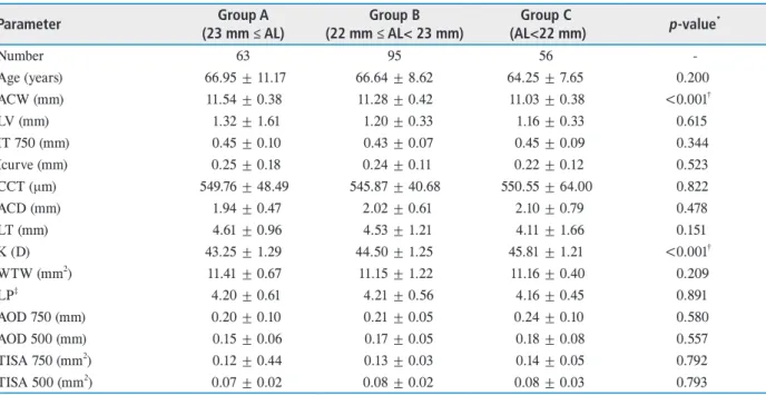
Table 1. Comparison of ocular anatomical parameters in acute primary angle closure among three AL groups
Parameter Group A
(23 mm ≤ AL)
Group B (22 mm ≤ AL< 23 mm)
Group C
(AL<22 mm) p-value*
Number 63 95 56 -
Age (years) 66.95 ± 11.17 66.64 ± 8.62 64.25 ± 7.65 0.200
ACW (mm) 11.54 ± 0.38 11.28 ± 0.42 11.03 ± 0.38 <0.001†
LV (mm) 1.32 ± 1.61 1.20 ± 0.33 1.16 ± 0.33 0.615
IT 750 (mm) 0.45 ± 0.10 0.43 ± 0.07 0.45 ± 0.09 0.344
Icurve (mm) 0.25 ± 0.18 0.24 ± 0.11 0.22 ± 0.12 0.523
CCT (μm) 549.76 ± 48.49 545.87 ± 40.68 550.55 ± 64.00 0.822
ACD (mm) 1.94 ± 0.47 2.02 ± 0.61 2.10 ± 0.79 0.478
LT (mm) 4.61 ± 0.96 4.53 ± 1.21 4.11 ± 1.66 0.151
K (D) 43.25 ± 1.29 44.50 ± 1.25 45.81 ± 1.21 <0.001†
WTW (mm2) 11.41 ± 0.67 11.15 ± 1.22 11.16 ± 0.40 0.209
LP‡ 4.20 ± 0.61 4.21 ± 0.56 4.16 ± 0.45 0.891
AOD 750 (mm) 0.20 ± 0.10 0.21 ± 0.05 0.24 ± 0.10 0.580
AOD 500 (mm) 0.15 ± 0.06 0.17 ± 0.05 0.18 ± 0.08 0.557
TISA 750 (mm2) 0.12 ± 0.44 0.13 ± 0.03 0.14 ± 0.05 0.792
TISA 500 (mm2) 0.07 ± 0.02 0.08 ± 0.02 0.08 ± 0.03 0.793
Values are presented as mean ± standard deviation.
AL = axial length; ACW = anterior chamber width; LV = lens vault; IT 750 = iris thickness 750 μm; Icurve = iris curvature; CCT
= central corneal thickness; ACD = anterior chamber depth; LT = lens thickness; K = keratometry; D = diopters; WTW = white to white; LP = lens position; AOD 750 = angle opening distance at 750 μm; AOD 500 = angle opening distance at 500 μm; TISA 750 = trabecular-iris space area at 750 μm; TISA 500 = trabecular-iris space area at 500 μm.
*By one way analysis of variance among the all groups (Group A vs. Group B vs. Group C); †p < 0.05; ‡ACD + 1/2 LT.
결과
총 214명의 급성원발폐쇄각 환자가 연구에 포함되었고, 안축장 에 따라서 세 군(Group A 63명, Group B 95명, Group C 56명) 으로 나누었다. 세 군(Group A vs. Group B vs. Group C)의 평 균 연령은 66.95 ± 11.17세 vs. 66.64 ± 8.62세 vs. 64.25 ± 7.65세 로 유의한 차이는 없었다. 안구 해부학적 지표 중 전방폭(ACW, 11.54 ± 0.38 mm vs. 11.28 ± 0.42 mm vs. 11.03 ± 0.38 mm) 과 각막곡률(K, 43.25 ± 1.29 diopters [D] vs. 44.50 ± 1.25 D vs. 45.81 ± 1.22 D)은 유의미한 차이(p<0.001)가 있었다. 그 외 의 전방깊이(ACD), 수정체원개(LV), 홍채두께(IT 750), 홍채곡 률(Icurve), 중심각막두께(CCT), 수정체두께(LT), 각막 크기 (WTW), 수정체위치(LP), 전방각계측치(AOD 750, AOD 500, TISA 750, TISA 500)는 세 군에서 유의한 차이를 보이지 않았다 (Table 1).
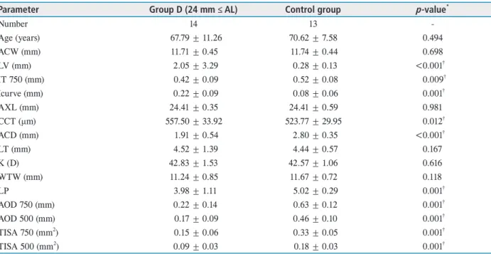
안축장이 24 mm 이상인 군(Group D) 14안과 정상 대조군 13안 사이의 해부학적 지표를 비교해 보았을 때(Group D vs.
control group) 수정체원개(LV, 2.05 ± 3.29 mm vs. 0.28 ± 0.13 mm, p<0.001), 홍채두께(IT 750, 0.42 ± 0.99 mm vs. 0.52 ±
0.08 mm, p=0.009), 홍채곡률(Icurve, 0.22 ± 0.09 mm vs. 0.08
± 0.06 mm, p=0.001), 중심각막두께(CCT, 557.50 ± 33.92 mm vs. 523.77 ± 29.95 mm, p=0.012), 전방깊이(ACD, 1.91 ± 0.54 mm vs. 2.80 ± 0.35 mm, p<0.001), 수정체 위치(LP, 3.98 ± 1.11 vs. 5.02 ± 0.29, p=0.001), 전방각계측치 중 AOD 750는 0.22 ± 0.14 mm vs. 0.63 ± 0.12 mm (p=0.001), AOD 500는 0.17 ± 0.09 mm. vs 0.46 ± 0.10 mm (p=0.001), TISA 750는 0.15 ± 0.06 mm vs. 0.33 ± 0.05 mm (p=0.001), TISA 500는 0.09 ± 0.03 mm vs.
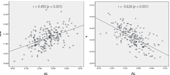
0.18 ± 0.03 mm (p=0.001)에서 유의한 차이를 보였다. 그 외의 전 방폭(ACW), 수정체두께(LT), 각막곡률(K), 각막 크기(WTW) 는 유의한 차이를 보이지 않았다(Table 2). 연구에 참여한 환자 모두를 대상으로 안축장(AL)과 안구 해부학적 지표의 상관관계 를 보았을 때 전방폭(ACW, r=0.474)과 각막곡률(K, r=-0.638)은 유의한 상관관계(p<0.001)를 보였으며(Fig. 3), 그 외의 안구 해부 학적 지표는 유의한 상관관계를 보이지 않았다(Table 3).
고찰
이전 연구에서 급성원발폐쇄각 환자의 해부학적 특징으로 짧은
Table 2. Comparison of ocular anatomical parameters between acute primary angle closure and control group
Parameter Group D (24 mm ≤ AL) Control group p-value*
Number 14 13 -
Age (years) 67.79 ± 11.26 70.62 ± 7.58 0.494
ACW (mm) 11.71 ± 0.45 11.74 ± 0.44 0.698
LV (mm) 2.05 ± 3.29 0.28 ± 0.13 <0.001†
IT 750 (mm) 0.42 ± 0.09 0.52 ± 0.08 0.009†
Icurve (mm) 0.22 ± 0.09 0.08 ± 0.06 0.001†
AXL (mm) 24.41 ± 0.35 24.41 ± 0.59 0.981
CCT (μm) 557.50 ± 33.92 523.77 ± 29.95 0.012†
ACD (mm) 1.91 ± 0.54 2.80 ± 0.35 <0.001†
LT (mm) 4.52 ± 1.39 4.44 ± 0.57 0.167
K (D) 42.83 ± 1.53 42.57 ± 1.06 0.616
WTW (mm) 11.24 ± 0.85 11.67 ± 0.72 0.118
LP 3.98 ± 1.11 5.02 ± 0.29 0.001†
AOD 750 (mm) 0.22 ± 0.14 0.63 ± 0.12 0.001†
AOD 500 (mm) 0.17 ± 0.09 0.46 ± 0.10 0.001†
TISA 750 (mm2) 0.15 ± 0.06 0.33 ± 0.05 0.001†
TISA 500 (mm2) 0.09 ± 0.03 0.18 ± 0.03 0.001†
Values are presented as mean ± standard deviation.
AL = axial length; ACW = anterior chamber width; LV = lens vault; IT 750 = iris thickness 750 μm; Icurve = iris curvature; AXL = axial length; CCT = central corneal thickness; ACD = anterior chamber depth; LT = lens thickness; K = keratometry; D = diopters;
WTW = white to white; LP = lens position; AOD 750 = angle opening distance at 750 μm; AOD 500 = angle opening distance at 500 μm;
TISA 750 = trabecular-iris space area at 750 μm; TISA 500 = trabecular-iris space area at 500 μm.
*By Mann Whitney U-test between Group D and Control group; †p < 0.05; ‡ACD + 1/2 LT.
안축장 길이, 얕은 전방, 두껍고 앞쪽에 위치한 수정체, 작은 각 막곡률이 있다고 보고했다.14-16 그리고 전방깊이가 2.6 mm 미만 인 경우 급성원발폐쇄각의 발병률이 급격하게 증가하며, 전방 깊 이가 2.6 mm 이상인 경우는 급성원발폐쇄각의 발병률이 낮았
다.12,17,18 따라서 안축장이 더 길수록 안구의 크기도 더 크며 더 깊
은 전방을 가지므로 급성원발폐쇄각의 발병률은 낮을 것으로 생 각하였다.19
하지만 안축장이 긴 환자에서도 급성원발폐쇄각은 발병하고, 따라서 안축장이 긴 급성원발폐쇄각 환자들은 다른 해부학적 구 조를 가질 것이라 생각되었다. 급성원발폐쇄각 환자들을 안축 장에 따라 세 군으로 나누어 각각 군 간의 안구해부학적 지표의 차이를 비교한 결과 안축장이 긴 군(Group A vs. Group B vs.
Group C)일수록 다른 군보다 더 큰 전방폭, 더 작은 각막곡률을 가지고 있었으며 다른 안구 해부학적 지표는 차이가 없었다. 그리 고 안축장에 따른 안구 해부학적 지표를 비교하여 보았을 때도 안축장이 길어질수록 전방폭은 증가하고 각막곡률은 감소하며 다른 안구 해부학적 지표는 차이가 없는 일치하는 결과를 보였 다. 이는 안구가 앞뒤로 길어지게 되면 안구가 그와 수평 방향으 로 증가하면서 각막이 평평해지며 전방폭은 넓어지지만, 전방깊 이와 각막 크기 등 다른 안구 해부학적 지표는 따라 변하지 않아 결과적으로 전방각이 좁아지게 되어 급성원발폐쇄각이 발생한다 고 생각해 볼 수 있다. 이전 연구들에서도 안축장 길이가 길어짐 에 따라서 각막이 평평해지고, 전방폭이 증가한다고 보인 바가 있 어 신뢰할 수 있었다.20-22
따라서 급성원발폐쇄각 환자에서 안축장이 길어진 것은, 안구 의 전안부와 후안부가 같은 비율로 안구 크기가 증가한 결과가 아
Table 3. Correlation between axial length and the other ocular anatomical parameters
Parameter r value p-value*
ACW (mm) 0.493 <0.001†
LV (mm) 0.187 0.07
IT 750 (mm) -0.045 0.515
Icurve (mm) 0.063 0.356
CCT (μm) 0.021 0.755
ACD (mm) -0.082 0.316
LT (mm) 0.160 0.06
K (D) -0.626 <0.001†
WTW (mm) 0.125 0.078
LP 0.038 0.639
AOD 750 (mm) -0.238 0.112
AOD 500 (mm) -0.183 0.224
TISA 750 (mm2) -0.066 0.663
TISA 500 (mm2) -0.143 0.343
r = correlation coefficient; ACW = anterior chamber width; LV = lens vault; IT 750 = iris thickness 750 μm; Icurve = iris curvature;
CCT = central corneal thickness; ACD = anterior chamber depth;
LT = lens thickness; K = keratometry; D = diopters; WTW = white to white; LP = lens position; AOD 750 = angle opening distance at 750 μm; AOD 500 = angle opening distance at 500 μm; TISA 750 = trabecular-iris space area at 750 μm; TISA 500 = trabecular-iris space area at 500 μm.
†p < 0.05.
Figure 3. Correlation plots between axial length (AL) and ocular anatomical parameters. Pearson’s correlation coefficients (r) and p-values were determined. ACW = anterior chamber width; K = keratometry.
r = 0.493 (p < 0.001) r = -0.626 (p < 0.001)
니라 전안부보다 후안부가 증가함에 따라서 안축장이 길어진 것 임을 생각해 볼 수 있다. 안축장이 길수록 전방폭은 증가하고 더 편평한 각막을 가지게 되지만 전방깊이, 수정체두께 등은 차이가 없어 전방각이 상대적으로 좁아지는 결과를 초래한다. 이로 인해 각 폐쇄가 발생할 수 있는 환경이 조성되어 급성원발폐쇄각의 발 병이 가능함을 생각해 볼 수 있었다. 이렇게 전안부는 작지만 후 안부만 증가하는 비정상적인 안구의 성장 요인으로 유전적 소인 이 관련되어 있다는 연구도 있다.13
비슷한 조건의 안축장이 24 mm 이상인 그룹(Group D)과 대조 군의 안구 해부학적 지표를 비교해 보았을 때 안축장이 24 mm 이상인 그룹(Group D)이 큰 수정체 원개, 얇은 홍채, 굽은 홍채, 두꺼운 중심각막두께, 얕은 전방, 앞쪽으로 위치된 수정체, 좁은 전방각 수치를 가진다. 그 외의 전방폭, 수정체두께, 각막곡률, 각 막 크기는 유의한 차이를 보이지 않았다.
급성원발폐쇄각 환자와 정상인의 안구 해부학적 차이는 비슷 한 각막 크기, 각막곡률 그리고 전방폭을 가진다. 하지만 수정체 가 더 앞쪽으로 위치하면서 발생한 전방깊이의 감소, 홍채곡률의 증가 그리고 전방각이 폐쇄됨으로써 발생한 안압상승으로 인하 여 중심각막두께의 증가가 유의한 차이로 나타났다. 홍채가 더 두 꺼울수록 급성폐쇄각녹내장이 더 잘 발생한다는 기존 연구와 달 리 본 연구에서는 급성원발폐쇄각 환자에서 홍채두께는 더 얇은 것으로 나타났다. 이는 표본수가 부족하거나, 측정 과정의 오류 로 인한 가능성이 있으며 이에 대하여 추가적인 연구가 필요하겠 다.23
본 연구는 후향적으로 실시되었기 때문에 연구자의 선입견이 개 입되었을 가능성이 있으며, 한 개인에게서 안축장이 길어짐에 따 라 안구 해부학적 지표의 변화를 관찰할 수 없었던 점은 제한점 으로 생각된다. 연구에 포함된 급성원발폐쇄각 환자들과 나이가 비슷한 국내 정상안의 평균 안축장의 길이는 23.30 ± 0.45 mm인 데 이보다 긴 안축장의 길이가 24 mm 이상인 환자는 총 14안으 로 전체 급성원발폐쇄각 214안 중 7%에 해당하는 적은 비율을 가지고 있었다.10 이는 안축장이 길면 급성원발폐쇄각 발병률이 낮아진다는 이전 연구들과 일치하는 결과는 보여주었지만, 안축 장이 평균보다 긴 환자들에게 발생한 급성원발폐쇄각을 설명하 기에는 표본 수가 부족하였다.19 또한, 환자들이 연구에 참여할 때 과거 외상력이나 거짓비늘녹내장 등 섬모체소대의 상태에 영향 을 줄 수 있는 상황에 대한 평가가 이루어지지 않아 이는 여러 계 측치에 영향을 미칠 수 있고 추가적인 연구가 진행된다면 주의해 야 할 점이다. 내원 당시 전방각이 완전히 폐쇄되어 전방각 계측치 를 측정할 수 없었던 경우도 있어 어느 정도 연구 결과에 영향이 있었다고 생각한다. 그러므로 향후 보다 많은 환자를 대상으로 한
대규모 연구가 시행된다면 더 신뢰할 수 있는 결과를 얻을 수 있 을 것이다.
실제 급성원발폐쇄각 환자들은 평균보다 안축장이 긴 경우를 포함해 다양한 안축장을 가지고 있었다. 그에 대한 연구를 시행 하여 만약 안축장이 긴 것이 전방의 해부학적 구조의 변화보다 주로 후방의 해부학적 구조의 변화에 의한 것이며, 다른 계측치의 유의한 차이는 없지만 전방폭이 넓고, 각막이 평평하다면 전방각 폐쇄가 일어나는 상황이 발생할 수 있어 안축장이 긴 사람에게서 발생한 급성원발폐쇄각을 설명할 수 있었다는 점이 본 연구의 의 의라고 할 수 있겠다.
따라서 안축장이 긴 사람에게서도 전방각경검사, 전안부 빛간 섭단층촬영 그리고 저간섭성 반사계 등을 통한 전방에 대한 평가 가 이루어진다면 향후 발병할 수 있는 급성원발폐쇄각에 대한 적 절한 예방적 처치가 이루어질 수 있을 것이다. 추후 비정상적인 안 구 크기 증가를 초래하는 원인에 대한 연구, 안축장 길이에 따른 급성원발폐쇄각 발병률에 대한 연구, 같은 안구에서 안축장이 증 가함에 따라서 변화하는 해부학적 지표에 대한 전향적인 연구 등 이 이루어진다면 급성원발폐쇄각을 이해하는 데 더욱 도움이 될 것으로 생각되며 이에 대한 연구가 필요하겠다.
References
1. Thylefors B, Négrel AD. The global impact of glaucoma.
Bull World Health Organ 1994;72:323-6.
2. Dandona L, Dandona R, Mandal P, et al. Angle-closure glaucomain an urban population in Southern India. The Andhra Pradesh Eye Disease Study. Ophthalmology 2000;107:1710-6.
3. Foster PJ, Johnson GJ. Glaucoma in China: how big is the problem? Br J Ophthalmol 2001;85:1277-82.
4. Chew PT, Aung T. Primary angle-closure glaucoma in Asia. J Glaucoma 2001;10(5 Suppl 1):S7-8.
5. Alsbirk PH. Anterior chamber depth, genes and environ- ment. A population study among long-term Greenland Eskimo immigrants in Copenhagen. Acta Ophthalmol (Copenh) 1982;60:223-4.
6. Prum BE Jr, Herndon LW Jr, Moroi SE, et al. Primary angle closure preferred practice pattern® guidelines. Oph- thalmology 2016;123:P1-40.
7. Sihota R, Lakshmaiah NC, Agarwal HC, et al. Ocular pa- rameters in the subgroups of angle closure glaucoma. Clin Exp Ophthal 2000;28:253-8.
8. Congdon NG, Youlin Q, Quigley H, et al. Biometry in pri- mary angle-closure glaucoma among Chinese, White, and Black populations. Ophthalmology 1997;104:1489-95.
9. Yong KL, Gong T, Nongpiur ME, et al. Myopia in Asian subjects with primary angle closure: implications for glau- coma trends in East Asia. Ophthalmology 2014;121:1566- 71.
10. Kim CS, Kim SY, Park YH, Lee YC. Change in ocular di- mensions with age in patients with emmetropia. J Korean Ophthalmol Soc 2008;49:425-32.
11. Nongpiur ME, Cheng CY, Duvesh R, et al. Evaluation of primary angle-closure glaucoma susceptibility loci in pa- tients with early stages of angle-closure disease. Ophthal- mology 2018;125:664-70.
12. Nolan WP, Baasanhu J, Undraa A, et al. Screening for pri- mary angle closure in Mongolia: a randomised controlled trial to determine whether screening and prophylactic treatment will reduce the incidence of primary angle clo- sure glaucoma in an East Asian population. Br J Ophthal- mol 2003;87:271-4.
13. Jin G, Ding X, Guo X, et al. Does myopia affect angle clo- sure prevalence. Invest Ophthalmol Vis Sci 2015;56:5714- 9.
14. Lowe RF. Aetiology of the anatomical basis for primary angle-closure glaucoma. Biometrical comparisons be- tween normal eyes and eyes with primary angle-closure glaucoma. Br J Ophthalmol 1970;54:161-9.
15. Nongpiur ME, He M, Amerasinghe N, et al. Lens vault, thickness, and position in Chinese subjects with angle clo- sure. Ophthalmology 2011;118:474-9.
16. Mak H, Xu G, Leung CK. Imaging the iris with swept-
source optical coherence tomography: relationship be- tween iris volume and primary angle closure. Ophthalmol- ogy 2013;120:2517-24.
17. Wensor MD, McCarty CA, Stanislavsky YL, et al. The prevalence of glaucoma in the Melbourne Visual Impair- ment Project. Ophthalmology 1998;105:733-9.
18. Foster PJ, Devereux JG, Alsbirk PH, et al. Detection of gonioscopically occludable angles and primary angle closure glaucoma by estimation of limbal chamber depth in Asians: modified grading scheme. Br J Ophthalmol 2000;84:186-92.
19. Pan CW, Boey PY, Cheng CY, et al. Myopia, axial length, and age-related cataract: the Singapore Malay Eye Study.
Invest Ophthalmol Vis Sci 2013;54:4498-502.
20. Moghimi S, Vahedian Z, Fakhraie G, et al. Ocular biom- etry in the subtypes of angle closure: an anterior segment optical coherence tomography study. Am J Ophthalmol 2013;155:664-73, 673.e1.
21. Chen H, Lin H, Lin Z, et al. Distribution of axial length, anterior chamber depth, and corneal curvature in an aged population in South China. BMC Ophthalmol 2016;16:47.
22. Nongpiur ME, Sakata LM, Friedman DS, et al. Novel as- sociation of smaller anterior chamber width with angle closure in Singaporeans. Ophthalmology 2010;117:1967- 73.
23. Wang BS, Narayanaswamy A, Amerasinghe N, et al. In- creased iris thickness and association with primary angle closure glaucoma. Br J Ophthalmol 2011;95:46-50.
국문초록
급성원발폐쇄각 환자에서 안축장과 안구 해부학적 지표의 관련성
목적: 급성원발폐쇄각 환자에서 전안부 빛간섭단층촬영과 저간섭성 반사계를 시행하여 안축장과 안구 해부학적 지표의 상관관계 및 안축장이 긴 급성원발폐쇄각의 발생 기전에 대하여 연구하고자 하였다.
대상과 방법: 2010년 1월부터 2019년 5월까지 전남대학교병원 안과에 내원하여 급성원발폐쇄각으로 진단된 환자의 214안에 대하여 의무기록을 통하여 후향적으로 조사하였다. 환자들에게서 전안부 빛간섭단층촬영과 저간섭성 반사계를 시행하여 전방깊이, 전방폭, 수정체원개, 홍채두께, 홍채곡률, 안축장, 중심각막두께, 수정체두께, 각막곡률, 각막 크기, 수정체 위치, 전방각 계측치를 측정하였다. 환자들은 안축장에 따라 A군(안축장 23 mm 이상), B군(안축장 23 mm 미만 그리고 22 mm 이상), 그리고 C군(안축장 22 mm 미만)으로 나누어 군 간의 안구 해부학적 지표를 비교하였다. 또한 안축장이 긴 D군(안축장 24 mm 이상)과 정상대조군 두 군 간의 안구 해부학적 지표를 비교하였다. 마지막으로 급성원발폐쇄각 환자의 안축장에 따른 안구 해부학적 지표의 관련성을 조사하였다.
결과: 안구 해부학적 지표는 세 군(A군 vs. B군 vs. C군)에서 전방폭과 각막곡률의 결과값은 세 군 간의 유의한 차이가 있었고(p<0.01), 그 외의 안구 해부학적 지표는 군 간 유의한 차이를 보이지 않았다. 두 군(D군 vs. 대조군)을 비교하였을 때, 안구 해부학적 지표는 수정체원개, 홍채두께, 홍채곡률, 전방깊이, 수정체두께, 수정체 위치 그리고 전방각 계측치에서 유의한 차이가 있었고(p<0.01), 그 외 안구 해부학적 지표는 군 간 유의한 차이를 보이지 않았다. 급성원발폐쇄각 환자에서 안축장과 안구 해부학적 지표의 상관관계를 보았을 때, 전방폭(r=0.47, p<0.01)과 각막곡률(r=-0.64, p<0.01)에서 유의한 상관관계를 보였으나, 다른 안구 해부학적 지표에서는 유의한 차이를 찾을 수 없었다.
결론: 급성원발폐쇄각 환자에서 안축장이 길어짐에 따라 전방폭과 각막곡률 외 다른 안구 해부학적 지표는 유의한 차이가 없었고, 정상인보다 수정체가 앞쪽에 위치하여 폐쇄각을 유발할 수 있었다. 이런 안구 해부학적 특징은 급성원발폐쇄각 발생 기전과 안축장이 긴 환자들에게 발생한 급성원발폐쇄각 기전을 설명할 수 있었다.