pISSN: 0378-6471 eISSN: 2092-9374 http://dx.doi.org/10.3341/jkos.2012.53.6.761
= 증례보고 =
ICL 삽입술 후 일시적인 엎드린 자세가 Vaulting과 전방각에 미치는 영향
김욱겸⋅류익희⋅김진국⋅양 훈
비앤빛 강남밝은세상안과목적: ICL (implantable Contact Lens) 삽입술을 받은 환자에서 일시적인 엎드린 자세가 vaulting및 전방각에 미치는 영향을 알아보고 자 하였다.
대상과 방법: ICL 삽입술을 시행 받은 지 최소 1개월 이상 된 20명 40안에 대하여 전안부 빛간섭단층촬영계를 이용하여 앉은 자세에서 그리고 상체를 90도 구부려 엎드린 자세에서 vaulting과 angle opening distance at 500 μm (AOD500)을 측정하여 비교하였다.
결과: Vaulting의 평균값은 앉은 자세에서 0.55 ± 0.21 mm였으며 엎드린 자세에서는 0.59 ± 0.21 mm로 증가하였다(p<0.0001).
비측 및 이측에서 측정한 AOD500은 앉은 자세에서 각각 0.26 ± 0.11 mm, 0.28 ± 0.08 mm로 엎드린 자세에서 모두 감소하여 각각 0.24 ± 0.10 mm, 0.26 ± 0.08 mm였으나 통계적으로 유의하지는 않았다(p=0.08, p=0.09).
결론: ICL 삽입술을 시행 후 일시적인 엎드린 자세는 vaulting을 증가시키는 것으로 나타났다.
<대한안과학회지 2012;53(6):761-766>
■ 접 수 일: 2011년 8월 2일 ■ 심사통과일: 2011년 11월 22일
■ 게재허가일: 2012년 4월 29일
■ 책 임 저 자: 양 훈
서울특별시 서초구 서초대로 77길 3 14층 비앤빛 강남밝은세상안과
Tel: 080-722-0202, Fax: 02-501-6435 E-mail: [email protected]
ICL (STAAR® Surgical AG, Nidan, Switzerland) 삽입 술은 후방 유수정체용인공수정체로서 고도근시나 각막혼탁 이나 원추각막 등에서도 굴절이상을 교정할 수 있는 유용 한 수술방법이다.1-3수정체의 조절력을 보존할 수 있으면 서도 전방 유수정체용인공수정체인 홍채 고정렌즈와는 달 리 내피세포를 손상시킬 위험이 적은 장점이 있다. 그러나 ICL의 크기선택이 적당하지가 않으면 수술 후 ICL과 수정 체 전낭과의 거리, 즉 vaulting이 너무 작거나 크게 될 수 있 다. Vaulting이 너무 작으면 수정체 전낭하혼탁이 발생할 수 있으며,4반대로 vaulting이 너무 크면 동공차단 및 전방 각폐쇄 기전에 의하여 안압상승이 발생할 수 있다.5,6
홍채와 ICL의 접촉에 의한 동공차단 안압상승을 예방하 기 위하여 수술 전 혹은 수술 중에 주변부 홍채절개술을 시 행하게 된다. 홍채절개가 충분하다고 하더라도 ICL의 크기 가 과도하면 전방각이 좁아지고 방수의 배출이 감소하여 안압상승의 위험이 있다.5 이러한 경우는 더 작은 크기의 ICL으로 교환을 고려하여야 한다. 하지만, 안압상승의 징후 나 증상이 없다면 우선 경과 관찰해 볼 수도 있는데 그것은
시간이 경과하면서 vaulting이 자연적으로 감소하는 경향이 있기 때문이다.7
ICL 삽입술 후 중요한 인자인 vaulting에 대해서 많은 연 구가 이루어져왔다. 수술 후 안정적으로 유지되며 수개월 내지 수년이 지나면서 약간 감소하는 것으로 보고되었다.8,9 조절이 vaulting에 미치는 영향으로는 감소한다는 보고와 영향을 미치지 않는다는 보고가 있다.10,11하지만 아직까지 환자의 자세 변화에 따른 vaulting에 대한 연구는 없었다.
이에 본 연구에서는 일시적인 엎드린 자세가 vaulting에 어 떤 영향을 미치는지 그리고 전방각에는 어떤 영향을 주는 지 알아보고자 하였다.
대상과 방법
2010년 2월부터 2010년 10월까지 본원에서 ICL 삽입술 을 시행 받은 환자 중 1개월 이상 추적 관찰한 환자 총 20 명(40안)을 대상으로 하였다.
전안부 빛간섭단층촬영계(VisanteTM, Carl Zeiss Meditec Inc., Dublin, CA, USA)를 이용하여 앉은 자세에서의 vaulting 과전방각을 angle opening distance at 500 μm (AOD500) 을 이용하여 측정하였다. 전안부 빛간섭단층촬영계는 후방 유수정체용인공수정체의 위치와 vaulting 그리고 전방각을 측정하는데 유용한 비침습적인 방법이다.12 Vaulting은 시 축과 일치하는 수정체 전낭의 가장 높은 곳에서 ICL 후면
Table 1. General characteristics of 20 patients
Characteristics Values
Sex (M:F) 7:13
Age (yr) 25.3 ± 3.7
Follow-up period (mon) 4.0 ± 3.1
Values are presented as number or mean ± SD.
Table 2. General characteristics of 40 eyes of 20 patients
Characteristics Values
Preoperative Spherical equivalent (diopters) -9.08 ± 1.72
Preoperative BCVA (log MAR) 0.0 ± 0.0
Preoperative IOP (mm Hg) 14.9 ± 2.7
Preoperative internal ACD (mm) 3.34 ± 0.22
Preoperative CCT (micron) 525 ± 27
Preoperative axial length (mm) 27.22 ± 0.91 White to white with topolyzer (mm) 11.75 ± 0.35 White to white with UBM (mm) 11.72 ± 0.29
Implanted ICL size 120.0 ± 2.8
Postoperative Spherical equivalent (diopters) -0.07 ± 0.49 Postoperative UCVA (log MAR) 0.0 ± 0.0
Postoperative IOP (mm Hg) 14.5 ± 3.3
Values are presented as mean ± SD.
ACD = anterior chamber depth; CCT = central corneal thickness.
Table 3. Comparison of parameters between sitting and prone position
Characteristics Sitting position Prone position p-value
Vault (mm) 0.55 ± 0.21 0.59 ± 0.21 <0.0001
AOD500 (nasal, mm) 0.26 ± 0.11 0.24 ± 0.10 0.08
AOD500 (temporal, mm) 0.28 ± 0.08 0.26 ± 0.08 0.09
Paired t-test was done.
Values are presented as mean ± SD.
AOD500 = angle opening distance at 500 μm.
의 최단거리를 측정하였다. 전방각은 비측과 이측에서 측정 하였으며 AOD500을 이용하였다. AOD500은 섬유주를 따 라서 접선을 그리고 공막극에서 앞쪽으로 500 μm 지점에 서 홍채까지의 수직길이를 나타내는 값으로 검사자가 공막 극을 정하면 기계에 내장된 소프트웨어에 의하여 자동으로 계산된다. Vaulting과 AOD500은 안구의 가운데를 수평으 로 스캔하여 얻은 이미지에서 측정하였다.
앉은 자세에서의 측정이 끝나면 전안부 빛간섭단층촬영 계를 90도 수직으로 돌려서 세웠다. 그리고 환자를 발판을 딛고 올라가게 하여서 상체를 숙여서 얼굴이 아래를 향하 게 하고 vaulting과 AOD500을 측정하였다. 환자의 자세에 대하여 검사자가 가질 수도 있는 편견을 제거하기 위하여, 전안부 빛간섭단층촬영계를 이용하여 환자의 이미지를 측 정하는 검사자와 측정된 이미지에서 vaulting과 AOD500을 계산하는 검사자를 다르게 하는 맹검법을 시행하였다.
그 외 환자에 대한 수술 전 및 수술 후 정보는 의무기록
을 리뷰하였으며 나이, 성별, 술 전 굴절이상 값, 술 전 교정 시력, 술 전 안압, 술 전 전방깊이, 각막지형도(OCULUS, Inc., USA)를 통해 산출된 각막직경(WTW diameter) 등이 포함되었다.
검사를 통하여 얻은 자료들을 바탕으로 자세에 따른 vaulting 및 AOD500값의 비교는 SPSS 15.0 프로그램의 paired
t
-test를 이용하였으며 상관분석은 동일 프로그램 의 Pearson correlation을 사용하여 분석하였으며,p
값이 0.05 미만인 경우를 통계학적으로 의미 있다고 정의하였다.결 과
대상 환자 20명 중 남자는 7명, 여자는 13명이었고 평균 나이는 25.3 ±3.7세였다. 수술 후 평균관찰기간은 4.0 ± 3.1개월이었다(Table 1). 전체 40안에 대하여 술 전 평균 구면렌즈 대응치는 -9.08 ±1.72D였으며 초음파생체현미 경으로 측정한 평균 각막직경은 11.72 ±0.29 mm였다. 삽 입술에 사용된 ICL의 크기는 평균 120.0 (범위 115-125) 이었으며 술 후 평균 구면렌즈 대응치는 -0.07 ±0.49D였 다(Table 2).
평균 vaulting은 앉은 자세에서 0.55 ±0.21 mm였으며 엎드린 자세에서는 0.59 ±0.21 mm로 엎드린 자세를 취할 때 더 큰 값을 보였다(
p
<0.0001). 비측 및 이측에서 측정 한 AOD500은 앉은 자세에서 각각 0.26 ±0.11 mm, 0.28±0.08 mm로 엎드린 자세에서 모두 감소하여 각각 0.24
±0.10 mm, 0.26 ±0.08 mm였으나 통계적으로 유의하지 는 않았다(
p
=0.08,p
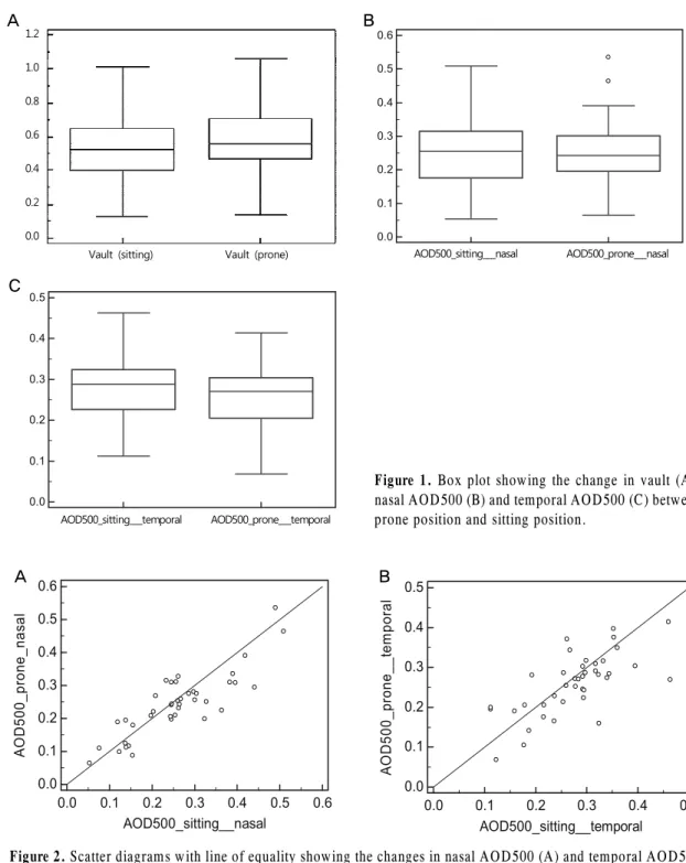
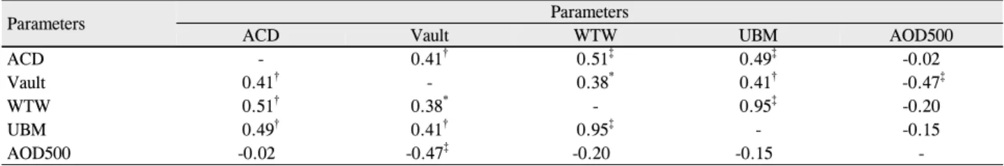
=0.09)(Table 3).자세 변화에 따른 vaulting과 평균 비측 및 이측에서 측 정한 AOD500의 변화를 그래프로 나타내어 보면 일시적인 엎드린 자세 후에 vaulting은 커지고 비측과 이측에서 측정 한 AOD500은 감소하는 것을 알 수 있다. 그러나 AOD500 의 변화는 통계적으로 유의하지는 않았다(Fig. 1). 산포도 에서도 비측과 이측의 AOD500이 엎드린 자세에서 감소하 는 경우가 더 많으나 증가하는 경우도 있었음을 볼 수 있다 (Fig. 2).
여러 변수들 간의 상관관계를 분석해 보면 AOD500과 가
1.2 1.0 0.8 0.6 0.4 0.2
0.0
Vault (sitting) Vault (prone)
0.0 0.1 0.2 0.3 0.4 0.5 0.6
AOD500_sitting___nasal AOD500_prone___nasal
0.0 0.1 0.2 0.3 0.4 0.5
AOD500_sitting___temporal AOD500_prone___temporal
Figure 1. Box plot showing the change in vault (A), nasal AOD500 (B) and temporal AOD500 (C) between prone position and sitting position.
0.0 0.1 0.2 0.3 0.4 0.5 0.6
0.0 0.1 0.2 0.3 0.4 0.5 0.6
AOD500_sitting__nasal
AOD500_prone_nasal
0.0 0.1 0.2 0.3 0.4 0.5
0.0 0.1 0.2 0.3 0.4 0.5
AOD500_sitting__temporal
AOD500_prone__temporal
Figure 2. Scatter diagrams with line of equality showing the changes in nasal AOD500 (A) and temporal AOD500 (B) induced by transient prone position.
장 상관관계가 높은 것은 vaulting으로 나타났다(r=-0.47,
p
<0.0024). 즉, vaulting이 클수록 AOD500은 감소하는 것 으로 나타났다(Table 4).엎드린 자세를 취할 때 vaulting이 증가하는 정도에 영향 을 주는 요인이 있는지 분석해 보았다. 전방깊이, 각막직경, 술 후 vaulting 등은 vaulting의 증가량과 유의한 상관관계 를 보이지 않았으며 vaulting 증가량에 영향을 주는 요소는 발견되지 않았다.
고 찰
ICL 삽입술 후 시력과 안압 외에 가장 중요하게 생각하 는 인자가 수정체와 ICL 사이의 거리인 vaulting이다. 이는 수술 후 백내장이나 안압 상승의 위험 정도를 가늠할 수 있 는 지표이기 때문이다. 하지만 수술 후 vaulting은 일정한 것이 아니라 여러 가지 상황과 조건과 시간에 따라서 변화 할 수 있는 값이다. 밝은 조명으로 동공이 작아지는 경우는
A B
C
A B
Table 4. Pearson correlation matrix of parameters
Parameters Parameters
ACD Vault WTW UBM AOD500
ACD - 0.41† 0.51‡ 0.49‡ -0.02
Vault 0.41† - 0.38* 0.41† -0.47‡
WTW 0.51† 0.38* - 0.95‡ -0.20
UBM 0.49† 0.41† 0.95‡ - -0.15
AOD500 -0.02 -0.47‡ -0.20 -0.15 -
ACD = anterior chamber depth; WTW = white to white by topography; UBM = sulcus to sulcus by UBM; AOD500 = angle opening distance at 500 μm.
*p < 0.05; †p < 0.01; ‡p < 0.005.
vaulting이 더 작게 되는 것을 관찰할 수 있으며 조절에 따라 서도 vaulting이 변할 수 있다.10,11이에 본 연구는 자세에 따 른 vaulting의 변화를 연구하였다는 점에서 그 의의가 있다.
ICL 삽입술을 받은환자 중에서 간혹 수술 초기에 고개 를 숙이면 오심을 느끼고 고개를 들면 이러한 증상이 해소 되었다고 이야기하는 경우가 있다. 물론 이러한 현상은 모 든 ICL 삽입술을 시행 받은 환자에서 나타나는 것은 아니 며 또한 수술 후 시간이 지나면서 없어지는 증상이나 이러 한 증상이 본 연구에서 나타난 엎드린 자세가 유발하는 vaulting과 전방각의 변화와 관련이 있을 가능성도 있다. 본 연구는 오직 일시적인 엎드린 자세를 취함으로써 유도되는 변화만을 연구하였으나 좀 더 오랜 시간 엎드린 자세를 취 한 후의 vaulting과 전방각의 변화는 더 클 수도 있기 때문 이다. 따라서 더 오랜 시간을 엎드린 자세를 취하고 난 후 의 vaulting 및 전방각의 변화와 이에 따른 안압에 미치는 영향에 대해서는 추가적인 연구가 필요할 것으로 생각한다.
ICL 수술 후 폐쇄각 녹내장이 발생하였다는 보고가 있으 며5이것은 동공폐쇄가 아닌 전방각 폐쇄로 방수의 유출이 안 되어서 발생하였으며 이런 경우는 술 전에 시행하는 주 변부 홍채절개술이 도움이 되지 못한다. 엎드린 자세가 유 발하는 전방각 감소는 통계적으로 유의한 차이가 없었다.
그러나 그 값은 감소하는 경향을 보였다. 그러므로 더 오랜 시간 동안 엎드린 자세를 취하게 된다면 전방각의 감소가 발생할 가능성도 완전 배제할 수는 없을 것으로 생각한다.
그렇게 된다면 전방각 폐쇄를 유발하거나 더 조장 가능할 것이다. 따라서 술 후 높은 vaulting을 보이거나 좁은 전방 각을 보이는 환자에서 더 작은 크기의 ICL으로 교환할 것 을 고려하고 있는 상황이라면 엎드린 자세를 취하지 않는 것이 전방각 폐쇄에 의한 안압상승을 예방하는 방법이 될 수 있을 것으로 생각되나 이는 추후 더 연구되어야 할 부분 으로 생각한다.
ICL은 홍채와 수정체 사이에 삽입되므로 엎드린 자세를 취하는 것이 중력의 작용에 의하여 ICL을 앞쪽으로 움직이 게 하여 vaulting은 크게 하고 전방각은 좁게 만드는 것으로
생각한다. ICL의 자세변화에 따른 위치이동을 최소로 하기 위해서는 ICL의 재질과 방수와의 비중이 가장 비슷하게 만 드는 것이 중요할 것으로 생각한다.
이러한 ICL의 위치변화는 이를 지지하는 홍채조직의 특 성이나 각막 직경의 크기에 따라서 그 정도가 다를 수도 있 다는 가정하에 vaulting의 변화량에 영향을 미치는 인자가 있는지 조사하였으나 이와 상관관계가 있는 술 전 및 술 후 인자는 발견되지 않았다.
본 연구에서 사용된 AOD500은 공막 돌기에서 500 μm 떨어져서 측정된 섬유주와 홍채 사이의 거리를 나타내는 것으로서 전방각을 정량적으로 보여줄 수 있는 지표 중의 하나로서 폐쇄각 녹내장이나 이런 위험이 있는 환자에서 유의하게 감소하였다는 보고가 있었다.13-16 본 연구에서도 이 지표를 사용하였으며 통계적으로 유의하지는 않았으나 일시적인 엎드린 자세가 미치는 전방각의 변화를 AOD500 의 미세한 감소로 나타났다.
ICL 삽입술 후 전방각의 크기와 가장 관련이 있는 인자 로는 수술 후 vaulting인 것으로 나타났다. 즉, vaulting이 높으면 AOD500은 작아지고 vaulting이 낮으면 AOD500이 증가하는 음의 상관 관계를 보였다(r=-0.47,
p
<0.0024).이것은 수술 후 백내장 및 녹내장의 위험성이 모두 vaulting 과 연관이 있다는 것이며 삽입할 ICL의 크기를 결정하는 것이 그만큼 중요하다고 할 수 있을 것이다.
결론적으로 본 연구는 ICL 삽입술을 받은 환자에서 일시 적인 엎드린 자세가 vaulting을 유의하게 변화시킨다는 것 을 발견하였으며 이것은 삽입된 ICL이 자세 변화에 의해서 미세하지만 위치가 변화하는 것을 의미한다고 할 수 있을 것이다. 이러한 결과들이 수술 후 발생할 수 있는 백내장 및 안압상승을 이해하고 예방하는 데 도움이 될 것으로 생 각한다.
참고문헌
1) Ieong A, Hau SC, Rubin GS, Allan BD. Quality of life in high my-
opia before and after implantable Collamer lens implantation.
Ophthalmology 2010;117:2295-300.
2) Rayner SA, Bhikoo R, Gray T. Spherical implantable collamer lenses for myopia and hyperopia: 126 eyes with 1-year follow up.
Clin Experiment Ophthalmol 2010;38:21-6.
3) Kamiya K, Shimizu K, Igarashi A, et al. Four-year follow-up of posterior chamber phakic intraocular lens implantation for moder- ate to high myopia. Arch Ophthalmol 2009;127:845-50.
4) Khalifa YM, Moshirfar M, Mifflin MD, et al. Cataract develop- ment associated with collagen copolymer posterior chamber phak- ic intraocular lenses: clinicopathological correlation. J Cataract Refract Surg 2010;36:1768-74.
5) Khalifa YM, Goldsmith J, Moshirfar M. Bilateral explantation of Visian Implantable Collamer Lenses secondary to bilateral acute angle closure resulting from a non-pupillary block mechanism. J Refract Surg 2010;26:991-4.
6) Chung TY, Park SC, Lee MO, et al. Changes in iridocorneal angle structure and trabecular pigmentation with STAAR implantable collamer lens during 2 years. J Refract Surg 2009;25:251-8.
7) Kojima T, Maeda M, Yoshida Y, et al. Posterior chamber phakic implantable collamer lens: changes in vault during 1 year. J Refract Surg 2010;26:327-32.
8) Alfonso JF, Lisa C, Abdelhamid A, et al. Three-year follow-up of subjective vault following myopic implantable collamer lens implantation. Graefes Arch Clin Exp Ophthalmol 2010;248:
1827-35.
9) Kamiya K, Shimizu K, Kawamorita T. Changes in vaulting and the
effect on refraction after phakic posterior chamber intraocular lens implantation. J Cataract Refract Surg 2009;35:1582-6.
10) Petternel V, Köppl CM, Dejaco-Ruhswurm I, et al. Effect of ac- commodation and pupil size on the movement of a posterior cham- ber lens in the phakic eye. Ophthalmology 2004;111:325-31.
11) Lindland A, Heger H, Kugelberg M, Zetterström C. Vaulting of myopic and toric Implantable Collamer Lenses during accom- modation measured with Visante optical coherence tomography.
Ophthalmology 2010;117:1245-50.
12) Bechmann M, Ullrich S, Thiel MJ, et al. Imaging of posterior chamber phakic intraocular lens by optical coherence tomography.
J Cataract Refract Surg 2002;28:360-3.
13) Mérula RV, Cronemberger S, Diniz Filho A, Calixto N. New com- parative ultrasound biomicroscopic findings between fellow eyes of acute angle closure and glaucomatous eyes with narrow angle.
Arq Bras Oftalmol 2008;71:793-8.
14) Marchini G, Pagliarusco A, Toscano A, et al. Ultrasound biomicro- scopic and conventional ultrasonographic study of ocular di- mensions in primary angle-closure glaucoma. Ophthalmology 1998;105:2091-8.
15) Ramani KK, Mani B, Ronnie G, et al. Gender variation in ocular biometry and ultrasound biomicroscopy of primary angle closure suspects and normal eyes. J Glaucoma 2007;16:122-8.
16) Friedman DS, Gazzard G, Foster P, et al. Ultrasonographic bio- microscopy, Scheimpflug photography, and novel provocative tests in contralateral eyes of Chinese patients initially seen with acute angle closure. Arch Ophthalmol 2003;121:633-42.
=ABSTRACT=
Effects of Transient Prone Position on Vault and Anterior Chamber Angle in ICL Implanted Patients
Wook Kyum Kim, MD, Ik Hee Ryu, MD, Jin Kuk Kim, MD, Hun Yang, MD
B&VIIT Eye Center, Seoul, Korea
Purpose: To evaluate the effects of transient prone position on vault and anterior chamber angle parameters in ICL im- planted patients.
Methods: 40 eyes of 20 ICL implanted patients with at least 1 month of follow-up were included in the present study. The central ICL vault and anterior chamber parameters including angle opening distance at 500 (AOD500) were measured with the Visante anterior segment optical coherence tomography (OCT) (Carl Zeiss, Dublin, CA) in both the sitting and prone positions by tilting the OCT 90 degrees in the vertical axis and having the patient fixate downwards towards the floor.
Results: The mean central vault was 0.55 ± 0.21 mm (SD) and 0.59 ± 0.21 mm (SD) in the sitting and prone positions, re- spectively (p < 0.0001). The nasal and temporal AOD500 were 0.26 ± 0.11 mm and 0.28 ± 0.08 mm, respectively in the sit- ting position, which decreased to 0.24 ± 0.10 mm and 0.26 ± 0.08 mm in the prone position, however, both were not statisti- cally significant (p = 0.08, p = 0.09). AOD500 was inversely correlated with vault (r = -0.47; p = 0.0024). There were no sig- nificant correlations between increase of vault and anterior chamber depth or white to white nor ICL vault.
Conclusions: Transient prone positioning of ICL implanted patients can induce a significant increase in ICL vault.
J Korean Ophthalmol Soc 2012;53(6):761-766
Key Words: Angle opening distance 500, Implantable collamer lens, Phakic intraocular lens, Position change, Vaulting
Address reprint requests to Hun Yang, MD B&VIIT Eye Center
#14F, 3 Seocho-daero 77-gil, Seocho-gu, Seoul 137-856, Korea
Tel: 82-80-722-0202, Fax: 82-2-501-6435, E-mail: [email protected]