pISSN: 0378-6471 eISSN: 2092-9374 DOI : 10.3341/jkos.2010.51.3.353
= 증례보고 =
전안부 빛간섭단층촬영으로 측정한 백내장 수술 후 전방깊이와 전방각의 변화
신철진⋅이주은⋅김재용⋅차흥원 울산대학교 의과대학 서울아산병원 안과학교실
목적: 전안부 빛간섭단층촬영을 이용해 초음파수정체유화술 및 인공수정체 낭내삽입술 후 전방깊이와 전방각의 변화를 측정하였다.
대상과 방법: 69명, 78안을 대상으로 합병증이 없는 백내장 수술을 시행하였다. 술 전, 술 후 1달 째 전안부 빛간섭단층촬영으로 Angle- referenced (ACD1), pupil-referenced (ACD2), lens-referenced (ACD3) 전방깊이, crystalline lens rise, angle opening distance at 500 μm (AOD500), trabecular-iris space at 750 μm (TISA750)를 측정하여, paired samples T-tests를 이용해 비교하였다.
결과: 평균 ACD1은 술 전 3.19±0.24, 술 후 3.22±0.21 (P=.21); ACD2는 술 전 2.99±0.40, 술 후 3.56±0.28 (P<.05); ACD3는 술 전 2.75±0.41, 술 후 4.00±0.27 (P<.05)였다(이상 mm). 비측과 이측의 전방각, AOD500, TISA750 모두 수술 후 유의하게 증가하였다 (P<.05).
결론: 백내장 수술 후 전방깊이와 전방각이 유의하게 증가됨을 전안부 빛간섭단층촬영을 이용해 확인할 수 있었다.
<대한안과학회지 2010;51(3):353-358>
■ 접 수 일: 2009년 6월 25일 ■ 심사통과일: 2009년 10월 29일
■ 책 임 저 자: 차 흥 원
서울특별시 송파구 풍납2동 388-1 울산대학교 서울아산병원 안과 Tel: 02-3010-3680, Fax: 02-470-6440 E-mail: [email protected]
* 본 논문의 요지는 2009년 대한안과학회 제101회 춘계학술대회에서 구연으로 발표되었음.
전안부의 단면영상기술은 지난 10년간 점점 발전해왔다.
최근 빛간섭단층촬영(OCT) 기술이 전안부 검사에 접목되 어, 전방의 단면구조를 비침습적으로 빠르고 쉽게 검사할 수 있게 되었다. 전안부 빛간섭단층촬영(AS-OCT) 은 고 해상도의 각막과 전방각(ACD) 소견을 초당 4000 axial scan의 속도로 구현해 준다. 따라서 전방의 깊이, 너비, 전 방각 등을 비교적 정확히 정량화할 수 있게 되었다.1
백내장 수술은 안구의 전방(AS) 내의 구조와 여러 지표 (parameter) 에 변화를 가져온다. 이전의 여러 연구들은 초 음파 생체현미경(Ultrasound biomicroscopy) 등 기존의 방 법(modality) 을 이용하여, 백내장 적출이 전방을 깊게 하 고, 전방각을 넓혀주는 것을 보여주었다.2
이에 본 연구에서는 새로운 영상기법인 전안부 빛간섭단 층촬영을 통하여 초음파 수정체유화술 및 인공수정체 낭내 삽입술 후, 전방깊이와 전방각의 변화를 살펴보았다.
대상과 방법
2008년 8월부터 2009년 4월까지 본원에서 노년성(senile cataract) 혹은 노년기 전 백내장(presenile cataract) 으로 합병증이 없는 백내장 수술을 시행 받은 69명, 78안을 대상 으로 하여 전향적인 연구를 시행하였다. 모든 환자들에게 설명에 근거한 동의를 받았으며, 이전에 안과적 수술이나 외상을 받았거나 백내장 이외에 안과적 질환을 가진 사람 은 연구에서 제외하였다.
동일한 술자가 점안마취로 2.75 mm의 투명윤부절개창 을 통하여 초음파 수정체유화술 및 인공수정체 낭내삽입술 을 시행하였다. 모든 환자들에게 시력, 세극등현미경, 안압, 안저검사 등의 기본 안과검사와 함께 전안부 빛간섭단층촬 영(Visante, Carl Zeiss Meditec) 을 수술 전 및 수술 후 한 달째에 시행하였다. 전안부 빛간섭단층촬영계는 Corneal scan, angle scan, pachymetry map 등 세 가지 종류(mode) 를 검사할 수 있으며, 본 연구에서는 angle scan 을 이용하 였다. 검사시의 조건은 무산동 상태로 동일하게 하였으며, 검사 및 측정(measurement) 모두 동일한 술자가 시행하였 다. 한 사람당 2번의 촬영을 시행하여 그 중 해상도가 좋은 것을 선택하였고 두 명의 관찰자에 의해 중복으로 검토되 었다. 기계에 내장된 소프트웨어(software)를 이용하여 총 8가지의 변수를 측정하였다.
전방 깊이(ACD)는 세 가지 방법으로 각각 측정하였다:
A B
Figure 1.(A) Anterior segment OCT image of the left eye of an 55-year-old man before phacoemulsification and IOL implantation; ACD1 is 3.02 mm (2.47+0.55), ACD3 is 2.47 mm, and CLR is 0.55 mm. (B) Postoperative image of the same eye; ACD1 is 3.06 mm (3.86-0.80). and ACD3 is 3.86 mm (ACD1=angle-referenced anterior chamber depth; ACD3=lens-referenced anterior chamber depth; CLR=crystalline lens rise).
A B
Figure 2. (A) Anterior segment OCT image of the right eye of an 51-year-old man before phacoemulsification and IOL implantation; ACD2 is 3.48 mm. (B) Postoperative image of the same eye; ACD2 is 3.81 mm (ACD2=pupil- referenced anterior chamber depth).
(1) angle-referenced ACD (ACD1)는 각막내피로부터 2 개의 전방각(iridocorneal angle)을 수평으로 연결한 선(3 to 9 o’clock) 중심까지의 거리로 정하였고, crystalline lens rise (CLR)는 이 선으로부터 수정체까지의 높이로 하 였다(Fig. 1); (2) pupil-referenced ACD는 각막내피로부 터 동공색소상피 (pupillary pigment epithelium) 즉, 동공 면(pupillary plane) 중심까지의 거리로 정하였다(Fig. 2);
(3) lens-referenced ACD는 각막내피로부터 백내장 수정 체의 전극(anterior pole)까지의 거리로 하였다(Fig. 1).
비측(nasal side) 및 이측(temopral side)의 전방각(ir- idocorneal angle)을 측정하였다. 섬유주(trabecular mes- hwork) 를 따라 접선을 그리고, 공막극(scleral sprur)에서 앞쪽으로 500 μm 지점에서 홍채까지의 수직길이를 구하여 angle opening distance 500 (AOD 500)을 측정하였다. 그 리고 공막극에서 앞쪽 750 μm 지점에서 홍채까지 수직선 을 그어서 섬유주, 각막내피, 홍채의 앞쪽 표면이 이루는 면
적을 구하여 trabecular-iris space at 750 μm (TISA750) 을 측정하였다. 검사자가 공막극을 정하면 angle opening distance at 500 μm (AOD500), trabecular- iris space at 750 μm (TISA750) 은 기계에 내장된 소프트웨어에 의하 여 자동으로 측정되었다(Fig. 3).
수술 전후의 측정치 비교는 paired samples T-tests를 이용하여 비교하였고, CLR과 ACD 사이의 상관관계는 Pe- arson correlation analysis를 이용하여 분석하였다. 모든 통계분석은 SPSS (version 15.0, software for Windows;
SPSS Inc., Chicago, IL, USA)를 이용하였다.
결 과
남자 41명(46안) 과 여자 28명(32안) 의 평균 나이는 65.45±13.38세(31세에서 88세까지)였다. 평균 술 전 cry- stalline lens rise는 평균 0.430±0.350 mm 였다.
A B
Figure 3.(A) Anterior segment OCT image of the left eye of an 70-year-old man before phacoemulsification and IOL implantation; the nasal iridocorneal angle is 29.5 degrees and the temporal iridocorneal angle is 26.0 degrees, the nasal AOD500 is 0.390 mm and the temporal AOD500 is 0.435 mm, the nasal TISA750 is 0.274 mm2 and the temporal TISA750 is 0.306 mm2. (B) Postoperative image of the same eye; the nasal iridocorneal angle is 34.2 degrees, and the temporal iridocorneal angle is 36.5 degrees, the nasal AOD500 is 0.698 mm and the temporal AOD500 is 0.460 mm, the nasal TISA750 is 0.458 mm2and the temporal TISA750 is 0.319 mm2. (AOD500=angle opening distance at 500 um; TISA750=trabecular-iris space at 750 μm).
-500.00 0.00 500.00 1000.00 1500.00 2000.00
CLR 2.50
3.00 3.50 4.00 4.50
ACD1
-500.00 0.00 500.00 1000.00 1500.00 2000.00
CLR 2.00
2.50 3.00 3.50 4.00
ACD2
-500.00 0.00 500.00 1000.00 1500.00 2000.00
CLR 1.50
2.00 2.50 3.00 3.50 4.00
ACD3
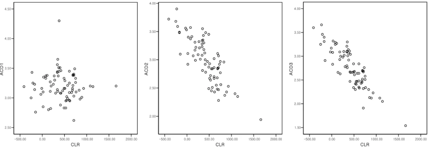
Figure 4. Correlation between CLR and preoperative ACD.
*Pearson correlation test (CLR=crystalline lens rise; ACD=anterior chamber depth; ACD1=angle-referenced anterior chamber depth; ACD2= pupil-referenced anterior chamber depth; ACD3=lens-referenced anterior chamber depth).
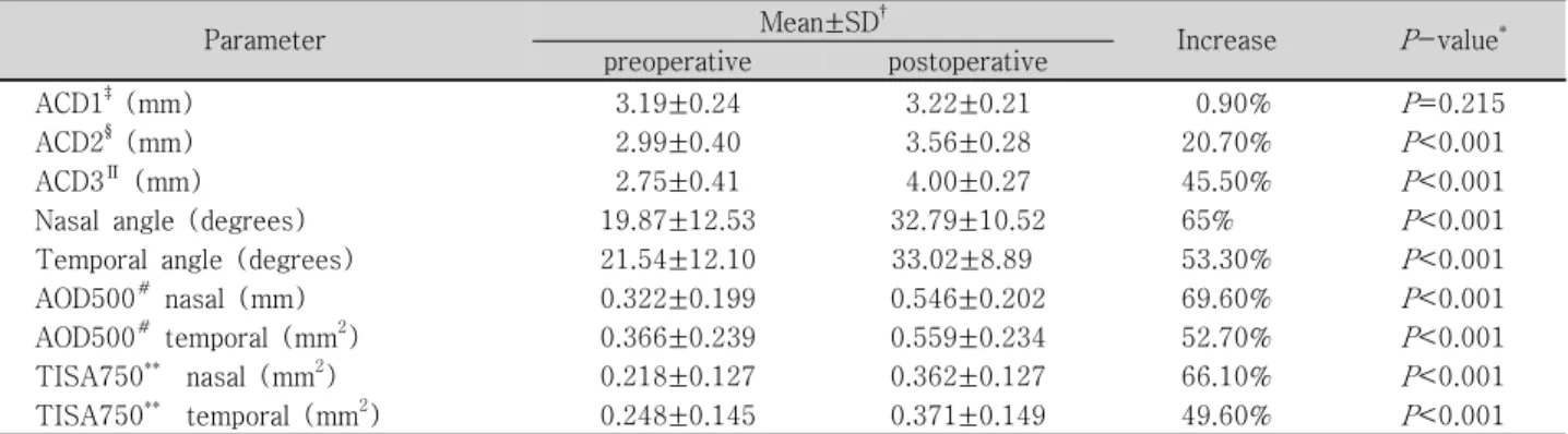
ACD1을 제외하고 ACD2, ACD3 모두 백내장 수술 후 통 계적으로 유의한 증가를 보였다. ACD2 (20.7 % 증가)에 비하여, ACD3 (45.5 % 증가)에서 두드러진 증가를 보였다 (이상 Table 1).
비측과 이측의 전방각(iridocorneal angle) 모두 수술 후 유의하게 넓어졌다. 비측과 이측의 AOD500, TISA750 모 두에서 통계적으로 유의한 증가를 보였다.
수술전의 crystalline lens rise와 anterior chamber depth parameter들 간의 상관관계를 pearson correlation으로 살 펴보았다(Fig. 4, Table 2). ACD3에서 crystalline lens rise와 가장 큰 음의 상관관계를 보였다(r=-0.864, P<.01).
고 찰
전안부 빛간섭단층촬영(anterior segment OCT)은 전안 부를 빠르고 쉽게 좋은 해상도로 구현해 준다. 그리고 비접 촉 방식(non-contact method)으로 협조가 부족하거나 개 방성 안구손상(open-globe injury) 환자에서도 안전하게 이용할 수 있다. 하지만 홍채 후방의 구조물들을 보여줄 수 없다는 점과, 영상(image) 에서 기준점들을 정하고 변수 (parameter) 들을 분석할 때 재현성 (reproducibility)이 떨어진 다는 점에서 제약이 있다.1
Kohnen et al은 빛간섭단층촬영이 여러 장점에도 불구하
Table 1. Changes in anterior chamber parameters before and after cataract surgery
Parameter Mean±SD† Increase P-value*
preoperative postoperative
ACD1‡ (mm) 3.19±0.24 3.22±0.21 0.90% P=0.215
ACD2§ (mm) 2.99±0.40 3.56±0.28 20.70% P<0.001
ACD3∏ (mm) 2.75±0.41 4.00±0.27 45.50% P<0.001
Nasal angle (degrees) 19.87±12.53 32.79±10.52 65% P<0.001
Temporal angle (degrees) 21.54±12.10 33.02±8.89 53.30% P<0.001
AOD500#nasal (mm) 0.322±0.199 0.546±0.202 69.60% P<0.001
AOD500#temporal (mm2) 0.366±0.239 0.559±0.234 52.70% P<0.001
TISA750** nasal (mm2) 0.218±0.127 0.362±0.127 66.10% P<0.001
TISA750** temporal (mm2) 0.248±0.145 0.371±0.149 49.60% P<0.001
*Paired samples T-tests; †SD=standard deviation; ‡ACD1=angle-referenced anterior chamber depth; §ACD2=pupil-referenced anterior chamber depth; ∏ACD3=lens-referenced anterior chamber depth; #AOD500=angle opening distance at 500 μm; **TISA750=
trabecular-iris space at 750 μm.
Table 2. Correlation between CLR and preoperative ant- erior chamber depth
Parameter CLR‡ P-value*
ACD1§ †r=-0.26 0.818
ACD2∏ r=-0.816 <0.001
ACD3# r=-0.864 <0.001
*Pearson correlation test; †r=Pearson correlation coefficient;
‡CLR=crystalline lens rise; §ACD1=angle-referenced anterior chamber depth; ∏ACD2=pupil-referenced anterior chamber ddepth; #ACD3=lens-referenced anterior chamber depth.
고, 그래픽(graphic) 을 이용하여 분석을 할 때 여러 오류가 발생할 수 있음을 보고한 바 있다.15이에 우리는 오류를 최 소화하기 위해 적어도 4번의 동일한 영상을 촬영하여 그 중 가장 해상도가 좋은 이미지를 택하였고, 이를 다시 두 명의 검사자가 중복으로 확인하여 변수를 측정하였다.
지금까지 백내장 수술 전후의 전안부 변화를 전안부빛간 섭단층촬영을 이용하여 정량적으로 분석한 연구는 많지 않 다. 본 연구를 통해 우리는 다수의 환자들에게 백내장 수술 후 전방이 깊어지고, 전방각이 넓어지는 것을 전안부 빛간 섭단층촬영을 이용하여 확인해 보고자 하였다.
과거의 연구들을 살펴보면 전방깊이(anterior chamber depth)에 관한 정의는 다양하게 내릴 수 있다. 그 중 본 연 구에서는 세 가지 방법으로 전방깊이를 측정하여 비교하였 다. Kucumen et al은 전안부 빛간섭단층촬영을 이용하여 백내장 수술 후 ACD1에 비하여 ACD2, ACD3가 유의하게 증가함을 보고하였고, Memarzadeh et al과 Rabsilber et al 도 비슷한 결과를 보고하였다.1,3,11 본 연구에서도 대체로 비슷한 결과를 보였지만, ACD3가 ACD2에 비해서 훨씬 유 의하게 증가하였다는 점에서 차이를 보였다.
Kucumen et al이 보고한 것처럼 Angle referenced ACD (ACD1) 는 전방 깊이의 변화를 잘 반영하지 못하므로 임 상적으로 큰 의미를 가지지 못하고, Pupil-referenced
ACD (ACD2) 는 전방깊이의 변화를 잘 반영하지만, 동공 연(pupillary plane) 등의 기준점(orientation point) 을 잡 는 것은 주관적일 수 있다는 점에서 제약이 따른다.1 본 연 구에서 보듯이 Lens-referenced ACD (ACD3)는 전방깊 이의 변화를 가장 잘 반영하고, 기준점을 잡는 것도 ACD2 에 비해 쉬우므로 임상적으로 쉽게 이용될 수 있다.
섬유주(trabecular meshwork) 의 실제적인 기능을 반영 하는 지표로 알려진 AOD500, TISA750 은 이측(temporal side) 보다 비측(nasal side)에서 유의하게 증가를 보였다 (paired t test, P<.05). Nolan et al2의 연구에서는 이를 검 사 시 어두운 조건(mesopic condition)과 밝은 조건(phot- opic condition)으로 나누어 비교하였는데, 밝은 조건에서 검사할 경우 비측에서 유의하게 증가하는 경향을 나타내었 다. 본 연구에서 나타난 이와 같은 비측과 이측의 AOD500, TISA750 값 차이에 대한 원인을 알아보기 위해, 수술 시 절개창의 방향에 따라 이측 절개창(35안), 비측 절개창(26 안), 그리고 상측(superior) 절개창(17안)의 세 군(group) 으로 구분하였다. 그러나 세 군 사이에 수술 전 후 유의한 차이는 관찰되지 않았다(ANOVA test, P>0.05). 비측에서 이측보다 AOD500, TISA750이 실제로 더 증가하는지, 증 가한다면 원인이 무엇인지에 대해서는 향후 추가적인 연구 가 필요하다.
Kucumen et al은 Crystalline lens rise (CLR)가 ACD2, ACD3와 유의한 음의 상관관계를 보이며, 따라서 CLR 이 백내장 수술 전 전방깊이를 나타내는 좋은 지표가 될 수 있 다고 보고하고 있다.1본 연구에서도 CLR이 ACD2, ACD3 와 유의한 음의 상관관계를 보였다.
전안부 빛간섭단층촬영은 다양한 목적의 전안부 평가에 있어서 중요한 정보를 제공해 주며, 향후 점차 그 이용이 늘어날 것이다. 현재 공막극(scleral spur) 등의 기준점을 잡는 데 있어서 재현성(reproducibility)이 떨어진다는 제
약이 있지만, 이는 향후 기술(technology)의 발전에 따라 극복될 것으로 보인다.
참고문헌
1) Kucumen RB, Yenerel NM, Gorgun E, et al. Anterior segment optical coherence tomography measurement of anterior chamber depth and angle changes after phacoemulsification and intraocular lens implantation. J Cataract Refract Surg 2008;34:1694-8.
2) Nolan WP, See JL, Aung T, et al. Changes in angle configuration after phacoemulsification measured by anterior segment optical coherence tomography. J Glaucoma 2008;17:455-9.
3) Memarzadeh F, Tang M, Li Y, et al. Optical coherence tomog- raphy assessment of angle anatomy changes after cataract surgery.
Am J Ophthalmol 2007;144:464-5.
4) Baikoff G, Lutun E, Ferraz C, Wei J. Static and dynamic analysis of the anterior segment with optical coherence tomography. J Cataract Refract Surg 2004; 30:1843-50.
5) Baikoff G, Jitsuo Jodai H, Bourgeon G. Measurement of the internal diameter and depth of the anterior chamber: IOLMaster versus anterior chamber optical coherence tomographer. J Cataract Refract Surg 2005; 31:1722-8.
6) Radhakrishnan S, Goldsmith J, Huang D, et al. Comparison of optical coherence tomography and ultrasound biomicroscopy for detection of narrow anterior chamber angles. Arch Ophthalmol 2005;123:1053-9.
7) Kurimoto Y, Park M, Sakaue H, Kondo T. Changes in the ante- rior chamber configuration after small-incision cataract surgery with posterior chamber intraocular lens implantation. Am J Op- hthalmol 1997;124:775-80.
8) Goldsmith JA, Li Y, Chalita MR, et al. Anterior chamber width measurement by high-speed optical coherence tomography.
Ophthalmology 2005; 112:238-44.
9) Leung CK, Chan W-M, Ko CY, et al. Visualization of anterior ch- amber angle dynamics using optical coherence tomography. Op-
hthalmology 2005; 112:980-4.
10) Hayashi K, Hayashi H, Nakao F, Hayashi F. Changes in anterior chamber angle width and depth after intraocular lens implant- ation in eyes with glaucoma. Ophthalmology 2006;107:698-703.
11) Rabsilber TM, Khoramnia R, Auffarth GU. Anterior chamber measurements using Pentacam rotating Scheimpflug camera. J Cataract Refract Surg 2006; 32:456-9.
12) Nonaka A, Kondo T, Kikuchi M, et al. Angle widening and alter- ation of ciliary process configuration after cataract surgery for primary angle closure. Ophthalmology 2006;113:437-41.
13) Pereira FA, Cronemberger S. Ultrasound biomicroscopic study of anterior segment changes after phacoemulsification and foldable intraocular lens implantation. Ophthalmology 2003;110:1799-806.
14) Pavlin CJ, Harasiewicz K, Foster FS. Ultrasound biomicroscopy of anterior segment structures in normal and glaucomatous eyes.
Am J Ophthalmol 1992;113:381-9.
15) Kohnen T, Thomala MC, Cichocki M, Strenger A. Internal ant- erior chamber diameter using optical coherence tomography compared with white-to-white distances using automated measu- rements. J Cataract Refract Surg 2006;32:1809-13.
16) Dada T, Sihota R, Gadia R, et al. Comparison of anterior segment optical coherence tomography and ultrasound biomicroscopy for assessment of the anterior segment. J Cataract Refract Surg 2007;
33:837-40.
17) Radhakrishnan S, Rollins AM, Roth JE, et al. Real-time optical coherence tomography of the anterior segment at 1310 nm. Arch Ophthalmol 2001;119:1179-85.
18) Nolan WP, See JL, Chew PT, et al. Detection of primary angle closure using anterior segment optical coherence tomography in Asian eyes. Ophthalmology 2007;114:33-9.
19) Radhakrishnan S, See JL, Smith SD, et al. Reproducibility of anterior chamber angle measurements obtained with anterior segment optical coherence tomography. Invest Ophthalmol Vis Sci 2007;48:3683-8.
20) Spaeth GL. The normal development of the human anterior cha- mber angle: a new system of descriptive grading. Trans Ophtha- lmol Soc U K 1971;91:709-39.
=ABSTRACT=
Changes in Anterior Chamber Depth and Angle After Phacoemulsification measured by Anterior Segment Optical
Coherence Tomography
Chul Jin Shin, MD, Joo Eun Lee, MD, Jae Yong Kim, MD, PhD, Hung Won Tchah, MD, PhD
Department of Ophthalmology, University of Ulsan College of Medicine, Asan Medical Center, Seoul, Korea
Purpose: To evaluate changes in anterior chamber depth (ACD) and angle after phacoemulsification and intraocular lens implantation using anterior segment optical coherence tomography (AS-OCT).
Methods: Seventy-eight eyes of 69 patients had uneventful phacoemulsification and IOL implantation using a clear corneal incision. Anterior segment OCT images of nasal and temporal angle quadrants were obtained before and at one month after surgery. The angle-referenced (ACD1), pupil-referenced (ACD2), lens-referenced (ACD3) ACDs, crystalline lens rise (CLR), nasal and temporal iridocorneal angles, angle opening distance at 500 μm (AOD500), and trabecular iris surface area at 750 μm (TISA750) were measured. Preoperative and postoperative measurements were compared using paired sample t-tests.
Results: The mean ACD1 was 3.19±0.24 mm preoperatively and 3.22±0.21 mm at one month postoperatively (P=0.21); ACD2 was 2.99±0.40 mm preoperatively and 3.56±0.28 mm at one month postoperatively (P<0.05); ACD3 was 2.75±0.41 mm pre- operatively and 4.00±0.27 mm at one month postoperatively (P<0.05). The mean iridocorneal angles, AOD500, and TISA750 for both nasal and temporal sides increased significantly at the postoperative examinations (P<0.05).
Conclusions: Changes in anterior chamber angle after cataract surgery can be objectively quantified by anterior segment OCT.
Deepening of the anterior chamber and widening of the nasal and temporal angles after cataract extraction were shown on AS-OCT.
J Korean Ophthalmol Soc 2010;51(3):353-358
Key Words: Angle, Anterior chamber depth, Anterior segment optical coherence tomography, Phacoemulsification
Address reprint requests to Hung Won Tchah, MD, PhD
Department of Ophthalmology, University of Ulsan, College of Medicine, Asan Medical Center
#388-1 Pungnap-2-dong, Songpa-gu, Seoul 138-736, Korea
Tel: 82-2-3010-3680, Fax: 82-2-470-6440, E-mail: [email protected]