After cataract surgery in eyes with angle closure glaucoma (ACG), declining intraocular pressure (IOP) and deepening of the anterior chamber has been reported to occur.1-3There are several reports that glaucoma patients who underwent filtration procedures with consequent lowering of IOP show a decrease in axial length.4,5Together, these reports surmise that the IOP lowering effect of cataract surgery also influences the axial length especially in ACG patients.
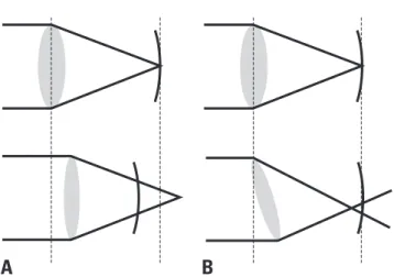
Because of post-operative deepening of the anterior chamber and decrease in axial length, preoperative calculation of intraocular lens (IOL) power may be inaccurate for eyes with ACG. Such an anatomic change after cataract surgery in eyes with ACG may lead to a hyperopic shift in the post-operative period (Fig. 1A).
However, shifting of the capsular apparatus and shortening of the axial length are not the only changes seen in ACG eyes. IOL power predictions for these eyes still encounter another challenge. Eyes with ACG display a propensity for a higher than normal intra- capsular volume.6-8This large capsular bag may result in tilting or even de-centering of an intra-capsular IOL (Fig. 1B). These deviated IOLs may cause unpredictable refractive outcomes and may be one of the reasons of poor IOL power prediction in ACG patients.
The present study aimed to determine the accuracy of preoperative IOL calculations in eyes with primary ACG undergoing cataract surgery.
Inaccuracy of Intraocular Lens Power Prediction for Cataract Surgery in Angle-Closure Glaucoma
Sung Yong Kang, Samin Hong, Jung Bin Won, Gong Je Seong, and Chan Yun Kim
Institute of Vision Research, Department of Ophthalmology, Yonsei University College of Medicine, Seoul, Korea.
Purpose:To assess the accuracy of intraocular lens (IOL) power predictions for cataract surgery in eyes with primary angle- closure glaucoma (ACG). Because of shifting of the capsular bag apparatus and shortening of the axial length, preoperative calculation of IOL power may be inaccurate for eyes with ACG. Materials and Methods:This retrospective comparative case series comprised of 42 eyes from 42 patients with primary ACG and 45 eyes from 45 subjects with normal open-angles undergoing uneventful cataract surgery. Anterior segment biometry including anterior chamber depth, lens thickness, and axial length were compared. Using the SRK-II formula, the powers of the implanted IOL and the actual postoperative spherical equivalent (SE) refractive errors were compared between the two groups. Also, the absolute values of differences between predicted and residual SE refractive errors were also analyzed for each group. Results:In ACG patients, anterior chamber depth and axial length were shorter and the lens was thicker than normal controls (all p < 0.001). Even though residual SE refractive error was not significantly different (p = 0.290), the absolute value of the difference between predicted and residual SE refractive error was 0.64
±
0.50 diopters in AGC patients and 0.39±
0.36 diopters in control subjects (p = 0.012). The number of eyes that resulted in inaccurate IOL power predictions of more than 0.5 diopters were 21 (50.00%) in the ACG group, but only 12 (26.67%) in the control group (p = 0.043). Conclusion:IOL power predictions for cataract surgery in ACG patients can be inaccurate, and it may be associated with their unique anterior segment anatomy.Key Words : Angle-closure, cataract, glaucoma, intraocular lens
Received: June 11, 2008 Revised: July 26, 2008 Accepted: August 20, 2008
Corresponding author: Dr. Chan Yun Kim, Institute of Vision Research,
Department of Ophthalmology, Yonsei University College of Medicine, 250 Seongsan-ro, Seodaemun-gu, Seoul 120-752, Korea.
Tel: 82-2-2228-3580, Fax: 82-2-312-0541 E-mail: [email protected]
© Copyright:
Yonsei University College of Medicine 2009
INTRODUCTION
After approval by the Institutional Review Board, 42 patients diagnosed with primary ACG and who had undergone extra- capsular cataract extraction by phacoemulsification with posterior chamber in-the-bag IOL implantation between March 2001 and December 2006 were identified from the patients’
database. All eyes had previously undergone laser peripheral iridotomy. Only those eyes with uneventful cataract surgery were included in the study, and in cases where capsular integrity was compromised were excluded. Those with a history of ocular trauma or any other ophthalmic disease other than angle closure were also excluded. All eyes had a clinical follow-up period of at least 3 months post-operatively. If both eyes met the entry criteria, only one eye was randomly selected.
The power of all inserted IOLs was calculated using the SRK-II formula for an emmetropic post-operative goal diopter. As a comparative control group, 45 patients confirmed with normal open angles and had uneventful cataract surgery were included in the study.
Retrospectively, the pre-operative data were collected from clinical records; uncorrected and best-corrected visual acuity (with Snellen chart), spherical equivalent (SE) refractive errors, IOP (with Goldmann applanation tonometry), visual field (with Humphrey Field Analyzer II, SITA standard 30-2 program, Carl Zeiss Meditec, Dublin, CL, USA), keratomety (with automated keratometer, RK-3, Canon, Tochigiken, Japan), central corneal thickness (with ultrasonic pachymetry, UP- 1000, Nidek, Fremont, CA, USA), and the number of topical anti-glaucoma medications. Anterior chamber depth, lens thickness, and axial length measured by ultrasonic A/B scan and biometer (UD-6000, Tomey Corporation, Nagoya, Japan), predicted SE refractive errors calculated by the SRK-II
formula, and power of the implanted IOL were also recorded.
Central corneal thickness and visual field were checked only in ACG patients.
Post-operatively, uncorrected and best-corrected visual acuity, residual SE refractive errors, IOP, visual field indices, and the number of topical anti-glaucoma medications were assessed. The difference between predicted and residual SE refractive errors was calculated.
Accurate IOL power prediction was defined as the residual post-operative SE refractive error within
±
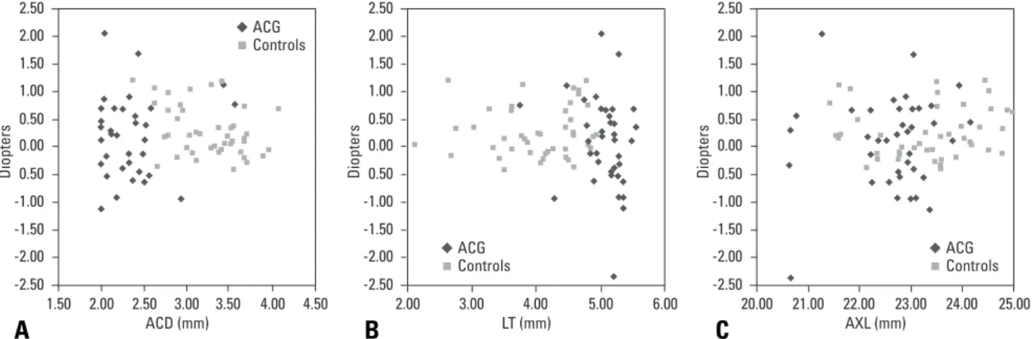
0.5 diopters from predicted SE refractive error. Patients who displayed inaccurate IOL power predictions were assessed for pre-operative anterior segment biometry including anterior chamber depth, lens thickness, and axial length to determine if any of these variables had an effect on post-operative prediction of refractive error.These correlations between anterior segment biometry and the accuracy of the IOL power calculations were sought. Scatter plots were constructed between anterior chamber depth, lens thickness, or axial length vs. the difference between predicted and residual SE refractive errors for each study group.
Comparisons between study groups were made with the Student t-test and the χ2test. All statistical analyses were performed with the SPSS for Windows, version 13.0 (SPSS Inc, Chicago, IL, USA). p value of less than 0.05 was defined as statistically significant.
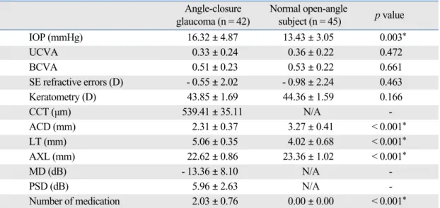
All pre-operative clinical data are summarized in Table 1. Eyes with ACG significantly showed both shorter anterior chamber depths and axial lengths than their control counterparts (both p < 0.001) while their lens thickness was significantly thicker (p < 0.001). Uncorrected and best-corrected visual acuity, SE refractive errors, and keratometry were not apparently different between both study groups.
The power of the implanted IOL, with target emmetropia calculated by the SRK-II formula, showed a significant diffe- rence between the groups with 22.88
±
2.10 diopters for ACG eyes and 20.45±
1.95 diopters for control eyes (p < 0.001).At postoperative 3 months, uncorrected and best-corrected visual acuity were both worse in the ACG group (both p = 0.001) (Table 2). There was no difference between predicted and residual SE refractive errors of the two study groups (predicted refractive errors, p = 0.777; residual refractive errors, p = 0.290).
However, the difference between them was much larger for the ACG group than the control group (p = 0.012) (Table 3).
Using the above definition of accurate IOL power prediction, a greater proportion of ACG patients showed inaccurate IOL power predictions compared to their controls. When using the SRK-II formula, 21 (50.00%) eyes showed inaccurate IOL power predictions in the ACG group, whilst only 12 (26.67%) eyes did in the normal control group (p = 0.043) (Table 3).
A B
Fig. 1. Possible anatomic changes after cataract surgery in eyes with angle closure glaucoma. (A) Hyperopic shift can be caused by deepening of the anterior chamber and shortening of the axial length. (B) Myopic and/or hyperopic shift by tilted or decentered intraocular lens due to the large capsular bag.
RESULTS
MATERIALS AND METHODS
Among those patients belonging to the inaccurate IOL power prediction group, 9 (42.86%) eyes showed a hyperopic shift while 12 (57.14%) eyes resulted in a myopic shift from the intended goal diopter in the ACG group. However, no eyes showed a hyperopic shift but all 12 (100.00%) eyes showed a myopic shift in the control group (Fig. 2). There were no
statistically significant differences in pre-operative anterior segment biometry eyes with ACG (Table 4), although eyes who demonstrated a post-operative myopic shift showed a trend to have a thinner lens.
When the correlation between anterior segment biometry and accuracy of IOL power calculation was sought, no significant
Table 3. Prediction of Intraocular Lens Power in Angle Closure Glaucoma Patients Angle-closure Normal open-angle
p value glaucoma (n = 42) subject (n = 45)
Predicted SE refractive errors (D) - 0.21
±
0.54 - 0.23±
0.35 0.777Residual SE refractive errors (D) - 0.31
±
1.00 - 0.51±
0.59 0.290Δ SE (D) 0.64
±
0.50 0.39±
0.36 0.012*Δ SE > 0.50 D 21 eyes (50.00%) 12 eyes (26.67%) 0.043* D, diopters; SE, spherical equivalent refractive errors.
Δ SE, | predicted SE refractive errors - residual SE refractive errors |.
Values given as means ±standard deviation.
*p < 0.05.
Table 1. Preoperative Data
Angle-closure Normal open-angle
p value glaucoma (n = 42) subject (n = 45)
IOP (mmHg) 16.32
±
4.87 13.43±
3.05 0.003*UCVA 0.33
±
0.24 0.36±
0.22 0.472BCVA 0.51
±
0.23 0.53±
0.22 0.661SE refractive errors (D) - 0.55
±
2.02 - 0.98±
2.24 0.463Keratometry (D) 43.85
±
1.69 44.36±
1.59 0.166CCT (µm) 539.41
±
35.11 N/A -ACD (mm) 2.31
±
0.37 3.27±
0.41 < 0.001*LT (mm) 5.06
±
0.35 4.02±
0.68 < 0.001*AXL (mm) 22.62
±
0.86 23.36±
1.02 < 0.001*MD (dB) - 13.36
±
8.10 N/A -PSD (dB) 5.96
±
2.63 N/A -Number of medication 2.03
±
0.76 0.00±
0.00 < 0.001* ACD, anterior chamber depth; AXL, axial length; BCVA, best corrected visual acuity; CCT, central corneal thickness; D, diopters;dB, decibels; IOP, intraocular pressure; LT, lens thickness; MD, mean deviation on visual field; N/A, not applicable; PSD, pattern standard deviation on visual field; SE, spherical equivalent; UCVA, uncorrected visual acuity.
Values given as means ±standard deviation.
*p < 0.05.
Table 2. Data at Postoperative 3 Month
Angle-closure Normal open-angle
p value glaucoma (n = 42) subject (n = 45)
IOP (mmHg) 12.97
±
2.73 11.23±
2.52 0.004*UCVA 0.56
±
0.26 0.75±
0.23 0.001*BCVA 0.82
±
0.17 0.94±
0.11 0.001*Corneal astigmatism (D) - 0.78
±
0.95 - 0.81±
0.55 0.847*MD (dB) - 13.65
±
8.26 N/A -PSD (dB) 6.22
±
2.90 N/A -Number of medication 0.78
±
0.89 0.00±
0.00 < 0.001* ACG, angle closure glaucoma; BCVA, best corrected visual acuity; dB, decibels; IOP, intraocular pressure; MD, mean deviation on visual field; N/A, not applicable; PSD, pattern standard deviation on visual field; UCVA, uncorrected visual acuity.Values given as means ±standard deviation.
*p < 0.05.
relationship was found for each group. Anterior chamber depth (Fig. 3A), lens thickness (Fig. 3B) and axial length (Fig. 3C) were not related to the difference between predicted and resid- ual SE refractive error.
The present study demonstrates the possibility of inaccurate IOL power calculations in eyes with ACG. Using the SRK-II
formula, the absolute value of the difference between predicted and residual SE refractive error was much larger in the ACG patients compared to controls. Among the ACG eyes showing inaccurate IOL power predictions, some had a hyperopic shift and the others had a myopic shift.
The relative position of the refractive lens between the cornea and macula in the ocular globe presents a stable refractive plane for entering light rays. This stable location of the lens plane is a pre-requisite for all IOL power calculation formulae. In the modern day era of cataract surgery becoming a refractive procedure, such a premise may not go unchallenged, especially in eyes where the relative position of the lens may post-opera- tively change. Such a change is often seen in eyes with ACG.
With the high prevalence of angle closure in Eastern Asia particularly with increasing age, and considering the increased life expectancy of this population, surgery for senile cataracts in eyes with angle closure are becoming more and more common.
Clearly, anatomical differences between eyes with ACG and normal eyes exist. Such differences are mainly found at the anterior segment, mainly at the lens-iris diaphragm due to a thicker than usual lens and subsequently at the anterior chamber angle which accounts for the angle closure in the first place.
Posterior shifting of the capsular bag after cataract removal will result in deepening of the anterior chamber,1-3a subsequent hyperopic shift in ocular power when an IOL is implanted in a more posterior plane than pre-operatively planned. Decrease in axial length due to IOP lowering after cataract extraction will
DISCUSSION
Table 4. Anterior Segment Biometry of Angle Closure Glaucoma Patients According to the Difference between Predicted and Residual Refractive Errors Using the SRK-II Formula
Hyperopic shift eyes Emetropic eyes Myopic shift eyes
p value
(n = 9) (n = 21) (n = 12)
ACD (mm) 2.30
±
0.33 2.22±
0.20 2.47±
0.54 0.203LT (mm) 5.12
±
0.35 5.12±
0.22 4.91±
0.47 0.254AXL (mm) 22.63
±
0.81 22.64±
0.89 22.58±
0.91 0.983ACD, anterior chamber depth; AXL, axial length; LT, lens thickness.
Values given as means ±standard deviation.
Hyperopic shift eyes Emetropic eyes Myopic shift eyes ACG
0.00 10.00 20.00 30.00 40.00 50.00 60.00 70.00 80.00
%
Controls
Fig. 2. Subgroups according to the accuracy of the intraocular lens power prediction after cataract surgery in angle closure glaucoma patients and normal controls. ACG, angle-closure glaucoma.
ACD (mm)
Diopters
ACG
-2.50 -2.00 -1.50 -1.00 0.50
2.00
1.50 2.50 3.00 3.50 4.00 4.50 0.00
0.50 1.00 1.50 2.00 2.50
Controls
LT (mm)
Diopters
ACG -2.50
-2.00 -1.50 -1.00 0.50
2.00 3.00 4.00 5.00 6.00
0.00 0.50 1.00 1.50 2.00 2.50
Controls
AXL (mm)
Diopters
ACG -2.50
-2.00 -1.50 -1.00 0.50
20.00 21.00 22.00 23.00 24.00 25.00 0.00
0.50 1.00 1.50 2.00 2.50
Controls
Fig. 3. Correlation between anterior segment biometry and accuracy of intraocular lens power calculation. (A) Anterior chamber depth (ACD), (B) Lens thickness (LT), (C) Axial length (AXL) in eyes with angle closure glucoma.
A B C
also predispose to a hyperopic shift in the post-operative period.4,5
However, in this study, post-operative myopic shift was noticed as much as the hyperopic shift. Post-operative myopic shift may be caused by the instability of the implanted in-the- bag IOLs. The large capsular volume and loosened lens zonules in ACG eyes has been reported to contribute to this IOL instability.6-8It may be presumed that a certain anatomical difference between eyes with ACG normal controls makesthis discrepancy in IOL prediction a reality. However, in this study, no pre-operative biometric factors closely related to the extent of inaccuracy of IOL power calculations were found. Preoper- ative anterior segment biometry such as anterior chamber depth, lens thickness, and axial length did not relate to extents of inaccuracy. In other words, hyperopic and myopic shifted eyes did not show any difference preoperatively. Therefore, a precise prediction of the accuracy of IOL calculations cannot be said to be determined from pre-operative biometric data in eyes with ACG.
Although the pre-operative factors which predict the inac- curate IOL calculation are not found in this study, it is certain that IOL power prediction can be incorrect in ACG patients.
Hyperopic or myopic shifting after cataract surgery in eyes with angle closure can be a significant problem presenting the patient with visual discomfort. Careful consideration of such inaccurate IOL power prediction must be entertained pre- operatively and appropriately managed.
1. Yang CH, Hung PT. Intraocular lens position and anterior chamber angle changes after cataract extraction in eyes with primary angle-closure glaucoma. J Cataract Refract Surg 1997;23:1109-13.
2. Hayashi K, Hayashi H, Nakao F, Hayashi F. Changes in anterior chamber angle width and depth after intraocular lens implantation in eyes with glaucoma. Ophthalmology 2000;107:698-703.
3. Nonaka A, Kondo T, Kikuchi M, Yamashiro K, Fujihara M, Iwawaki T, et al. Angle widening and alteration of ciliary process configuration after cataract surgery for primary angle closure. Ophthalmology 2006;113:
437-41.
4. Francis BA, Wang M, Lei H, Du LT, Minckler DS, Green RL, et al.
Changes in axial length following trabeculectomy and glaucoma drainage device surgery. Br J Ophthalmol 2005;89:17-20.
5. Law SK, Mansury AM, Vasudev D, Caprioli J. Effects of combined cataract surgery and trabeculectomy with mitomycin C on ocular dimensions. Br J Ophthalmol 2005;89:1021-5.
6. Lowe RF. Aetiology of the anatomical basis for primary angle-closure glaucoma. Biometrical comparisons between normal eyes and eyes with primary angle-closure glaucoma. Br J Ophthalmol 1970;54:161-9.
7. Markowitz SN, Morin JD. Angle-closure glaucoma: relation between lens thickness, anterior chamber depth and age. Can J Ophthalmol 1984;19:300-2.
8. Marchini G, Pagliarusco A, Toscano A, Tosi R, Brunelli C, Bonomi L.
Ultrasound biomicroscopic and conventional ultrasonographic study of ocular dimensions in primary angle-closure glaucoma. Ophthalmology 1998;105:2091-8.