돌발성 시력 저하는 노인 인구에서 흔하게 접할 수 있는 임 상 증상이다. 명확한 원인을 밝힐 수 없는 경우가 많으나, 혈류 공급 장애가 중요한 원인 중 하나이며, 전구 인자로 고혈압, 당 뇨, 고지질혈증 등이 알려져 있다(1).
돌발성 시력 저하의 원인 질환의 진단 및 감별에는 주로 검 안경(ophthalmoscopy)과 형광안저촬영(fluorescein angiography)이 유용한 것으로 알려져 있으며, 이외에도 시 력검사 및 시야검사, 망막전기측정(electroretinography, ERG), 구심성동공이상(relative afferent pupillary defect, RAPD)검사 등이 사용된다. 돌발성 시력 저하의 원인 질환 감 별을 위해 경동맥 협착 유무 및 정도를 파악하거나, 색전성 망 막동맥폐쇄의 경우 색전의 원인을 찾으려면 경동맥 초음파가 도움 될 수 있다(1). 또한, 도플러 기법을 이용한 초음파 (Doppler Ultrasonography, DUS)는 안구 및 안구 혈관의 평가에도 이용된다. 안구의 구조뿐만 아니라 안구후방혈관
(retrobulbar vasculature)의 평가를 통해 안혈관질환, 망막 중심동맥폐쇄, 경동맥해면정맥동류(carotid cavernous fistula), 상안정맥혈전증(superior ophthalmic vein thrombosis), 일과성 흑암시(amaurosis fugax) 등을 진단하 고 평가하는데 유용한 것으로 알려졌다(2). 또한, 10분 미만의 짧은 검사시간에 비침습적 방법으로 안구 후방의 혈관을 관찰 할 수 있을 뿐만 아니라 속도정보를 얻을 수 있다는 장점도 있 다(2-4).
본 연구에서는 혈관 폐쇄에 의한 시력 손상이 의심되는 환자 에서 의미 있는 시력감소와 도플러 지수의 연관성을 살펴봄으 로써, 돌발성 시력 저하 환자에서 망막중심동맥(central retinal artery, CRA) 도플러 지표에 대한 유용성을 알아보 고자 하였다.
대상과 방법
2003년 3월부터 2007년 12월까지 돌발성 시력 저하를 주소 로 본원에 내원한 환자 중 비외상성 혈류 공급 장애가 시력 저 하의 원인으로 추정되는 환자 86명을 대상으로 환자군 망막중
망막혈관 폐쇄에 의한 돌발성 시력 저하 환자군에서 망막중심동맥 도플러 초음파의 유용성
1박 지 원∙이 희 중∙이 종 민
목적: 망막혈관 폐쇄에 의한 돌발성 시력 저하 환자에서 망막중심동맥 도플러 초음파를 이용한 혈류 지표 측정의 유용성에 대해 알아보고자 하였다.
대상과 방법: 돌발성 시력 저하 환자 86명을 대상으로 망막중심동맥 도플러 초음파를 시행하여
혈류속도와 저항지수 및 박동지수를 구하였다. 원인 질환이 확인된 환자 65명의 환측안과 건측 안, 그리고 정상 대조군 33명의 양안을 비교하였다. 또한, 스넬렌 시력검사상의 유의한 시력 상 실 (<20/400)과 상관성이 높은 도플러 초음파 지표 인자를 검사하였다.
결과: 망막중심동맥 도플러 상 환자군의 환측안의 최대수축기혈류속도, 말기이완기혈류속도는 대조군의 양안과 환자군의 건측안 보다 유의하게 낮았다(p < 0.05). ROC 곡선에서 도플러 초 음파 지표 중 평균속도가 유의한 시력 상실을 예측하는데 가장 유용하였으며(estimated area 0.958,p-value < 0.01), 절단 값을 4.3 cm/sec으로 할 경우 민감도 89%, 특이도 95%, 양성 예측률 95%, 음성예측률 88%, 정확도는 92%이었다(p < 0.05).
결론: 돌발성 시력 저하 환자에서 망막중심동맥 도플러 초음파를 이용한 혈류 지표 측정 결과는 유의한 시력 상실을 예측하는데 유용하였다. 따라서 허혈성 시력 장애를 검사하고 잠재성 시력 상실을 예측함에 도플러 초음파는 기존의 안과적 검사에 추가적인 정보를 제공할 수 있을 것으 로 생각된다.
1경북대학교병원 영상의학과
“본 연구는 지식경제부 및 정보통신연구진흥원의 대학 IT연구센터 지원사업 의 연구결과로 수행되었음”(IITA-2008-C1090-0804-0004 )
이 논문은 2008년 9월 10일 접수하여 2008년 12월 26일에 채택되었음.
심동맥 도플러 초음파 검사를 시행하였다. 그 중 시력 저하의 원인 질환에 대한 진단이 불명확한 21명은 배제하고 나머지 65명만을 본 연구의 대상에 포함하였다. 환자군의 나이는 40 에서 88세로 평균 61세이었으며 남성 대 여성의 비율은 45:20 이었다. 또한, 정상치를 설정하기 위하여 성별과 나이의 분포 가 환자군과 유사하게 안과적 증상이 없는 건강한 33명으로 대조군을 구성하였다. 대조군의 나이 분포는 43에서 87세, 평 균나이는 60세이었으며 남성 대 여성의 비율은 24:9이었다.
내원 시 모든 환자에게 안과적 이학적 검사, 시력 검사, 검안 경, 형광안저촬영을 시행하였으며 임상적 진단은 숙련된 안과 전문의에 의해 행해졌다. 시력은 스넬렌 시력검사(Snellen test)에 의해 NLP(non light perception), LP(light perception), HM(hand motion), CF(count finger), 20/400, 20/100, 20/80, 20/60, 20/40, 20/30, 20/20, 20/15 중 한 가지 항목으로 측정되었다. 본 연구에서는 20/400 이하 즉 NLP, LP, HM, CF, 20/400의 시력을 유의 한 시력 저하로 가정하였다.
망막중심동맥에 대한 도플러 초음파 검사에 사용된 초음파 기기는 HDI 5000 scanner(Advanced Technology Laboratories, Bothell, WA, U.S.A.)이었고, 4-7 MHz의 선형 탐촉자를 이용하였다. 11년간의 도플러 초음파 경험이 있는 영상의학 전문의가 환자의 환측 안을 포함한 양측 안(65 안)과 대조군의 양측 안(65안)에 대하여 도플러 초음파를 시 행하였다. 도플러 초음파 검사는 앙와위에서 눈을 감은 채로 시선을 정면에 고정하도록 한 다음 눈꺼풀 위에 탐촉자를 접촉 시킨 상태로 시행하였다. 혈류 영상에서의 인공물과 환자의 불 편감을 줄이고자 탐촉자가 안구에 가해지는 압력을 최소화 하 였다. 반면 탐촉자의 안정성을 위해 4번과 5번 손가락을 이마
나 뺨에 접촉하여 검사자의 손을 지지하였다. 먼저 회색조 (gray scale)영상으로 시신경을 확인하고 나서 색 도플러 영 상으로 시신경 내부 중심에 있는 망막중심동맥을 확인하였다.
이어서 도플러 커서(Doppler sampling cursor)을 망막중심 동맥에 위치시키고 펄스 도플러 영상을 획득하였다. 검사 시간 은 안구당 약 5분이 소요되었다. 혈역학 지표는 최대수축기혈 류속도(peak systolic velocity, PSV), 말기이완기혈류속도 (end-diastolic velocity, EDV), 평균속도(mean velocity, MV=(PSV+2EDV)/3) 및 저항지수(resistive index, RI = PSV-EDV/PSV), 박동지수(pulsatile index, PI = PSV- EDV/MV)가 포함되었다.
환자군의 환측안과 건측안 간의 혈역학 지표의 차이를 paired t-test를 통하여 구하였으며, 환자군의 환측안과 정상 대조군 간의 혈역학 지표의 차이를 independent t-test를 통 하여 구하였다. 그리고 유의한 시력 저하를 보였으면 이를 예 측하는 혈역학 지표를 알아보기 위하여 ROC(receiver operating characteristic) 곡선 분석법을 이용하였으며, 유 의한 시력 저하 예측에 있어서의 민감도, 특이도, 양성 예측률, 음성 예측률, 및 정확도를 산출하였다. 통계학적 평가는 SPSS (SPSS for Windows, version 14.0; Chicago, IL)를 이용하 였으며, 유의성은p 값이 0.05미만일 경우로 정하였다.
결 과
대상 환자 65명은 망막중심동맥폐쇄(n=22), 망막분지동맥 폐쇄(n=3), 망막중심정맥폐쇄(n=6), 망막분지정맥폐쇄
Table 1. Comparison of Doppler Ultrasonographic Parameters Measured in Affected and Unaffected Central Retinal Artery
Affected (n=65) Unaffected (n=65) p-value PSV 4.63 3.27 10.89 1.610 <0.001
EDV 1.59 1.01 2.93 0.74 <0.001
CRA RI 0.50 0.34 0.73 0.04 <0.001
MV 2.56 1.56 4.72 2.22 <0.001
PI 1.04 0.91 1.44 0.15 <0.001
CRA: central retinal artery, PSV: peak systolic velocity; EDV: end diastolic velocity; RI: resistive index, MV: mean velocity, PI: pul- satile index, Numbers are average values in each group.
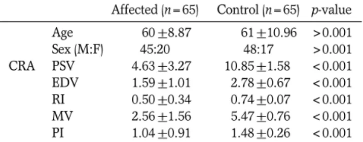
Table 2. Comparison of Doppler Ultrasonographic Parameters Measured in Affected and Healthy Central Retinal Artery
Affected (n=65) Control (n=65) p-value Age 00.60 8.87 00.61 10.96 >0.001
Sex (M:F) 45:20 48:17 >0.001
CRA PSV 04.63 3.27 10.85 1.58 <0.001 EDV 01.59 1.01 02.78 0.67 <0.001 RI 00.50 0.34 00.74 0.07 <0.001 MV 02.56 1.56 05.47 0.76 <0.001 PI 01.04 0.91 01.48 0.26 <0.001 CRA: central retinal artery, PSV: peak systolic velocity; EDV: end diastolic velocity; RI: resistive index, MV: mean velocity, PI: pul- satile index, Numbers are average values in each group.
Table 3. The Feasibility of Ultrasonographic Parameters in Predicting Significant Visual Loss
TP TN FP FN SN SP PPV NPV Accuracy p-value ROC
curve area
PSV<8.0 57 59 6 8 0.877 0.908 0.905 0.881 0.892 <0.001 0.944
EDV<2.5 59 42 23 6 0.908 0.646 0.720 0.875 0.777 <0.001 0.822
RI<6.9 45 54 11 20 0.692 0.831 0.804 0.730 0.762 <0.001 0.748
MV<4.3 62 57 3 8 0.886 0.950 0.954 0.877 0.915 <0.001 0.958
PI<1.35 45 47 18 20 0.692 0.723 0.714 0.701 0.708 <0.001 0.739
TP: true positive, TN: true negative, FP: false positive, FN: false negative, SN: sensitivity, SP: specificity, PPV: positive predictive value,
(n=2), 안허혈증후군(n=32)으로 진단되었다. 시력 20/400 이하를 의미 있는 시력 저하로 가정할 때, 51예에서 유의한 시 력 저하를 보였다.
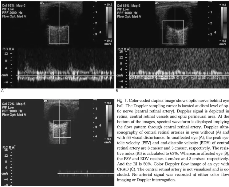
환자군의 환측안 망막중심동맥 도플러에서 혈류가 측정되지 않아 망막중심동맥폐쇄로 판정한 경우는 5예 이었으며, 60예 의 경우는 혈류를 측정할 수 있었다(Fig. 1). 환자군의 환측안 과 건측안의 망막중심동맥에서 시행한 도플러 초음파 지수의 비교에서 PSV(4.63 cm/sec vs 10.89 cm/sec), EDV (1.59 cm/sec vs 2.93 cm/sec), MV (2.56 cm/sec vs 4.72 cm/sec), RI(0.50 vs 0.73) 및 PI(1.04 vs 1.44) 의 평균치 는 유의한 차이를 보였다(Table 1).
환자군의 환측안과 대조군의 망막 중심 동맥에서 측정한 도 플러 지수의 비교에서, PSV (4.63 cm/sec vs 10.85 cm/sec), EDV (1.59 cm/sec vs 2.78 cm/sec), MV (2.56 cm/sec vs 5.47 cm/sec), RI (0.50 vs 0.74) 및 PI (1.04 vs 1.48) 의 평균치도 유의한 차이를 보였다(Table 2).
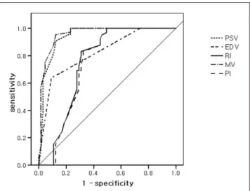
ROC 곡선의 estimated area를 이용하여 예측인자를 평가 할 경우, 유의한 시력 저하의 예측에 가장 유용한 인자는 MV
(estimated area of ROC curve=0.958)였으며, 그다음으로 PSV와 EDV의 순서로 분석되었다. MV의 경우 절단 값을 4.3 cm/sec으로 설정할 경우 민감도 89%, 특이도 95%, 양성예측 률 95%, 음성예측률 88%, 정확도 92%로 산출되었다 (estimated area 0.958, p-value < 0.01)(Table 3, Fig. 2).
고 찰
안구에 혈류 공급 장애를 가져오는 질환을 망막혈관폐쇄질 환(retinal vascular occlusive disorders)으로 정의할 수 있 으며, 망막중심동맥폐쇄와 망막분지동맥폐쇄(central and branch retinal artery occlusion, CRAO and BRAO), 안 허혈증후군(ocular ischemic syndrome, OIS), 망막중심정 맥폐쇄와 망막분지정맥폐쇄(central and branch retinal vein occlusion, CRVO and BRVO)등이 포함된다(5).
CRAO는 색전, 강내혈전, 동맥경화반(Atherosclerotic plaque)등에 의해 발생하여 급성 시력 상실을 가져오는 안과 적 응급 질환이다. 환자 대부분은 통증의 수반 없이 급격한 안
A B
Fig. 1. Color-coded duplex image shows optic nerve behind eye ball. The Doppler sampling cursor is located at distal level of op- tic nerve (central retinal artery). Doppler signal is depicted in retina, central retinal vessels and optic perineural area. At the bottom of the images, spectral waveform is displayed implying the flow pattern through central retinal artery. Doppler ultra- sonography of central retinal arteries in eyes without (A) and with (B) visual disturbance. In unaffected eye (A), the peak sys- tolic velocity (PSV) and end-diastolic velocity (EDV) of central retinal artery are 8 cm/sec and 3 cm/sec, respectively. The resis- tive index (RI) is calculated to 63%. Whereas,in affected eye (B), the PSV and EDV reaches 4 cm/sec and 2 cm/sec, respectively.
And the RI is 50%. Color Doppler flow image of an eye with CRAO (C). The central retinal artery is not visualized and is oc- cluded. No arterial signal was recorded at either color flow imaging or Doppler interrogation.
C
전지수(count fingers) 또는 광각맹(non light perception) 단계의 심각한 시력감퇴를 보인다(6, 7). OIS는 혈역학적으로 유의한 경동맥 협착 후 안구의 혈류장애에 의해 발생하며, 단 안의 충혈 및 각막부종, 방수플레어, 홍채위축, 홍채혈관신생, 신생혈관녹내장, 백내장 변화, 시력감소, 안통, 수명 및 일과성 흑암시 등이 나타나며, 형광안저 소견이 특징적이다(7).
CRAO의 가장 진단적 가치가 있는 도플러초음파 소견은 CRA 에서 혈류를 발견하지 못하는 것이며, 망막 부종을 부가적으로 관찰할 수 있다(6, 8). 그러나 재관류가 있으면 CRA에서 혈 류를 관찰할 수 있으며(8), 이럴 때 OIS와 감별이 어려운 것 으로 알려져 있다. 혈역학 지표 역시 OIS의 경우, PSV의 의 미 있는 감소와 RI의 증가가 유용한 소견으로 알려져 있지만 재개통된 CRAO와는 감별이 어렵다.
본 연구에서는 망막 혈관 폐쇄 질환에 이환된 환자의 혈류 역학 지표와 유의한 시력 저하와의 연관성에 대해 알아보고자 하였다. Yang 등(9)에 의하면 정상인에서 DUS 지수의 정상 치는 PSV가 11.5 +/- 2.45 cm/sec, EDV가 3.73 +/- 1.06 cm/sec, RI가 0.68 +/- 0.04로 보고되고 있다. 본 연구에서 대조군에 포함된 33명 정상인의 도플러 초음파 소견은 보고된 정상치와 비교해 조금 낮았다. 이는 본 연구에 포함된 대조군 의 평균 연령이 61세인 고연령 군에 의한 것으로 생각된다. 실 제로 여러 연구에서 망막 혈류 속도와 나이와의 유의한 상관성 에 대해 보여주고 있다(9, 10). 반면 환자군의 환측안은 환자 군의 건측안 및 대조군의 양안과 비교해서 망막중심동맥의 혈 류 속도 및 도플러 수치의 유의한 감소세를 보였다. 특히 MV 가 4.3 cm/sec 이하를 보일 경우 20/400 이하의 시력을 예측 하는데 유용한 것으로 보여, 도플러 초음파의 혈류 정보가 시 력 상실을 예측하는데 유용한 것으로 나타났다.
혈관성 안질환의 DUS 지표는 이환 기간과 밀접한 연관성이 있을 뿐만 아니라, 당뇨, 고지혈증 등 직접적으로 안동맥에 영 향을 주는 인자, 안동맥의 근위 동맥인 내경동맥의 측부 순환
에 영향을 주는 다양한 인자에 의해 영향을 받는 것으로 알려 져 있다(11, 12). 따라서 도플러 초음파를 시력 상실 시 원인 질환의 초기 감별 진단 검사로 이용하는 데에는 제한점이 있어 안저 검사를 포함한 임상적 검사가 초기 진단 기법으로 이용된 다. 그러나 임상적으로 혈류 장애에 의한 시력손상이 예상되더 라도 재개통된 CRAO나 가벼운 OIS의 경우 안저검사 등의 임 상적 진단이 부적절 할 수 있다. 이 경우 보조적 진단 기법으로 써 경동맥 및 망막중심동맥 도플러 초음파가 이용될 수 있을 것으로 생각된다. 당뇨병이나, 베체트씨병 등 다양한 만성 혈 관성 안질환에서도 도플러 초음파 지표와 병태생리의 연관성 이 보고되고 있으며, 도플러 초음파의 장점인 비침습적 반복 검사 가능성으로 말미암아 선별검사 및 추적조사에서의 유용 성이 제안될 수 있다.
본 연구의 제한점으로는 첫째, 후향적 연구이므로 선택의 바 이어스(selection bias)가 존재할 수 있다. 즉, 본 연구에서는 당뇨병성 혈관 질환 등의 기저질환에 의한 추가적 혈류장애를 고려하지 않고 시력저하군과 정상군을 단순 비교하였다. 둘째, 환자군에서 추적조사가 이루어지지 않았다는 점이다. 1년 경 과 관찰을 시행한 연구 결과 CRA의 혈류 속도는 시력이 회복 함에 따라 증가하는 추세를 보였으며, 이는 CRA DUS가 추적 조사에 유용한 검사법임을 시사한다(1). 셋째, 증상 발현 시기 와 DUS 검사시기가 1일에서 13일로 다양한 차이를 보이고 있 다. 혈류의 폐쇄에 따른 혈역학의 변화는 매우 역동적이기 때 문에 기간에 따른 DUS 지표역시 다양한 결과를 보일 수 있으 며, 일정한 기간에 따른 검사가 필요하다고 하겠다.
결론적으로, 돌발성 시력 저하를 동반한 환자군에서 시행한 도플러 초음파에서 망막중심동맥의 DUS 지수는 시력 상실을 예측하는데 유용한 검사법으로 생각되며 DUS의 비침습성과 간편성을 고려한다면, 안혈관의 혈류 공급 장애가 예측되는 환 자의 선별 검사 및 치료 경과를 살펴보는데 유용할 것으로 생 각된다. 또한, 돌발성 시력 저하는 잠재성 시력 상실을 예측함 에 도플러 초음파는 기존의 안과적 검사에 추가적인 정보를 제 공할 수 있을 것으로 생각된다.
참 고 문 헌
1. Baxter GM, Williamson TH. The value of serial Doppler imaging in central retinal vein occlusion: correlation with visual recovery.
Clin Radiol 1996;51:411-414
2. Baxter GM, Williamson TH. Color Doppler flow imaging in central retinal vein occlusion: a new diagnostic technique? Radiology 1993;187:847-850
3. Mendivil A, Cuartero V, Mendivil MP. Color Doppler imaging of the ocular vessels. Graefes Arch Clin Exp Ophthalmol 1995;233:135- 139
4. Rojanapongpun P, Drance SM. Velocity of ophthalmic arterial flow recorded by Doppler ultrasound in normal subjects. Am J Ophthalmol 1993;115:174-180
5. Hayreh SS. Prevalent misconceptions about acute retinal vascular occlusive disorders. Prog Retin Eye Res 2005;24:493-519
6. Belden CJ, Abbitt PL, Beadles KA. Color Doppler US of the orbit.
Fig. 2. ROC curve of Doppler ultrasonographic parameters in predicting significant visual loss.
7. Foncea Beti N, Mateo I, Diaz La Calle V, Ruiz J, Gomez Beldarrain M, Garcia-Monco JC. The ocular ischemic syndrome. Clin Neurol Neurosurg 2003;106:60-62
8. Williamson TH, Baxter GM, Dutton GN. Color Doppler velocime- try of the optic nerve head in arterial occlusion. Ophthalmology 1993;100:312-317
9. Yang H, Wu Z. Color Doppler imaging in the study of normal or- bital vessels and its hemodynamics. Yan Ke Xue Bao 1993;9:208- 212, 202
10. Greenfield DS, Heggerick PA, Hedges TR, 3rd. Color Doppler
imaging of normal orbital vasculature. Opthalmology 1995;102:
1598-605
11. Kerty E, Eide N, Horven I. Ocular hemodynamic changes in pa- tients with high-grade carotid occlusive disease and development of chronic ocular ischaemia. II. Clinical findings. Acta Ophthalmol Scand 1995;73:72-76
12. Orihashi K, Matsuura Y, Sueda T, Shikata H, Morita S, Hirai S, et al. Flow velocity of central retinal artery and retrobulbar vessels during cardiovascular operations. J Thorac Cardiovasc Surg 1997;
114:1081-1087
J Korean Soc Radiol 2009;60:153-157
Address reprint requests to : Jongmin Lee, M.D., Department of Radiology, Kyungpook National University Hospital 52 Samduk-dong, 2ga Daegu, 700-721, Korea.
Tel. 82-53-420-5390 Fax. 82-53-422-2677 E-mail: [email protected]
The Feasibility of Central Retinal Artery Doppler Ultrasonography in Patients with Sudden Visual Disturbance that’s Caused
by Retinal Vascular Occlusion
1Jiwon Park, M.D., Hui Joong Lee, M.D., Ph.D., Jongmin Lee, M.D., Ph.D.
1Department of Radiology, Kyungpook National University Hospital
Purpose: We evaluated the usefulness of central retinal artery Doppler ultrasonography for assessing the pa- tients with sudden visual disturbance that’s caused by retinal vascular occlusion.
Materials and Methods: The blood flow velocity and the resistive and pulsatile indexes were measured at the central retinal arteries of 65 patients with sudden visual disturbance. For all the subjects, the patient’s affected eye was compared with the non-affected eye. We investigated the efficacy of the Doppler ultrasonography pa- rameters to predict a significant visual loss on the Snellen test (<20/400).
Results: A significantly lower central retinal arterial flow velocity was measured in the affected eye as com- pared with the unaffected normal eye for all the subjects (p < 0.05). The ROC curves showed that the mean velocity was the best parameter to predict significant visual loss (p < 0.05). If a mean velocity of 4.3 cm/sec was chosen as the cutoff value to predict significant visual loss, then the sensitivity, specificity, positive predic- tive value, negative predictive value and accuracy were 89%, 95%, 95%, 88% and 92%, respectively (estimat- ed area = 0.958, p-value <0.01).
Conclusion: Central retinal artery Doppler ultrasonography was helpful for predicting significant visual loss in patients who complain of sudden visual disturbance. In addition to the other ophthalmologic diagnostic modalities, Doppler ultrasonography may be a feasible modality to depict impending further visual loss of an ischemic origin.
Index words :Vision, low Retinal artery
Ultrasonography, Doppler