상완골 간부 골절 시 골수강 내 금속정 고정술에 영향을 미칠 수 있는
상완골 원위부 후방 만곡의 단순방사선 사진 분석
염재광 • 부경환 • 성민규 • 장지석
인제대학교 의과대학 상계백병원 정형외과학교실
Plain Radiograph Analysis of the Distal Humerus Posterior Bowing That May Affect Interlocking Intramedullary Nailing
for Humerus Shaft Fracture
Jaekwang Yum, M.D., Ph.D., Kyunghwan Boo, M.D. , Minkyu Sung, M.D., and Jiseok Jang, M.D.
Department of Orthopaedic Surgery, Sanggye Paik Hospital, Inje University of College of Medicine, Seoul, Korea
서 론
상완골 간부 골절에 대한 수술적 치료 방법으로는 골수강 내 금 속정 삽입술, 금속판과 나사 고정술 등이 있다. 골수강 내 금속정
삽입술은 비교적 짧은 수술 시간 및 작은 피부 절개로 출혈이 적 고, 간부의 분쇄 또는 분절 골절에 유리하다는 장점으로 인해1,2) 현재까지 사용되고 있다. 골수강 내 금속정 삽입술을 이용한 상 완골 간부 골절의 치료 시 유의해야 할 점 중 하나는 상완골 원위 부 약 1/3 지점에 후방 만곡이 있다는 점이나3) 이에 대한 영상의 학적 분석이 국내에서는 이루어진 바가 없다. 상완골 간부 골절 환자에서 원위부에 존재하는 후방 만곡에 대하여 고려하지 않고 강제로 골수강 내 금속정을 원위 골편까지 삽입하는 경우 골절 원위부 전방 각형성 또는 전방 전위가 발생할 수 있으며, 골수강 이 좁고 후방 만곡의 각도가 큰 경우에서는 상완골 원위부에 추
Copyright © 2015 by The Korean Orthopaedic Association
“This is an Open Access article distributed under the terms of the Creative Commons Attribution Non-Commercial License (http://creativecommons.org/licenses/by-nc/3.0/) which permits unrestricted non-commercial use, distribution, and reproduction in any medium, provided the original work is properly cited.”
The Journal of the Korean Orthopaedic Association Volume 50 Number 1 2015 Received July 18, 2014 Revised September 15, 2014
Accepted December 18, 2014
Correspondence to: Kyunghwan Boo, M.D.
Department of Orthopaedic Surgery, Sanggye Paik Hospital, 1342 Dongil-ro, Nowon- gu, Seoul 139-707, Korea
TEL: +82-2-950-1114 FAX: +82-2-950-1429 E-mail: [email protected]
Purpose: No research on posterior bowing of the distal humerus in the sagittal plane requiring evaluation during performance of
intramedullary nailing has been reported in Korea. This study is designed to evaluate the location and angle of distal humeral posterior bowing in the sagittal plane through analysis of true lateral radiographs of humerus and discusses key points when performing intramedullary nailing.Materials and Methods: A retrospective study was conducted on 99 people with a simple lateral radiograph of the humerus and the
authors analyzed total length of humerus, the angle and location of maximum posterior bowing in the distal shaft of the humerus.Results: The mean length of the humerus was 319.7 mm, and the mean angle of the distal posterior bowing was 8.8 degrees. The mean
point of posterior bowing was 221.6 mm from the proximal end, which was 69.3% of the total length of the humerus.Conclusion: The average posterior angulation of humerus existed at the point of 69.3% from the proximal humerus. Careful assessment is
needed during intramedullary nailing in order to prevent complications.Key words: humeral fracture, posterior bowing of humerus, intramedullary nailing
가 골절이 발생할 수 있어 주의를 요한다.
이에 저자들은 상완골 원위부의 후방 만곡에 대하여 진성 측면 영상 분석을 시행하고 골수강 내 금속정 삽입술을 이용한 상완골 간부 골절 치료에 후방 만곡이 영향을 미칠 수 있는지에 대해 분 석하고자 하였다.
대상 및 방법
2011년 1월부터 2013년 8월까지 인제대학교 상계백병원에서 상 완골 측면 사진 촬영을 시행한 총 99명(남자43명, 여자 56명)의 환 자를 대상으로 연구를 시행하였다. 진성 측면 사진이 아닌 경우, 병적 골절 및 감염의 경우, 15세 미만의 성장판이 닫히지 않은 소 아 환자, 상완골 전장이 포함되지 않은 경우는 연구 대상에서 제 외하였다. Maro view 5.4 version (Infinitt, Seoul, Korea)을 사용하 여 상완골 전장의 길이를 측정하였고, 상완골 간부 내측 피질골 의 연장선과 상완골 원위부 후방 내측 피질골이 이루는 후방 만 곡의 최대 각도를 측정하였다(Fig. 1). 상완골 근위부에서부터 후 방 만곡이 최대 각도를 이루는 지점까지의 거리를 측정하여 근위 부로부터 전체 길이의 몇 % 지점에서 각형성이 있는지 확인하였 다(Fig. 2). 동일한 관찰자에 의해 2회씩 측정되었고, 2회 측정한
값의 평균값을 사용하였다.
통계적 분석은 PASW Statistics ver. 18.0 (IBM Co., Armonk, NY, USA)을 이용하여 성별에 따른 후방만곡의 정도(Student t-test), 연령에 따른 후방만곡의 상관분석 및 후방만곡을 이루는 지점에 대한 정규성 검정을 시행하였다.
본 연구는 후향적 연구로 계획되었으며 인제대학교 상계백병 원의 연구윤리심의위원회의 심사를 통과하였다.
결 과
총 99명의 평균 연령은 46세(16-90세)였다. 좌측이 55예, 우측이 44예였으며 남자 43예, 여자 56예였다. 성별에 따른 후방 만곡 각 도의 차이는 보이지 않았으며(p=0.788), 연령 차이에 따른 후방 만곡 각도의 상관관계는 보이지 않았다(Pearson correlation r=
-0.156, p=0.123). 상완골의 평균 길이는 319.7±24.7 mm였고, 후 방 만곡을 이루는 지점까지의 평균 거리는 221.6±21.2 mm였으 며, 평균적으로 근위부로부터 69.3%±3.3% 지점의 위치에서 후 방 만곡이 있음을 확인하였다(Kolmogorov-Smirnov test p=0.143,
Figure 1. Measurement of posterior angulation of the distal humerus.
Figure 2. Measurement of the distance between the tip of the proximal humerus and the maximal point of posterior angulation of the distal humerus.
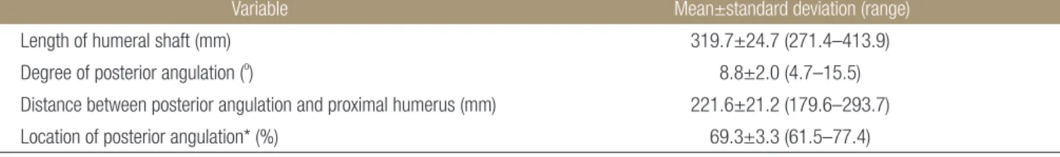
Table 1. Results of Measurement according to True Lateral Radiograph of Humerus
Variable Mean±standard deviation (range)
Length of humeral shaft (mm) 319.7±24.7 (271.4–413.9) Degree of posterior angulation (o) 8.8±2.0 (4.7–15.5) Distance between posterior angulation and proximal humerus (mm) 221.6±21.2 (179.6–293.7) Location of posterior angulation* (%) 69.3±3.3 (61.5–77.4)
*Distance between posterior angulation and proximal humerus/length of humeral shaft×100.
Shapiro-Wilk test p=0.652). 계측한 후방 만곡은 평균 8.8±2.0도 (4.7-15.5도)였다(Table 1).
고 찰
저자들은 상완골 원위부에 존재하는 후방 만곡에 대한 영상의학 적 계측을 통해 기존의 원위부 1/3에 존재하는 대략적인 각형성 측정 방법이 아닌 최대 각형성을 이루는 지점에서의 각도 및 각 형성을 이루는 지점까지의 거리를 분석하고자 하였다.
상완골 간부 골절 시 시행하는 골수강 내 금속정 삽입술은 금 속판과 나사 고정술과 함께 많이 사용하는 치료로, 여러 저자들 이 좋은 결과를 보고한 바 있다.4,5) 골수강 내 금속정 삽입술이 감 염이나 요골 신경 마비 등의 합병증에서 타 치료법과 큰 차이가 없으나 고령의 환자에서 견관절의 기능 제한이 더 발생할 수 있 다는 연구6) 및 술 후 발생할 수 있는 합병증 및 관절기능 면에서 불리하다는 것 등을 이유로 선호하지 않다고 보고된 연구가 있 다.7-9)
그러나 금속정 삽입술이 상완골 간부를 침범한 근위부 복합 골 절 등에서 좋은 결과를 얻었다는 연구10) 및 종양의 전이로 인한 증상이 있는 병적 골절의 증상 호전에 도움이 된다는 연구11)가 발 표된 바 있다. 골수강 내 금속정 삽입술을 시행한 경우 견관절의 움직임 제한이나 견관절의 충돌 위험이 높은 것으로 나타났지만 기능적 결과에서 타 치료법과 큰 차이가 없었다는 보고가 있고,12) Changulani 등13)은 비교연구에서 골수강 내 금속정 삽입술이 더 나은 치료법이라 보고하기도 하였다. Ristić 등14)은 상완골 간부 골절 환자를 4군으로 나누어 비교 연구한 결과 주관절 기능에서 골수강 내 금속정 삽입술이 더 나은 결과를 보였다고 보고하기도 하였다.
상완골 원위부의 해부학적인 후방 만곡에 대한 국내 연구가 보 고된 바 없고, 국외에서는 1개의 연구가 보고된 바 있다. Akpinar 등3)은 상완골의 대결절로 부터 원위부 1/3 지점까지의 거리와 평 균 각도를 측정한 결과 거리는 평균 21 mm, 후방 만곡의 각도는 9도였다고 보고하였다. 본 연구의 결과도 비슷한 결과를 보였다.
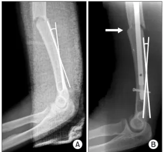
다만 Akpinar 등3)의 연구에서는 원위 2/3 지점을 정하고, 정해진 지점에서의 각도를 측정하였으나, 본 연구에서는 최대 각도를 먼 저 측정한 다음 최대 각도를 이루는 지점까지의 거리를 측정하는 방법을 사용하였다. 상완골의 원위부 2/3 지점을 먼저 지정한 다 음 각도를 측정할 경우, 후방 만곡의 최대 각도보다 적게 측정되 Figure 3. (A) True lateral radiograph of elbow before surgery shows
10 degrees of posterior angulation and 16 mm width of intramedullary canal. The fracture occurred approximately 70 mm proximally from the point of maximal posterior angulation. (B) Postoperative radiograph shows anterior angulation and displacement of the fracture site (arrow).
A B
Figure 4. (A) Insertion of an intramedullary nail should be done with caution when site of fracture (imaginary dashed line) is at the distal humeral shaft. Lateral view of the humeral shaft shows that the intramedullary nail impinges with the posterior cortex of the distal humeral shaft.
(B) Immediate postoperative radiograph shows posterior cortical breakage (arrow) resulting from impingement of the intramedullary nail with the distal humeral shaft due to posterior angulation of the humeral shaft.
는 오류를 줄이기 위해 본 연구에서는 최대 각도를 먼저 측정하 였다.
상완골 간부 골절 환자에서 본원 통계치인 평균 8.8도의 상완 골 원위부 후방 만곡은 골수강 내 금속정 삽입술을 시행함에 있 어 골절부에 존재하는 각형성 및 전위에 의해 큰 문제가 되지 않 을 것으로 예상되었다. 그러나 본 연구에서 측정된 후방 만곡의 정도가 15.5도로 큰 경우 골수강 내 금속정 삽입술 및 확공술 시 주의가 필요하며, 후방 만곡이 비교적 크고 골수강이 넓은 환자 에서 상완골 간부 골절이 발생하여 골수강 내 금속정 삽입술을 시행할 때 상완골 원위 골편이 전방 전위 및 전방 각형성을 이루 면서 고정되는 경우가 발생할 수 있다(Fig. 3). 술 전 주관절 진성 측면 사진에서 상완골 원위부 후방 만곡의 정도와 예상되는 금속 정의 길이를 분석함으로써 추가 발생할 수 있는 합병증을 예방할 수 있을 것으로 생각된다(Fig. 4).
본 연구의 임상적 의의는 상완골 간부 골절 환자에서 골수강 내 금속정 삽입술을 상완골 원위부의 후방 만곡을 고려하여 시 행하는 경우 상완골 원위 골편의 후방 피질골과 금속정 원위단 의 충돌 또는 추가 골절을 예방하고 골절부의 전위, 각형성 및 신 연을 예방하는 데 도움이 될 것으로 생각되며 수술적 방법에 있 어 후방만곡의 각도가 큰 경우 골수강 내 금속정 외에 금속판 고 정술을 선택함에 도움이 될 수 있겠다. 본 연구의 제한점으로는 Maro view 5.4 version을 이용한 영상의학적 방법을 사용한 계측 으로 실제 길이와 차이가 날 수 있으며 환자들이 가지고 있는 후 방 만곡에 대해 3차원적인 분석이 아닌 2차원적 분석, 만곡이라는 특징을 각 형성으로 단순화하여 측정한 점, 실제 골수강 내 금속 정 삽입술 시에는 골절부위의 각형성으로 인해 후방 만곡이 일부 상쇄되어 금속정의 끝단이 후방 피질골에 충돌하는 정도를 예상 하기 힘들다는 점을 들 수 있다.
결 론
본 연구의 결과, 평균적으로 상완골 근위부로부터 69.3% 지점의 위치에서 후방 만곡이 있음을 확인할 수 있었다. 이에 저자들은 상완골 간부 골절 환자에서 골수강 내 금속정 삽입술을 시행할 때 최대 각형성 포인트보다 짧은 골수정을 사용하여 금속정 원위 단의 충돌로 인한 추가 골절, 전위, 각형성 및 신연을 예방하는 데 도움이 될 것으로 생각된다.
CONFLICTS OF INTEREST
The authors have nothing to disclose.
REFERENCES
1. Crates J, Whittle AP. Antegrade interlocking nailing of acute humeral shaft fractures. Clin Orthop Relat Res. 1998;350:40- 50.
2. Lin J. Treatment of humeral shaft fractures with humeral locked nail and comparison with plate fixation. J Trauma.
1998;44:859-64.
3. Akpinar F, Aydinlioğlu A, Tosun N, Doğan A, Tuncay I, Unal O. A morphometric study on the humerus for intramedullary fixation. Tohoku J Exp Med. 2003;199:35-42.
4. Park SR, Lee TJ, Kim RS, Moon KH, You DS. Result of in- terlocking intramedullary nailing for humeral shaft fracture evaluation of post-operative shoulder function. J Korean Fract Soc. 2007;20:166-71.
5. Verdano MA, Pellegrini A, Schiavi P, Somenzi L, Concari G, Ceccarelli F. Humeral shaft fractures treated with antegrade intramedullary nailing: what are the consequences for the ro- tator cuff? Int Orthop. 2013;37:2001-7.
6. Khan AS, Afzal W, Anwar A. Comparison of shoulder func- tion, radial nerve palsy and infection after nailing versus pla- ting in humeral shaft fractures. J Coll Physicians Surg Pak.
2010;20:253-7.
7. Cox MA, Dolan M, Synnott K, McElwain JP. Closed interlock- ing nailing of humeral shaft fractures with the Russell-Taylor nail. J Orthop Trauma. 2000;14:349-53.
8. Denies E, Nijs S, Sermon A, Broos P. Operative treatment of humeral shaft fractures. Comparison of plating and intramed- ullary nailing. Acta Orthop Belg. 2010;76:735-42.
9. Raghavendra S, Bhalodiya HP. Internal fixation of fractures of the shaft of the humerus by dynamic compression plate or intramedullary nail: a prospective study. Indian J Orthop.
2007;41:214-8.
10. Garnavos C, Lasanianos N. Intramedullary nailing of com- bined/extended fractures of the humeral head and shaft. J Orthop Trauma. 2010;24:199-206.
11. Spencer SJ, Holt G, Clarke JV, Mohammed A, Leach WJ, Rob- erts JL. Locked intramedullary nailing of symptomatic metas- tases in the humerus. J Bone Joint Surg Br. 2010;92:142-5.
12. Kurup H, Hossain M, Andrew JG. Dynamic compres- sion plating versus locked intramedullary nailing for hu- meral shaft fractures in adults. Cochrane Database Syst Rev.
2011;6:CD005959.
13. Changulani M, Jain UK, Keswani T. Comparison of the use
of the humerus intramedullary nail and dynamic compres- sion plate for the management of diaphyseal fractures of the humerus. A randomised controlled study. Int Orthop.
2007;31:391-5.
14. Ristić V, Maljanoviv M, Arsić M, Matijević R, Milankov M.
Comparison of the results of treatment of humeral shaft frac- tures by different methods. Med Pregl. 2011;64:490-6.
상완골 간부 골절 시 골수강 내 금속정 고정술에 영향을 미칠 수 있는
상완골 원위부 후방 만곡의 단순방사선 사진 분석
염재광 • 부경환 • 성민규 • 장지석
인제대학교 의과대학 상계백병원 정형외과학교실
목적: 상완골 간부 골절에서 골수강 내 금속정 고정술을 시행할 때 고려해야 할 시상면에서의 상완골후방 만곡에 대한 연구는 국내 에 알려진 바가 없다. 상완골 진성 측면 방사선 사진의 분석을 통해 상완골 원위부의 후방 만곡의 위치와 정도 그리고 골수강 내 금속 정 고정술 시 유의할 점에 대해 분석하고자 하였다.
대상 및 방법: 상완골 단순 방사선 사진을 촬영한 99명의 환자를 대상으로 후향적 연구를 시행하였다. 진성 측면 사진에서 상완골의 전체 길이, 후방 피질골의 후방 만곡의 각도 및 각을 이루는 지점을 측정하여 분석하였다.
결과: 상완골의 평균 길이는 319.7 mm, 원위부 후방 피질골의 후방 만곡 각도는 평균 8.8도였다. 각을 이루는 지점은 근위부로부터 평균 221.6 mm 지점이었고 전체 길이의 69.3% 지점이었다.
결론: 상완골의 영상의학적 분석에서 평균적으로 상완골 근위부로부터 69.3% 지점의 위치에서 후방 만곡이 있음을 확인할 수 있었 다. 상완골에서의 골수강 내 금속정 삽입술 시 합병증을 예방하기 위하여 후방 만곡에 대한 고려가 필요하다.
색인단어: 상완골 골절, 상완골 후방만곡, 골수강 내 금속정 삽입술
접수일 2014년 7월 18일 수정일 2014년 9월 15일 게재확정일 2014년 12월 18일 책임저자 부경환
서울시 노원구 동일로 1342, 인제대학교 상계백병원 정형외과
TEL 02-950-1114, FAX 02-950-1429, E-mail [email protected]
Copyright © 2015 by The Korean Orthopaedic Association
“This is an Open Access article distributed under the terms of the Creative Commons Attribution Non-Commercial License (http://creativecommons.org/licenses/by-nc/3.0/) which permits unrestricted non-commercial use, distribution, and reproduction in any medium, provided the original work is properly cited.”