- 155 -
Allergic Fungal Sinusitis
- - -
- A Report of Two Cases - - - -
Sea-Yuong Jeon, M.D., Jong-Pil Byun, M.D., Jae-Yong Kang, M.D. and Jae-Hong Chon, M.D.
ABSTRACT
Allergic fungal sinusitis (AFS) is a benign, noninvasive form of fungal sinusitis. Histologically, AFS is characterized by all- ergic mucin, which consists of eosinophilic mucinous material with occasional laminar deposits of eosinophils. A diagnosis of AFS can be made when there is a demonstration of characteristic allergic mucin and an appearance of fungal hypae scattered within the mucin with no evidence of tissue invasion, or when fungi cultures yield positive results. Until now, no cases of AFS have been reported in Korea, though there have been many cases of mycetomas and a few cases of invasive fungal sinusitis. We present the first two cases of AFS in Korea. The patients demonstrated characteristic allergic mucin, fungal hypae scattered wit- hin the mucin, and no evidence of tissue invasion on histopathology. Nasal polyps, involvement of the unilateral sinuses, perip- heral eosinophilia, an elevated total IgE, and hyperattenuated masses observed in the CT supported the diagnosis of AFS. The patients recovered and did not display recurrence after surgery and treatment with topical steroids and saline irrigation.
KEY WORDS:Fungal sinusitis·Allergic fungal sinusitis·Allergic mucin·Eosinophils·Total IgE.
INTRODUCTION
Fungal sinusitis is classified into four distinct clinicopath- ologic forms:1) acute/fulminant invasive, 2) chronic/indolent invasive, 3) mycetoma, and 4) allergic fungal sinusitis (AFS).1) AFS is the most recently described,2) and there remains con- troversy over its classification.
AFS was originally termed“allergic aspergillus sinusitis”
because of its histopathologic similarity to“allergic bronch- opulmonary aspergillosis”(ABPA).2) However, it is now clear that most cases of AFS are caused by non-aspergillus species.
Consequently, the term allergic aspergillus sinusitis has been replaced by the more general term, AFS.3)4) Histologically, AFS is characterized by allergic mucin, which consists of eo- sinophilic mucinous material with occasional laminar deposits of eosinophils. Scattered through the mucin are Charcot-Ley- den crystals.4) A diagnosis of AFS can be made when the pat- ient demonstrates characteristic allergic mucin and one of the following:1) fungal hypae within the allergic mucin with no evidence of tissue invasion or 2) positive results in the fungi culture.4-6)
Until now, no cases of AFS have been reported in Korea, though there have been reports of many cases of mycetomas and a few cases of invasive fungal sinusitis in the country.7-10) We present two cases of AFS and review the pertinent lite- rature.
CASE REPORTS
Case 1A 40-year-old woman had a four-year history of nasal ob- struction and discharge. Examination showed mucopurulent discharge and polyps in the left nasal cavity. There was no history of previous nasal surgery or asthma. A CT showed a soft-tissue mass occupying the left nasal cavity, the maxillary, the anterior ethmoid, and the frontal sinus. There was evidence of bony erosion in the ostium and high densities in the opa- cified sinuses (Fig. 1). The peripheral eosinophil count was elevated up to 9%, and the total IgE was class 3 on a MAST CLA allergen specific IgE assay (MAST). However, no pos- itive antigens were determined by a MAST or by a skin prick test (with TORII allergens). The patient underwent an endo- scopic intranasal frontoethmoidectomy and a left Caldwell- Lucs operation. During surgery, it was found that a dark-brown, wet clay-like material filled the maxillary antrum. Polyps, antral content, and mucosa were taken for histopathologic examin- ation. Hematoxylin and eosin (HE) stained sections showed eosinophilic mucinous material with laminar aggregates of Department of Otolaryngology, GyeongSang National Univer-
sity Hospital, Chinju, Korea
Address correspondence and reprint requests to Sea-Yuong Jeon, M.D., Department of Otolaryngology, GyeongSang National University Chilamdong 90, Chinju 660-702, Korea
Tel:82-591-750-8174, Fax:82-591-759-0613 Accepted for publication on October 13, 1998
eosinophils and necrotic debris (Fig. 2A and B). Gomori me- thenamine-silver (GMS) stained sections showed a few iso- lated fungal hypae scattered through the allergic mucin. The hypae were not aggregated into mycetoma, and showed dic- hotomous branching (Fig. 2C). The fragments of antral mu- cosa were edematous and contained inflammatory cells, pre- dominantly eosinophils. No fungal hypae were observed in the mucosal fragment. The patient recovered after surgery and treatment with saline irrigation. A follow-up of three years showed no evidence of recurrence.
Case 2
A 48-year-old man had a three-year history of nasal obst- ruction. Examination showed mucoid discharge and minute polyps in the right middle meatus. There was no history of previous nasal surgery or asthma. A CT showed opacity in the right maxillary and the anterior ethmoid sinus. There was ev- idence of bony erosion in the ostium and high densities in the
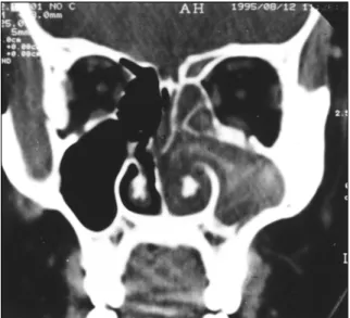
Fig. 1. A coronal CT scan of patient 1 showing a soft-tissue mass occupying the left nasal cavity, maxillary, anterior ethmoid. Bony erosion in the ostium and high densities in the opacified antrum are seen.
Fig. 2. A hematoxylin and eosin stained section of patient 1 showing eosinophilic mucinous ma- terial with laminar aggregates of inflammatory cells and necrotic cellular debris (A, ×200). Ag- gregated inflammatory cells (arrow) are dege- nerated eosinophils (B, ×400). A Gomori methe- namine-silver stained section shows a few isolated fungal hypae, some of which show dichotomous branching (C, ×400).
A A A A
BBB
B CCCC
opacified sinuses (Fig. 3). The peripheral eosinophil count was elevated up to 6.4%, and the total IgE was MAST class 2. A MAST revealed a positive IgE to white oak, rye, mugwort, and short ragweed. However, no fungal antigen-specific IgE were determined. The patient underwent endoscopic sinus surgery (ESS). During surgery, a dark-brown, thick pus-like material was observed filling the maxillary antrum. Polyps, antral co- ntent, and mucosa were taken for histopathologic examination.
HE-stained sections showed characteristic allergic mucin. In- tense infiltration of eosinophils, laminar aggregates of necrotic debris, and Charcot-Leyden crystals, which appeared as hex- agonal or rectangular crystals with the HE stain and which were stained purple by the GMS, were observed (Fig. 4A and B). The GMS-stained sections showed focal aggregates of fu- ngal hypae within the allergic mucin. Most of the hypae were fragmented and degenerated, but some of the hypae showed dichotomous branching (Fig. 4C). The fragments of mucosa showed eosinophilic infiltration, but no evidence of fungal in-
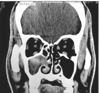
Fig. 3. A coronal CT of patient 2 showing opacity in the right maxillary and anterior ethmoid sinus. Bony erosion in the ostium and high densities in the opacified antrum are seen.
Fig. 4. A hematoxylin and eosin stained section of patient 2 showing characteristic allergic mu- cin with intense infiltration of eosinophils, and laminar aggregates of necrotic cellular debris (A, ×200) Charcot-Leyden crystals, hexagonal or rectangular in shape (arrowheads), are seen.
The cellular components on the background are degenerated eosinophils (B, ×400). A Gomori methenamine-silver stained section shows focal aggregates of weakly stained fungal hypae, most of which are fragmented and degenera- ted. (C, ×400)
A A A A
B B
BB CCCC
vasion. The patient recovered after surgery and a month-long treatment with a topical budesonide spray. A follow-up of ten months showed no evidence of recurrence.
DISCUSSION
We present the first two cases of AFS reported in Korea.
Seventeen cases of allergic fungal sinusitis and twelve cases of nonspecific inflammation of the unilateral sinuses from our department registry were reviewed. These represented posto- perative pathologic diagnoses made between 1987 and 1997 at the Gyeong-Sang National University Hospital. All histologic sections were reviewed. The clinical, radiological, and labor- atory findings were correlated. A diagnosis of AFS was made for all patients demonstrating characteristic allergic mucin, and fungal hypae within the mucin with no evidence of tissue invasion on histopathologic examination. Nasal polyps, unil- ateral involvement, peripheral eosinophilia, an elevated total IgE, and hyperattenuated masses found with CT examinations supported the diagnosis of AFS in our cases.
The diagnostic criteria of AFS are controversial. Strictly sp- eaking, the following criteria should be met for a diagnosis of AFS:1) type 1 hypersensitivity confirmed by history, skin tests, or serology, 2) nasal polyposis, 3) characteristic CT fi- ndings, 4) allergic mucin without fungal invasion into sinus tissue, and 5) positive fungal stain of sinus contents removed during surgery.3)11) However, most authors agree that a diag- nosis of AFS can be made with the following:1) demonstr- ation of characteristic allergic mucin, 2) fungal hypae within the allergic mucin with no evidence of tissue invasion, or cu- ltures for fungi showing positive results, and 3) the absence of immunodeficiency or diabetes.4-6)12) Immunologic evidence of type 1 hypersensitivity to specific fungal antigens is also thought to support a diagnosis of AFS. There are two reasons for this. First, in AFS, the majority of the causative fungal ag- ents is dematiaceous, and commercial detection of a specific antibody to dematiaceous fungi has not yet been made. Seco- ndly, there is still controversy in the literature about the role of hypersensitivity versus true infection in the pathogenesis of AFS.4) In our cases, a MAST failed to demonstrate the sp- ecific IgE of fungal antigens (Alternaria, Aspergillus, Clado- sporium, Fusarium, Candia, Stemphylium or Penicillium). A skin prick test for mold mix antigens (Alternaria, Phoma, Ho- rmodondrum, Helminthosporum, Aspergillus mix, Penicillium mix, Fusarium, Rhizopus, Mucor, and Pullularia) generated negative results in case 1. However, the peripheral eosinop- hilia and the elevated total IgE found in the patients of this study suggest them to be atopic, and these findings may evi- dence type 1 hypersensitivity.
Allergic mucin, a pathognomonic finding of AFS, is defined
as eosinophilic mucinous material with occasional laminar de- posits of eosinophils. Scattered through the mucin are Charcot- Leyden crystals, the degradation product of eosinophils.3)4) Samples taken from the patients in this study showed typical allergic mucin in HE-stained sections and scattered fungal hypae in the allergic mucin in GMS-stained sections. However, Charcot-Leyden crystals, appearing hexagonal in a longitud- inal section or rectangular or bipyramidal in a longitudinal section,3)13) were observed in case 2. Charcot-Leyden crystals are not observed in every case of AFS.3)
The radiographic characteristics of AFS are CT findings of central areas of hyperattenuation that correspond to hypointe- nse signals on a T1-weighted MR and signal void with a T2- weighted MR. CT findings showing bony erosion and hyper- attenuation are identical to those demonstrating mycetomas.14) CTs conducted on the patients in this study revealed findings that corresponded with previous reports of AFS.3)4)14) An MRI was not performed in either of these cases.
The surgical treatment of AFS consists of removing the fungus and allergic mucin and restoring sinus drainage and ventilation. Although systemic steroids have been used in a few cases, nasal topical steroids are a mainstay of treatment.
Antifungal agents are not considered necessary when fungus has not invaded the tissue. Unlike with ABPA, in which cases surgical extirpation of all of the allergic mucin is not possible, a surgical cure for AFS is possible. If all of the allergic mu- cin can be removed, either surgically or with surgery and ste- roids, the patient is cured of AFS.4)13) In case 1, the maxillary antrum was accessed by a Caldwell-Lucs operation, at which time the antral content was removed completely and mucosal specimens were taken. In case 2, however, the thick pus-like antral content could be removed completely with a wide mid- dle meatal antrostomy performed via ESS. In cases where AFS is suspected, the antral content should be removed completely and collected for histopathologic examination. It has been reported that one characteristic of AFS is the recurrence of symptoms, so topical steroids are used extensively.3)4) In case 1, we did not apply postoperative topical steroids because we had no concept of AFS at that time. In case 2, we applied po- stoperative topical steroids. No systemic antifungal agents were indicated in either cases. However, there has been no recurr- ence in our cases.
In summary, two cases of AFS are presented. The patients demonstrated characteristic allergic mucin, scattered fungal hypae within the mucin, and no evidence of tissue invasion on histopathology. Nasal polyps, involvement of the unilate- ral sinuses, peripheral eosinophilia, an elevated total IgE, and hyperattenuated masses in the CT were supportive of the di- agnosis of AFS. The patients recovered after surgery and tre- atment with topical steroids and saline irrigation, and there has
been no recurrence.
REFERENCES
1) Morrpeth JF, Rupp NT, Dolen WK, Bent JP. Kuhn FA. Fungal si- nusitis: An update. Ann Allergy Asthma Immunol 1996;78:128-39.
2) Katzenstein ALA, Sale SR, Greenberger PA. Allergic aspergillus sinusitis: A newly recognized form of sinusitis. J Allergy Clin Im- munol 1983;72:89-93.
3) Bent III JP, Kuhn FA. Diagnosis of allergic fungal sinusitis. Otol- aryngol Head Neck Surg 1994;111:580-8.
4) Corey JP, Delsupehe KG, Ferguson BJ. Allergic fungal sinusitis:
Allergic, infectious, or both? Otolaryngol Head Neck Surg 1995;
113:110-9.
5) Cody II DT, Neel III HB, Ferreiro JA, Roberts GD. Allergic Fu- ngal Sinusitis: The Mayo Clinic Experience. Laryngoscope 1994;
104:1074-9.
6) Waxman JE Spector JG, Sale SR. Allergic Aspergillus Sinusitis:
Concepts in Diagnosis and Treatment of a New Clinical Entity.
Laryngoscope 1987;97:261-6.
7) Min YG, Kang MK, Lee JW, Choo MJ, Lee KS. A clinical study
of mycotic sinusitis. Korean J Otolaryngol 1993;36:292-301.
8) Ma YW, Hong SK, Jeon SY, Hwang EG, Kim CS, Kim JP. A clinical study on aspergillus sinusitis. Korean J Otolaryngol 1993;
26:727-33.
9) CG Kim, Park JJ, Kim HS, Jeon SY. A case of frontal invasive aspergillus sinusitis with intracranial involvement. J Clin Otolar- yngol Head Neck Surg 1997;8:317-20.
10) Lee BJ, Kim H, Kim YJ. Fungal sinusitis: Clinical features and treatment outcomes with emphasis on endoscopic sinus surgery.
Korean J Otolaryngol 1998;41:318-22.
11) Manning SC Mabry RL, Schaefer SD, Close LG. Evidence of IgE- mediated Hypersensitivity in Allergic Fungal Sinusitis. Laryngos- cope 1993;103:717-21.
12) deShazo RD, Swain RE. Diagnostic criteria fro allergic fungal si- nusitis. J Allergy Clin Immunol 1995;96:244-35.
13) Friedmann GC, Hartwick RWJ, Ro JY, Saleh GY, Taarrand JJ, Ayala AG. Allergic fungal sinusitis. Report of three cases assoc- iated with Dematiaceous fungi. Am J Clin Pathol 1991;96:368-72.
14) Manning SC, Merkel M, Krrriesel K, Vuitch F, Marrple B. Comp- uted tomography and magnetic resonance diagnosis of allergic fu- ngal sinusitis. Laryngoscope 1997;107:170-6.