Open Access
Dummy Run of Quality Assurance Program before Prospective Study of
Hippocampus-Sparing Whole-Brain Radiotherapy and Simultaneous Integrated Boost for Multiple Brain Metastases from Non-small Cell Lung Cancer:
Korean Radiation Oncology Group (KROG) 17-06 Study
Original Article
Purpose
Lung Cancer Subcommittee of Korean Radiation Oncology Group (KROG) has recently launched a prospective clinical trial (KROG 17-06) of hippocampus-sparing whole brain radiotherapy (HS-WBRT) with simultaneous integrated boost (SIB) in treating multiple brain metastases from non-small cell lung cancer. In order to improve trial quality, dummy run studies among the participating institutions were designed. This work reported the results of two-step dummy run procedures of the KROG 17-06 study.
Materials and Methods
Two steps tested hippocampus contouring variability and radiation therapy planning com- pliance. In the first step, the variation of the hippocampus delineation was investigated for two representative cases using the Dice similarity coefficients. In the second step, the par- ticipating institutions were requested to generate a HS-WBRT with SIB treatment plan for another representative case. The compliance of the treatment plans to the planning protocol was evaluated.
Results
In the first step, the median Dice similarity coefficients of the hippocampus contours for two other dummy run cases changed from 0.669 (range, 0.073 to 0.712) to 0.690 (range, 0.522 to 0.750) and from 0.291 (range, 0.219 to 0.522) to 0.412 (range, 0.264 to 0.598) after providing the hippocampus contouring feedback. In the second step, with providing additional plan priority and extended dose constraints to the target volumes and normal structures, we observed the improved compliance of the treatment plans to the planning protocol.
Conclusion
The dummy run studies demonstrated the notable inter-institutional variability in delineating the hippocampus and treatment plan generation, which could be decreased through feed- back from the trial center.
Key words
Hippocampus-sparing whole brain radiotherapy, Dummy run, Multi-institutional study, QA program, Non-small cell lung cancer
Eunah Chung, PhD1 Jae Myoung Noh, MD, PhD1 Kyu Chan Lee, MD, PhD2 Jin Hee Kim, MD, PhD3 Weon Kuu Chung, MD, PhD4 Yang-Gun Suh, MD5 Jung Ae Lee, MD6 Ki Ho Seol, MD7
Hong Gyun Wu, MD, PhD8 Yeon Sil Kim, MD, PhD9 O Kyu Noh, MD, PhD10 Jae Won Park, MD11 Dong Soo Lee, MD12 Jihae Lee, MD13 Young Suk Kim, MD14 Woo-Yoon Park, MD, PhD15 Min Kyu Kang, MD, PhD16 Sunmi Jo, MD17
Yong Chan Ahn, MD, PhD1,18
+ + + + + + + + + + + + + + + + + + + + + + + + + + + + + + + + + + + + + + + + + + + + + + + + + + + + + + + + + + + + + + + + + + + + + + + + + + + + + + + + + + + + + + + + + + + + + + + + + + + + + + + + + + + + + + + + + + + + + + + + + + + + + + + + + + + + + + + + + + + + + + + + + + + + + + + + + + + + + + + + + + + + + + + + + + + + + + + + + + + + + + + + + + + + + + + + + + + + + + + + + + + + + + + + + + + + + + + + + + + + + + + + + + + + + + + + + + + + + + + + + + + + + + + + + + + + + + + + + + + + + + + + + + + + + + + + + + + + + + + + + + + + + + + + + + + + + + + + + + + + + + + + + + + + + + + + + + + + + + + + + + + + + + + + + + + + + + + + + + + +
Correspondence: Yong Chan Ahn, MD, PhD Department of Radiation Oncology, Samsung Medical Center, Sungkyunkwan University School of Medicine, 81 Irwon-ro, Gangnam-gu, Seoul 06351, Korea Tel: 82-2-3410-2602
Fax: 82-2-6190-5332 E-mail: [email protected]
Received July 21, 2018 Accepted October 12, 2018 Published Online October 15, 2018
*Eunah Chung and Jae Myoung Noh contributed equally to this work.
*A list author’s a!liations appears at the end of the paper.
Introduction
Whole-brain radiation therapy (WBRT) has long been one of the standard treatment options in treating the patients with brain metastasis from various malignancies [1,2]. Stereo- tactic radiosurgery (SRS) is an aggressive local treatment option and has been recommended, either alone or in con- junction with WBRT, to the selected patients. The indications for SRS usually include the limited number of metastatic lesions (! 3 to 4 lesions), relatively small metastatic tumor size (! 4 cm), well-controlled extracranial disease, and good performance status. Compared with SRS alone, WBRT in addition to SRS, though not associated with survival impro- vement, has the advantage of less frequent new metastatic lesions in the brain in addition to improved local control [3-6]. Neurocognitive dysfunction attributable relation to WBRT, meanwhile, has remained one of the major concerns.
To ameliorate WBRT-related neurocognitive dysfunction, hippocampus-sparing WBRT (HS-WBRT), which can achieve conformal dose reduction to the hippocampus, has been sug- gested as a solution [7,8] and demonstrated improved mem- ory preservation thorough the Radiation Therapy Oncology Group (RTOG) 0933 prospective trial [9].
Simultaneous integrated boost (SIB) during HS-WBRT, which can deliver higher dose to the grossly metastatic lesi- ons while keeping the hippocampus from high radiation dose, has recently been tried. A few planning studies tested the technical feasibility of volumetric-modulated arc therapy (VMAT) in HS-WBRT with SIB [10,11]. Moreover, a few clin- ical studies reported the efficacy and safety of this approach in treating the patients with multiple brain metastases, and reported no grade " 3 toxicities [12,13]. These studies, unlike the RTOG 0933 trial, have the weakness of lacking routine neurocognitive function evaluation, which can reflect the effect of HS-WBRT. Based on these backgrounds, the Korean Radiation Oncology Group (KROG) has recently launched a prospective phase II multi-institutional trial of HS-WBRT with SIB in treating the patients with multiple brain metas- tases from non-small cell lung cancer, which incorporates the Seoul-Verbal Learning Test as the neurocognitive function evaluation (KROG 17-06, NCT03366376).
As Gondi et al. [14] previously described, the pre-treat- ment centralized review in the RTOG 0933 trial was able to prevent the unacceptable protocol deviations significantly.
The necessity of the pre-treatment review process was agreed on among the members of the KROG’s lung cancer research section, and we conducted a series of dummy run studies before launching the main trial. Authors would report the results of the variations and variabilities in the target and hippocampus delineation, radiation dose distribution, and treatment technique.
Materials and Methods
1. Treatment planning protocol
The treatment planning protocol is summarized in Table 1, which was primarily based on the RTOG 0933 protocol [9]
and has been modified through a preliminary planning exercise by the participating institutions. The planning gross tumor volume (P-GTV) was defined by 2 mm expansion of the gross tumor volume (GTV), each of which has a dimen- sion greater than 3 mm. The planning clinical target volume (P-CTV) was defined as the whole brain parenchyma down to the bottom of C1 spine body excluding the hippocampus region, which was the 5 mm expansion of the actual hip- pocampus 3-dimensionally. Using the current protocol, a preliminary planning exercise was performed by the partic- ipating institutions to assess the variability in the treatment planning and protocol compliance. Following this prelimi- nary exercise, we recognized fairly large inter-institutional diversity in the planning techniques, even though all were to be based on the proposed protocol. To improve the com- pliance to the protocol, we designed two-step dummy run procedures and analyzed (1) hippocampus contouring vari- ability and (2) radiation therapy planning compliance and variability with contour delineation among the participating institutions.
2. Step 1: variation of hippocampus contouring
Computed tomography (CT) and magnetic resonance (MR) images taken on two anonymized patients (patients A and B), who actually had received HS-WBRT without SIB, were provided to all participants. The CT images were obtained using 120 kVp, 400 mA, 1.25 mm slice thickness.
The axial T1-weighted multi-planar reconstructed MR images with a 1.0 mm slice thickness were acquired using 3T magnetic flux. Both CT and MR images were obtained with contrast enhancement. For each case, the participants were asked to delineate the hippocampus, P-CTV and other sur- rounding normal structures on the CT image and to generate the treatment plans according to the protocol (Table 1).
Fusion of the MR images to the CT images was mandatorily requested in contouring the hippocampus. The CT-MR image fusion was performed semi-empirically using non- deformable image registration method. The treatment plan- ning priority was proposed by the following order: (1) optic chiasm, (2) left and right optic nerve, (3) hippocampus, (4) left and right lens, and (5) P-CTV. The structure set and radiation dose of each plan were saved as Digital Imaging and Communications in Medicine RT format and then imported into the MIM workstation (MIM Software Inc.,
Cleveland, OH). When transporting the plan data between different treatment planning systems (TPS) and MIM work- station, we did not observe any significant image or contour distortion which may impact to calculate the dose distribu- tion. The inter-institutional variation of the hippocampus delineation was evaluated using the Dice similarity coeffi- cients (DSC) [15,16] 3-dimensionally. The DSC ranges from 0 to 1, where 0 means that two contours are not similar at all and 1 means that two contours are perfectly matched. The DSC value between 0 and 1 indicates that two contours are partially overlapped. The dose distribution to the hippocam- pus contours was compared with the reference radiation dose file. The grid size of dose calculation was 2!2!2 mm3. The difference in the hippocampus delineation before and after providing the hippocampus contouring feedback was also analyzed.
3. Step 2: treatment plan compliance
CT and corresponding MR images of another anonymized patient (patient C), who actually had received HS-WBRT with SIB, was distributed to the participating institutions.
The participants were requested to delineate the GTV and the normal structures including the hippocampus, and to generate the treatment plans according to the planning pro- tocol as summarized in Table 1. These plans were uploaded in the same method as in step 1 and the inter-institutional variation of the GTV and hippocampus delineations as well as the radiation dose distributions to the target and normal structures were evaluated.
4. Ethical statement
This protocol was approved by KROG (KROG 17-06) and the Institutional Review Board of Samsung Medical Center (2017-08-070) and performed in accordance with the princi- ples of the Declaration of Helsinki. The informed consent was waived.
Results
1. Step 1: variation of hippocampus contouring
During the step 1, the contoured structure sets and radia- tion dose files were submitted by ten and nine institutions before and after the provision of feedback on hippocampus contouring. Figs. 1 and 2 summarize the inter-institutional variation of the hippocampus contouring among the partic- ipants on patients A and B, respectively. The structure set from the trial center was selected as the reference in calcu- lating the DSC of the hippocampus contouring. The radiation dose file from the same institution was also selected as the reference in evaluating the radiation dose distribution. For patient A, before providing the hippocampus contouring feedback, the median volume and DSC of the hippocampus were 4.21 cm3(range, 2.23 to 8.41 cm3) and 0.669 (range, 0.073 to 0.712), respectively. After providing the feedback, these values were 5.04 cm3 (range, 2.95 to 6.03 cm3) and 0.690 (range, 0.522 to 0.750), respectively. For patient B, before pro- viding the hippocampus contouring feedback, the median
Target/Normal structure Constraint
Per protocol Acceptable
P-GTVa) V95%" 40 Gy V93%" 40 Gy
P-CTVb) V95%" 25 Gy V90%" 25 Gy
Hippocampus Maximum # 20 Gy Maximum # 22 Gy
Maximum to 0.1 cm3 Maximum to 0.1 cm3
# 16 Gy # 18 Gy
Mean # 12 Gy Mean # 14 Gy
Optic chiasm Maximum # 30 Gy N/A
Optic nerve, both Maximum # 30 Gy N/A
P-Lensc), both Maximum # 5 Gy Maximum # 6 Gy
Eyeball, both Maximum # 10 Gy Maximum # 12 Gy
Table 1. The treatment planning protocol of KROG 17-06 study
a)P-GTV: planning gross tumor volume=gross target volume (GTV)+2 mm margin, b)P-CTV: planning clinical target vol- ume=whole brain–hippocampal avoiding region (hippocampus+5 mm margin), c)P-Lens: planning volume for lens=lens+5 mm margin.
volume and DSC of hippocampus were 5.10 cm3(range, 2.43 to 10.19 cm3) and 0.291 (range, 0.219 to 0.522), respectively.
After providing the feedback, these values were 5.00 cm3 (range, 2.52 to 9.72 cm3) and 0.412 (range, 0.264 to 0.598), respectively. After providing the hippocampus contouring
feedback, the median DSC were greater in both cases, which meant that the conformity of hippocampus delineation among the participants was improved. For patient A, the inter-institutional variation of hippocampus dose distribu- tion was notably decreased after providing the contouring Fig. 1. Hippocampus delineation in a computed tomography image for patient A before (A) and after providing the hip- pocampus contouring feedback (B). (C) Change of hippocampus volume-to-reference volume ratio delineated by each par- ticipating institution. (D) Dice similarity coefficients before and after providing the feedback in the scatter plots with median lines. (E, F) The inter-institutional variation of the dose distributions to the delineated hippocampus contours from reference dose distribution before and after providing the feedback, respectively.
A
C
E
Relative volume (%)
110100
0 20 40 60
0
Dose (Gy) 4
2 6 8 10121416 1820222426 2830 8090
70 50 30 10
Volume to reference volume ra
tio 3.0 2.5
0 0.5 1.0 1.5
After Before
2.0
B
D
Dice similarity coefficient 1.0
0.8
0 0.2 0.4
After Before
0.6
AF B
G C
H D
I E
J
F
Relative volume (%)
110100
0 20 40 60
0
Dose (Gy) 4
2 6 8 10121416 1820222426 2830 8090
70 50 30 10
AF B
G C
H D
I E
feedback as shown in Fig. 1E and F. However, for patient B, the dose distribution variation for modified hippocampus contours among the participating institutions was not reduced significantly as shown in Fig. 2E and F. As summa- rized in Fig. 2C, the scatter degree of volume to reference vol-
ume ratio of the hippocampus contours for patient B was similar before and after providing the feedback, while it was reduced greater for patient A as plotted in Fig. 1C. We spec- ulate that the greater variation of hippocampus volume delineation for patient B might induce the smaller change in Fig. 2. Hippocampus delineation in a computed tomography image for patient B before (A) and after providing the hip- pocampus contouring feedback (B). (C) Change of hippocampus volume-to-reference volume ratio delineated by each par- ticipating institution. (D) Dice similarity coefficients before and after providing the feedback in the scatter plots with median lines. (E, F) The inter-institutional variation of the dose distributions to the delineated hippocampus contours from an iden- tical radiation dose distribution before and after providing the feedback, respectively.
A
C
E
Relative volume (%)
110100
0 20 40 60
0
Dose (Gy) 4
2 6 8 10121416 1820222426 2830 8090
70 50 30 10
Volume to reference volume ra
tio 3.0 2.5
0 0.5 1.0 1.5
After Before
2.0
B
D
Dice similarity coefficient 1.0
0.8
0 0.2 0.4
After Before
0.6
AF B
G C
H D
I E
J
F
Relative volume (%)
110100
0 20 40 60
0
Dose (Gy) 4
2 6 8 10121416 1820222426 2830 8090
70 50 30 10
AF B
G C
H D
I E
the inter-institutional variation of the hippocampus dose dis- tribution before and after providing the contouring feedback.
2. Step 2: treatment plan compliance
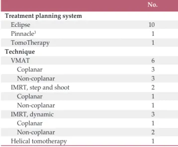
A total of 12 institutions participated in the step 2. Table 2 summarizes the TPS and techniques; ten institutions used Eclipse (Varian Medical Systems, Palo Alto, CA) as their TPS;
one did Pinnacle3(Philips Health Care, Andover, MA); and one did TomoTherapy (Accuray Inc., Sunnyvale, CA), res- pectively. Various techniques, including VMAT and step- and-shoot or dynamic intensity-modulated radiotherapy (IMRT) with or without couch rotation (non-coplanar or coplanar), were used. These TPS were used for generating actual patient treatment plans in the participating institu- tions. Therefore, the accuracy of TPS commissioning has been already established before participating in this dummy run study.
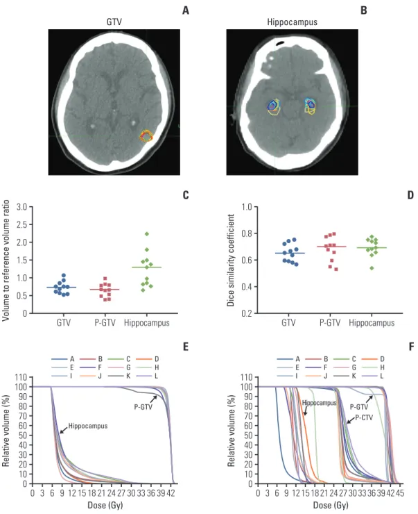
Similar to the step 1, one structure set from the trial center, was chosen as the reference structure set and the plan from the same institution was selected as the reference dose dis- tribution. Fig. 3A-E shows the GTV and hippocampus delin- eations on the step 2 (patient C), the contoured volume-to- reference volume ratio, and the corresponding DSC for the GTV, P-GTV and hippocampus, and the inter-institutional variations of the radiation dose distribution to the P-GTV and hippocampus when compared to the reference dose dis- tribution. The median volume and DSC of the GTV were 8.49 cm3(range, 6.02 to 12.1 cm3) and 0.653 (range, 0.567 to 0.752),
respectively. For the P-GTV, the median volume and DSC were 22.2 cm3(range, 12.8 to 32.2 cm3) and 0.708 (range, 0.533 to 0.801), respectively. For the hippocampus, the median vol- ume and DSC were 4.46 cm3(range, 2.56 to 8.68 cm3) and 0.692 (range, 0.538 to 0.777), respectively. Even though the hippocampus volume had wider range, the range of the DSC was similar to those of the GTV and P-GTV. When using the reference dose distribution, it was shown that the dose dis- tribution to the P-GTV defined by one institution (institution K) was much lower than the others (Fig. 3E). This was because institution K did not follow the planning protocol and delineated all 12 brain metastases including three metas- tases smaller than 3 mm as the GTV.
Fig. 3F and Table 3 summarize the radiation dose distribu- tion to the P-GTV, P-CTV and hippocampus and the number of the treatment plans that satisfied the dose constraints to the P-GTV, P-CTV, hippocampus and surrounding normal structures for patient C. Each dummy run treatment plan was generated using the contours delineated by each partic- ipant. Seven out of 12 institutions satisfied the dose con- straint to P-GTV when expanding the dose constraint to the variation acceptable limit. One institution (institution I), which did not meet the dose constraint to the P-GTV, gener- ated the plan by optimizing the dose delivery to 30 Gy instead of 40 Gy for one of the 12 brain metastases. This was because this contour partially overlapped with the hip- pocampal avoiding region, which was 5 mm extended from the hippocampus. Most of the dummy run treatment plans (11 out of 12) satisfied the dose constraint to the P-CTV with the acceptable variation limit. The variation of the dose dis- tribution to the hippocampus was greater than those to P-GTV and P-CTV due to the relatively small size of the hip- pocampus. For the hippocampus, nine out of 12 treatment plans satisfied the suggested planning protocol with the acceptable variation limit. For the optic chiasm and right and left optic nerves, most of the plans satisfied the dose con- straints. For the planning volume for lens and eyeballs, except for one institution for each structure, all institutions generated the treatment plans satisfying the dose constraints in the acceptable variation limit. Compared to the prelimi- nary planning experiment (data not shown), we found that the compliance of the treatment plans to the planning proto- col were improved by applying the plan priority and exten- ded dose constraints to the treatment planning.
Discussion
Unacceptable deviations from the radiation therapy pro- tocol have been demonstrated to have a negative impact on Table 2. Summary of the treatment planning systems and
techniques for the dummy run study
VMAT, volumetric-modulated arc therapy; IMRT, inten- sity-modulated radiotherapy.
No.
Treatment planning system
Eclipse 10
Pinnacle3 1
TomoTherapy 1
Technique
VMAT 6
Coplanar 3
Non-coplanar 3
IMRT, step and shoot 2
Coplanar 1
Non-coplanar 1
IMRT, dynamic 3
Coplanar 1
Non-coplanar 2
Helical tomotherapy 1
Fig. 3. Gross tumor volume (GTV) (A) and hippocampus delineation (B) in a computed tomography image for patient C.
(C) Contoured volume-to-reference volume ratio delineated by each participating institution. (D) Dice similarity coefficients of GTV, planning gross tumor volume (P-GTV) and hippocampus in the scatter plots with median lines. (E) Inter-institutional variation of the dose distribution to the P-GTV and hippocampus contours from a reference radiation dose distribution pro- vided from the trial center. (F) Dose-volume histograms for P-GTV, planning clinical target volume (P-CTV) and hippocam- pus contours from the 12 dummy run treatment plans for patient C. Each treatment plan was generated based on the two target volumes (P-GTV and P-CTV), hippocampus and other normal structures which were delineated by each participating institution.
A
C
Volume to reference volume ra
tio 3.0 2.5
0 0.5 1.0 1.5
Hippocampus P-GTV
Hippocampus
P-GTV
GTV 2.0
B
D
Dice similarity coefficient 1.0
0.8
0.2 0.4 0.6
E
Relative volume (%)
110100
0 20 40 60
0
Dose (Gy) 6
3 9 12 15182124 2730333639 42 8090
70 50 30 10
AE I
BF J
CG K
DH L
F
Relative volume (%)
110100
0 20 40 60
0
Dose (Gy) 6
3 9 12 15 2118 24 27 3330 3639 4245 8090
70 50 30 10
AE I
BF J
CG K
DH L
GTV Hippocampus
Hippocampus P-GTV
GTV
Hippocampus P-GTV P-CTV
the clinical outcomes [17-19]. Since HS-WBRT with SIB involves a complex technique of IMRT requiring steep dose gradient around the gross metastatic lesions and hippocam- pus, the protocol deviation could affect the doses to the target volumes and hippocampus and clinical outcomes subse- quently. The primary endpoint of the KROG 17-06 study is the intracranial progression-free survival and the secondary endpoints are the verbal neurocognitive function, overall survival, toxicity, and quality of life. As these endpoints could be influenced by protocol deviations, the comprehen- sive pre-treatment dummy run as a quality assurance pro- gram seems quite essential in conducting this kind of pros- pective study.
In these dummy run procedures, we found several varia- tions in terms of delineating the target volumes, hippocam- pus and the radiation therapy planning. First of all, the delineation of the hippocampus was quite diverse, although the participants were advised to refer to the contouring atlas for hippocampal delineation on the RTOG website [20]. For re-orientation of the participants, an example case of hip- pocampus contouring on CT/MR fused images was pro- vided from the trial center as a feedback, which helped decreasing the degree of hippocampus delineation diversity.
In addition to sparing the hippocampus, our protocol would involve SIB and IMRT technique to improve the local control of the gross metastatic lesions. The eligibility criteria for the KROG 17-06 study include the patients with (1) three or more brain metastases, (2) with ! 3 mm in size, and (3) lesions located outside the 5-mm margin around the hippocampi on both sides. Therefore, the great variation in GTV delineation was induced by one institute’s delineating the small enhanc- ing lesion smaller than 3 mm. Furthermore, the closest dis- tance between the GTV and hippocampus was almost 5 mm in patient C, which made it difficult to fulfill the planning protocol. Authors, however, would expect that this rather
tough dummy run study could have enhanced the quality of planning procedures by the participants.
In the pilot experiment, we found the considerable varia- tions in the radiation dose to the target volume and hip- pocampus and the real-time transport protocol at the partici- pating institutions seemed more or less preliminary and lacked detail. Through these pilot studies, we could develop more detailed planning protocol and improve the compli- ance to the treatment plan protocol, though tough and diffi- cult cases were selected (Fig. 3). Application of the plan priority and extended dose constraints were quite important in these processes.
Our dummy run studies have a few potential limitations.
There were still variations in delineating the target volumes and hippocampus and patient C might not have been an ideal case for this type of dummy run practice. It might be helpful to repeat a third dummy run procedure in order to further reduce the variations. As shown in Table 2, the majority of the planning techniques were linear accelerator (LINAC)-based IMRT including VMAT, while helical tomo- therapy technique was used in only one institution. There- fore, we could not compare LINAC-based IMRT and helical tomotherapy as Gondi et al. [7] did.
In conclusion, there were notable inter-institutional differ- ences in planning HS-WBRT with SIB, including hippocam- pus contouring and target volume delineation. Feedback and re-orientation could reduce the variations, and detailed dose specification with plan priority is quite essential before launching multi-institutional study involving complex IMRT planning.
Conflicts of Interest
Conflict of interest relevant to this article was not reported.
Target/Normal structure No. of treatment plans satisfying the dose constraints
Per protocol Acceptable
P-GTV 3 7
P-CTV 6 11
Hippocampus, maximum 6 9
Hippocampus, maximum to 0.1 cm3 5 10
Hippocampus, mean 6 10
Optic chiasm, maximum 11 -
Optic nerve (right, left), maximum 12, 12 -
P-Lens (right, left), maximum 8, 8 12, 11
Eyeball (right, left), maximum 9, 9 11, 11
Table 3. Summary of the results of 12 dummy run treatment plans for patient C
P-GTV, planning gross tumor volume; P-CTV, planning clinical target volume; P-Lens, planning volume for lens.
Author Details
1Department of Radiation Oncology, Samsung Medical Center, Sungkyunkwan University School of Medicine, Seoul, 2Department of Radiation Oncology, Gachon University Gil Medical Center, Incheon, 3Department of Radiation Oncology, Keimyung University Dongsan Medical Center, Daegu, 4Department of Radiation Oncol- ogy, Kyung Hee University Hospital at Gangdong, Seoul, 5Proton Therapy Center, National Cancer Center, Goyang, 6Department of Radiation Oncology, Korea University Guro Hospital, Seoul,
7Department of Radiation Oncology, Catholic University of Daegu School of Medicine, Daegu, 8Department of Radiation Oncology, Seoul National University Hospital, Seoul, 9Department of Radia- tion Oncology, College of Medicine, The Catholic University of
Korea, Seoul, 10Department of Radiation Oncology, Ajou University School of Medicine, Suwon, 11Department of Radiation Oncology, Yeungnam University College of Medicine, Daegu, 12Department of Radiation Oncology, Uijeongbu St. Mary's Hospital, College of Med- icine, The Catholic University of Korea, Uijeongbu, 13Department of Radiation Oncology, Ewha Womans University School of Medicine, Seoul, 14Department of Radiation Oncology, Jeju National Univer- sity School of Medicine, Jeju, 15Department of Radiation Oncology, Chungbuk National University College of Medicine, Cheongju,
16Department of Radiation Oncology, School of Medicine, Kyung- pook National University, Daegu, 17Department of Radiation Oncology, Inje University School of Medicine, Busan, 18Department of Medical Device Management and Research, SAIHST, Sungkyun- kwan University, Seoul, Korea
1. Gaspar LE, Mehta MP, Patchell RA, Burri SH, Robinson PD, Morris RE, et al. The role of whole brain radiation therapy in the management of newly diagnosed brain metastases: a sys- tematic review and evidence-based clinical practice guideline.
J Neurooncol. 2010;96:17-32.
2. Tsao MN, Rades D, Wirth A, Lo SS, Danielson BL, Gaspar LE, et al. Radiotherapeutic and surgical management for newly diagnosed brain metastasis(es): an American Society for Radi- ation Oncology evidence-based guideline. Pract Radiat Oncol.
2012;2:210-25.
3. Aoyama H, Shirato H, Tago M, Nakagawa K, Toyoda T, Hatano K, et al. Stereotactic radiosurgery plus whole-brain radiation therapy vs stereotactic radiosurgery alone for treat- ment of brain metastases: a randomized controlled trial. JAMA.
2006;295:2483-91.
4. Chang EL, Wefel JS, Hess KR, Allen PK, Lang FF, Kornguth DG, et al. Neurocognition in patients with brain metastases treated with radiosurgery or radiosurgery plus whole-brain irradiation: a randomised controlled trial. Lancet Oncol.
2009;10:1037-44.
5. Kocher M, Soffietti R, Abacioglu U, Villa S, Fauchon F, Baumert BG, et al. Adjuvant whole-brain radiotherapy versus observation after radiosurgery or surgical resection of one to three cerebral metastases: results of the EORTC 22952-26001 study. J Clin Oncol. 2011;29:134-41.
6. Aoyama H, Tago M, Shirato H; Japanese Radiation Oncology Study Group 99-1 (JROSG 99-1) Investigators. Stereotactic radiosurgery with or without whole-brain radiotherapy for brain metastases: secondary analysis of the JROSG 99-1 ran- domized clinical trial. JAMA Oncol. 2015;1:457-64.
7. Gondi V, Tolakanahalli R, Mehta MP, Tewatia D, Rowley H, Kuo JS, et al. Hippocampal-sparing whole-brain radiotherapy:
a "how-to" technique using helical tomotherapy and linear accelerator-based intensity-modulated radiotherapy. Int J Radiat Oncol Biol Phys. 2010;78:1244-52.
8. Gondi V, Tome WA, Mehta MP. Why avoid the hippocampus?
A comprehensive review. Radiother Oncol. 2010;97:370-6.
9. Gondi V, Pugh SL, Tome WA, Caine C, Corn B, Kanner A, et al. Preservation of memory with conformal avoidance of the hippocampal neural stem-cell compartment during whole- brain radiotherapy for brain metastases (RTOG 0933): a phase II multi-institutional trial. J Clin Oncol. 2014;32:3810-6.
10. Hsu F, Carolan H, Nichol A, Cao F, Nuraney N, Lee R, et al.
Whole brain radiotherapy with hippocampal avoidance and simultaneous integrated boost for 1-3 brain metastases: a fea- sibility study using volumetric modulated arc therapy. Int J Radiat Oncol Biol Phys. 2010;76:1480-5.
11. Giaj Levra N, Sicignano G, Fiorentino A, Fersino S, Ricchetti F, Mazzola R, et al. Whole brain radiotherapy with hippocam- pal avoidance and simultaneous integrated boost for brain metastases: a dosimetric volumetric-modulated arc therapy study. Radiol Med. 2016;121:60-9.
12. Oehlke O, Wucherpfennig D, Fels F, Frings L, Egger K, Weyer- brock A, et al. Whole brain irradiation with hippocampal spar- ing and dose escalation on multiple brain metastases: Local tumour control and survival. Strahlenther Onkol. 2015;191:
461-9.
13. Kim KH, Cho BC, Lee CG, Kim HR, Suh YG, Kim JW, et al.
Hippocampus-sparing whole-brain radiotherapy and simul- taneous integrated boost for multiple brain metastases from lung adenocarcinoma: early response and dosimetric evalua- tion. Technol Cancer Res Treat. 2016;15:122-9.
14. Gondi V, Cui Y, Mehta MP, Manfredi D, Xiao Y, Galvin JM, et al. Real-time pretreatment review limits unacceptable devia- tions on a cooperative group radiation therapy technique trial:
quality assurance results of RTOG 0933. Int J Radiat Oncol Biol Phys. 2015;91:564-70.
15. Dice LR. Measures of the amount of ecologic association between species. Ecology. 1945;26:297-302.
16. Zou KH, Warfield SK, Bharatha A, Tempany CM, Kaus MR,
References
Haker SJ, et al. Statistical validation of image segmentation quality based on a spatial overlap index. Acad Radiol. 2004;11:
178-89.
17. Ohri N, Shen X, Dicker AP, Doyle LA, Harrison AS, Showalter TN. Radiotherapy protocol deviations and clinical outcomes:
a meta-analysis of cooperative group clinical trials. J Natl Can- cer Inst. 2013;105:387-93.
18. Weber DC, Tomsej M, Melidis C, Hurkmans CW. QA makes a clinical trial stronger: evidence-based medicine in radiation
therapy. Radiother Oncol. 2012;105:4-8.
19. Fairchild A, Straube W, Laurie F, Followill D. Does quality of radiation therapy predict outcomes of multicenter cooperative group trials? A literature review. Int J Radiat Oncol Biol Phys.
2013;87:246-60.
20. Gondi V, Tome WA, Rowley H, Mehta MP. Hippocampal con- touring: a contouring atlas for RTOG 0933 [Internet]. Bethesda, MD: RTOG Foundation; 2010 [cited 2018 Sep 1]. Available from: http://www.rtog.org.