—70—
INTRODUCTION
A muddy loach Misgurnus mizolepis (Cobitidae) is widely distributed in Korea and China, and they prefer swamp, middle stream, rice canal and ricefield that are mainly composed of muddy bottom (Kim et al., 2005).
Its skin consisted of mucus cells and epithelia is subject to change by seasonal fluctuation of temperature both ricefield (Oh and Park, 2009) and natural stream (Oh and Park, 2010). Ricefield’s environments undergo more drastic change than those of natural streams. The ricefiled repeatedly suffer unstable condition throughout a year such as irrigation for cultivation, extraordinarily high water temperature, injection of chemicals, and drainage in late autumn for harvest. In particular, during the winter season the ricefields undergo severe environ- mental change that as its surface is directly exposed to the air, the loach begin to dig a burrow, due to a lack of water. In this period, the size and number of mucus cell on the skin rapidly increased and reached a peak in De- cember (Oh and Park, 2009). Those are closely related to a cold temperature and aerial exposure in the winter.
Regarding the possibility of cold temperature it was reported by Oh and Park, 2010, while little is known about aerial exposure untill now. So we investigated tem- perature-controlled experiment for an aerial exposure of skin of the muddy loach and compared with ricefield- dwelling muddy loach.
MATERIALS AND METHODS
Every three specimens of Misgurnus mizolepis used in present study were monthly collected from the ricefield in Jangsu-gun, Jeonlabuk-do, Korea from March 2008 to February 2009 (Oh and Park, 2009). Among them, six specimens that collected in summer (n==3) (June 2008;
Fig. 1A) and winter (n==3) (February 2009; Fig. 1B) were used as control groups. For experiment groups, three loach were collected at the same region in June 2009.
They transferred to a small tank (47×29×28 cm) with a muddy soil and they were forced to be exposed to air all day long for a month without supply of oxygen and water. As the tank evaporates, the mud was getting dry and hard, which finally the loach began to make burrows and enter there (Fig. 1C) as in wild habitats.
All specimens for the control and experiment group ABSTRACT During the winter, the rice field-dwelling muddy loach Misgurnus mizolepis is buried in burrows constructed of mud and are subjected to exposure to air at times of shortage of water. To investigate the environmental factors that lead to changes of the skin mucus cells of the muddy loach in rice fields, we carried out an experiment where we artificially exposure the fish to air, duplicating as close as possible winter conditions in nature. During the summer, a water tank containing M. mizolepis was filled with mud, and the water was allowed to evaporate. After a month of evaporation, the loach constructed burrows similar to those in a winter rice field. The epidermis in the experimental fish was mostly occupied by large elongated mucus cells, whose numbers drastically increased in all observed regions of the dorsum, lateral region, and the occiput. Such features are typically seen in fishes in wild habitats during the winter season.
Key words : Mucus cell, Misgurnus mizolepis, rice field, aerial exposure
*Corresponding author: Jong-Young Park Tel: 82-63-270-3344, Fax: 82-63-270-3362, E-mail: [email protected]
http://www.fishkorea.or.kr
were fixed in 10% formalin solution and skin tissues were taken from three regions of dorsum, lateral and occiput (Fig. 2). The tissues were dehydrated in ethanol, cleared in xylene, and embedded in paraffin. The pre- parations of 5μm thickness were deparaffined in xylene and stained with Hematoxylin-Eosin dye (Presnell and Schreibman, 1997). The tissues were analyzed by an Axio imager A1 microscope (Carl Zeiss, Germany) and an Axio Vision (ver. 4.5, Germany) as follows: The ratio of surface area of mucus cells layer (mcl) and mucus cells (mc) in surface area of the epidermis layer per 1 mm length; the number of mucus cells in epidermis layer per 1 mm length (Oh and Park, 2009, 2010). One-way analy- sis of variance (ANOVA) followed by least significant different (LSD) test (P==0.05) were conducted on the va- riations between the control and experiment group using SPSS ver. 12.0.
RESULTS AND DISCUSSION
1. General structure of skin
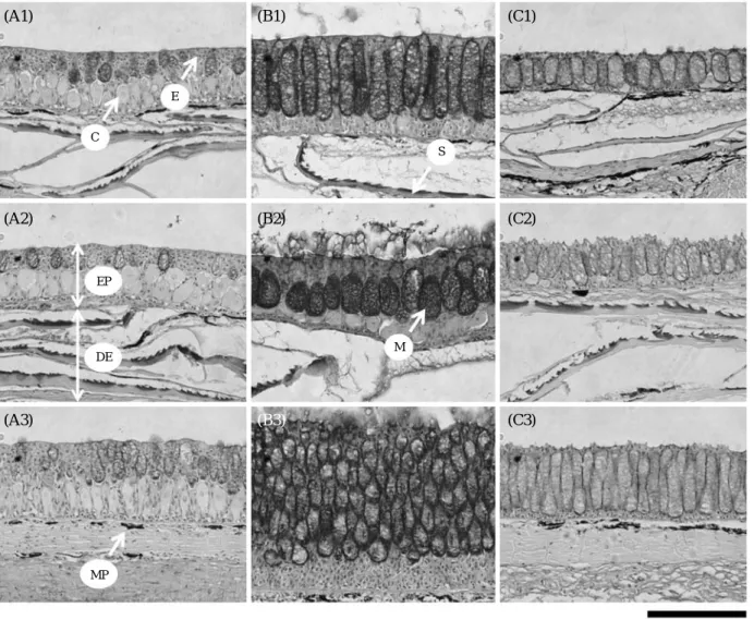
Skin of Misgurnus mizolepis in ricefields can be divid- ed into two layers, epidermis and dermis. The epidermis is composed mainly of basophilic mucus cells (Hemato- xylin positive), relatively small epithelial cells and unde- fined club cells. The dermis is characterized by acido- philic connective tissue, a lot of melanin elements and scales except for occiput region (Fig. 3). However, the mucus cell showed seasonal variations in its size and
number in accordance with a temperature. They were mostly small and few during the summer (Fig. 3A), whereas during the winter they were transformed into an enlarged form and its number also increased predo- minantly (Fig. 3B). Such results showed the same ten- dency as reported by Oh and Park (2009).
2. Aerial exposure of skin
On experimental group, the mucus cell remarkably increased in its size and number, even in the normal temperature (Fig. 3C). It looked just like a typical pheno- menon of winter season (Oh and Park, 2009, 2010). In the dorsal region, the ratio of surface area of mucus cells layer and mucus cells were 86.0±3.3 and 66.2±7.8, respectively. It means that the values are maximum and significantly different from two control group (P⁄0.05, Table 1, Fig. 4A). The lateral region showed relatively high values in the ratio of surface area of mucus cells layer and mucus cells and the number of mucus cell, showing a significant different from the control group of summer (P⁄0.05), whereas there was not any differ- ent between the control group of winter and experiment group (P¤0.05, Table 1, Fig. 4B). Especially, the size and number of mucus cells in the occiput region showed the greatest development that is quitely different from two control groups (P⁄0.05, Table 1, Fig. 4C). Oh and Park (2010) reported that the mucus cell on the occiput region of ricefield-dwelling muddy loach is well res- ponding to the change of temperature, hinting that it is more sensitive than other regions. In fact, the epidermis
Fig. 1. A wild ricefiled-habitat in June 2008 (A) and February 2009 (B), and after artificial evaporation in June 2009 (C), a mass of mud got dried and hardened.
Fig. 2. Tissue sampling regions of a ricefield-dwelling muddy loach Mis- gurnus mizolepis. (A) occiput region, (B) dorsal region, (C) lateral region.
(A)
(B)
(C)
of the occiput region was entirely packed with lots of elongated large mucus cell which is similar with a con- trol group of winter (Fig. 3B), and its volume on epider- mis was being the greatest.
Interestingly, the values were somewhat higher in the experiment group than the control group of winter (Fig.
4). In addition to several functions for gas exchange,
physical and chemical protection, ion and water regula- tion (Shephard, 1994), the differentiation of an extraor- dinarily large mucus gland is also true that is undoubted- ly correlated with burrowing and amphibious habit that can lead to a cutaneous respiration (Liem, 1967; Park et al., 2001). The experiment group started digging burrows as a water level lowered due to evaporation. Finally they
Fig. 3. Variations of skin of Misgurnus mizolepis dwelling in a ricefield under temperature-controlled experiment. (A1~C1) dorsal region, (A2~
C2) lateral region, (A3~C3) occiput region. C, club cell; DE, dermis; E, epithelial cell; EP, epidermis; M, mucus cell; MP, melanophore; S, scale.
Bar indicates 100μm. H-E staining.
Table 1. Comparison of ratio of volume and number of skin mucus cell between control and experiment groups of Misgurnus mizolepis
Dorsal region Lateral region Occiput region
mcl mc N. mcl mc N. mcl mc N.
Control (summer) 50.6±4.1 22.8±4.2 30.5±3.5 30.6±5.6 9.8±4.6 19.2±3.8 55.5±3.9 29.3±1.2 44.5±9.5 Control (winter) 75.3±3.2 49.5±3.3 41.3±2.1 61.3±5.2 38.5±5.1 41.5±4.2 80.8±2.7 73.1±4.6 52.5±5.0 Experiment 86.0±3.3 66.2±7.8 44.3±2.9 66.3±9.7 45.1±9.8 39.3±2.7 90.6±1.1 77.6±3.2 62.8±3.7 mcl: mucus cell layer, mc: mucus cell, N: number of mucus cell
EP
DE
MP
M
(A2) (B2) (C2)
(A3) (B3) (C3)
made burrows reaching to a bottom of breeding chamber.
Even though the burrows were formed both the experi- ment and control group, there was a little difference: the control group left burrows some humid in the wild rice- field, whereas the experimental group’s one made those quitely dry throughout a gradual evaporation process.
Considering a fact that the respiration rate increased in
the high temperature and vice versa in the cold tempera- ture (Oh et al., 2006; So et al., 2008), the increase of the mucus cell in the experiment group would be right for their physiological demands.
It become clear that an aerial exposure over skin cause any change of mucus cell of M. mizolepis. In many fishes, mucus (or mucus coat) has been known as being served as a intermedium for diffusion of oxygen in Misgurnus, Monopterus, Anguilla (Liem, 1967; Park et al., 2001;
Sayer, 2005; Oh and Park, 2008, 2009, 2010). Being revealed in this study, therefore, an drastic increase of mucus cell followed by an aerial exposure over skin seems considered as an evidence for cutaneous respira- tion. We are likely to reach a possible conclusion that two environmental factors, a cold temperature (Oh and Park, 2010) and an aerial exposure of skin (in present study), may be closely related to bi-modal respiration of M. mizolepis. Moreover, at least the aerial exposure is regarded as more effective factor than the cold tempera- ture with regard to the differentiation of the mucus cell.
To verify this hypothesis, however, further improved ex- periments is required.
REFERENCES
Kim, I.S., Y. Choi, C.R. Lee, Y.J. Lee, P.J. Kim and J.H.
Kim. 2005. Illustrated book of Korean fishes. Kyo- Hak Publishing Co., Ltd., Seoul, 615pp. (in Korean) Liem, K.F. 1967. Functional morphology of the integumen-
tary, respiratory, and digestive systems of the syn- branchoid fish Monopterus albus. Copeia, 1967: 375- 388.
Oh, M.K. and J.Y. Park. 2008. Seasonal change of the skin morphology of muddy loach, Misgurnus anguillicau- datus (Cobitidae) from Korea. Korean J. Ichthyol., 20:
90-96.
Oh, M.K. and J.Y. Park. 2009. Seasonal variation of skin structure in a ricefield-dwelling mud loach Misgurnus mizolepis (Cobitidae) from Korea. Korean J. Ichthyol., 21: 87-92.
Oh, M.K. and J.Y. Park. 2010. Seasonal change of skin mu- cus cells of Misgurnus mizolepis (Cobitidae) dwelling in a natural stream in Korea. Korean J. Ichthyol., 22:
230-237.
Oh, S.Y., C.H. Shin, J.Y. Jo, C.H. Noh, J.G. Myoung and J.M. Kim. 2006. Effects of water temperature and salinity on the oxygen consumption rate of juvenile spotted sea bass, Lateolabrax maculatus. Korean J.
Ichthyol., 18: 202-208. (in Korean)
Park, J.Y., I.S. Kim and S.Y. Kim. 2001. Morphology and histochemistry of the skin of the mud loach, Misgurns mizolepis, in relation to cutaneous respiration. Korean Fig. 4. Variations of the size and number of skin mucus cell in Misgur-
nus mizolepis. (A) dorsal region, (B) lateral region, (C) occiput region.
Same letters on the bars mean not significant difference (P¤0.05).
Mean value(%), numberMean value(%), numberMean value(%), number
100 90 80 70 60 50 40 30 20 10 0
100 90 80 70 60 50 40 30 20 10 0
100 90 80 70 60 50 40 30 20 10 0
Control (Summer) Control (Winter) Experiment
Control (Summer) Control (Winter) Experiment
Control (Summer) Control (Winter) Experiment
(A)
(B)
(C)
Surface area of mcl Surface area of mc Number of mc
c
c b
b
b
b
b b
c b
c
a a
a b
a
a a
a
a a b
b a
a
a
a
논에 서식하는 미꾸라지, Misgurnus mizolepis의 공기노출에 의한 피부 점액세포의 변화
오민기∙박종영
전북대학교 자연과학대학 생물과학부∙생물다양성연구소
요 약 :논에 서식하는 미꾸라지는 겨울철 수분증발에 의한 토굴을 형성하였고 피부는 공기 중에 노출되었다.
이때 미꾸라지 피부의 점액세포 변화를 유발하는 환경요인을 분석하기 위하여 진흙과 물을 수조에 채운 후 약 1개월 동안 자연증발시켜 인위적인 토굴형성에 의한 미꾸라지 피부의 공기노출 실험을 여름철에 실시하였다.
그 결과, 공기 중에 피부가 노출된 미꾸라지의 등, 체측, 후두부의 피부 점액세포의 형태는 대부분 크고 길다란 원주세포형으로 변화하였고, 수량 역시 급격히 증가하였는데, 이러한 특징은 겨울철 자연상태에서 나타나는 전 형적인 현상이었다.
찾아보기 낱말 :점액세포, 미꾸라지, 논, 공기노출