간에서 지방산 분할에 대한 지방산결합 단백질 5의 역할
박재승
서해대학 임상병리과 조교수
The role of Fatty acid binding protein 5 ( Fabp5 ) in fatty acid partitioning in the liver
Jae-Seung Park
Assistant Professor, Department of Clinical Pathology, Sohae College
요 약 간성 지방생성과 지방 대사에 있어서 FABP5의 역할에 대하여 알아보고자 하였다. 마우스에 바이러스 입자를 이용하여 간에서 FABP5를 과발현 그리고 침묵시켜 이용하였다. 마우스에 서양식 또는 일반 사료를 1주일간 섭취시키 고 24시간 동안 금식한 후 희생하였다. 간을 기계적으로 분쇄하여 웨스턴 블뢋으로 단백질 농도를 측정하였고, mRANA 분석에는 RT-PCR을 이용하였다. 간과 혈청의 지질 분석에는 박층 크로마토그래피를 이용하였다. 서양식이나 고농도 포화 지방식을 급식한 쥐에서 FABP5 발현이 높은 증가를 보였으나, FABP5 mRNA 발현은 급식에 비해 단식 조건에서 급격히 감소하였다. FABP5를 과발현 시킨 상태에서 급식과 금식을 실시한 결과 간성 TG가 중요하게 증가하 였다. 간성 유리 콜레스테롤은 급식상태에서 현저히 감소하였다. FABP5 발현 억제 시 간성 TG가 상당히 감소하였다.
결과들에서 보듯이, FABP5가 간지질 생성에 중요한 역할을 하며, 정상 상태 및 탄수화물 유도 조건에서 간성 TG 저장 형성에 관여할 것으로 사료된다. FABP5는 비알코올성 지방간 질환, 대사증후군, 비만 치료에 활용될 수 있을 것으로 사료된다. 더 나아가, FABP5 발현에 어떤 전사인자가 관여하는지에 대한 연구가 필요하다.
주제어 : 지방산 결합 단백질 5(FABP5), 간성 지질생성, 비알코올성 지방간 질환, 대사증후군, 비만.
Abstract The aim of investigated the role of FABP5 in the hepatic lipogenesis and lipid metabolisms.
Mice were overexpressed and silenced liver FABP5 using virus particles. Mice were fed a Western-type diet or regular chow for 1week and then sacrificed mouse after 24hr fasted. Liver homogenates were used for protein analysis by Western blot and mRNA levels by RT-PCR. Hepatic and serum lipids were analysed by thin-layer chromatography. Mice fed a Western-type or high saturated fat diet revealed large increases in FABP5 expression. However, FABP5 mRNA levels were drastically reduced under fasted. Hepatic TG was significantly increased FABP5-OEAV mice, but a significantly decreased hepatic free cholesterol under fed. The discovered a substantial decrease in hepatic TG mass with FABP5 silencing. In these data, presented evidence for an important role of FABP5 in hepatic lipogenesis and hepatic TG storage. FABP5 may also be a potential target in the treatment of NAFLD, metabolic syndrome, and obesity. Furthermore, studies to which transcription factors are involved in FABP5 expression and regulation.
Key Words : Fatty acid binding protein 5(FABP5), hepatic lipogenesis, non-alcoholic fatty liver disease(NAFLD), metabolic syndrome, obesity.
*Corresponding Author : Jae-Seung Park([email protected])
Received May 23, 2019 Revised June 10, 2019
Accepted August 20, 2019 Published August 28, 2019
1. 서론
지방산(fatty acids)은 세포에 에너지를 제공할 뿐만 아니라 핵 전사 인자(nuclear transcription factor) 활 성 조절, 세포질 구조(cellular structure), 그리고 세포 자살(cell apoptosis)과 같은 여러 가지 대사 기능들 (metabolic functions)에 관여하고 있다. 긴 사슬 지방 산(long chain fatty acids, LCFA)은 세포 염증 (cellular inflammation) 과정에 관여하는 에이코사노 이드(eicosanoids)의 합성에 중요한 요소이다. 지방산 결합 단백질들(fatty acid binding proteins, FABPs)은 14-16 KDa의 세포질 단백질이고 지방산과 높은 친화성 으로 결합하며 지방산 수송(trafficking)에 중요한 역할 을 한다. FABPs는 9개의 동형(isoform) 단백질이 있으 며, 이는 처음에 분리된 조직에 따라 이름이 지어졌고, 이 들은 15-70%의 아미노산 서열을 공유하고 있다[1]. 모 든 FABPs는 보통 삼차 구조(tertiary structure)로 되어 있고 열 개의 역평형 베타-가닥(ten antiparallel β -strands)으로 구성된 하나의 베타-통형(a β-barrel)을 이루고 있다. 지방산의 결합(binding)은 동형들 내부의 다양한 구멍(cavity)들과 각각의 결합 특이성에 의한다 [2-4]. FABPs는 지질 대사경로의 효소 조절, 막내 지방 산의 농도 유지, 미토콘드리아 그리고 페로시좀 (peroxisomes)에서 지방산 산화 (oxidation), 그리고 핵 (nuclear) 수용체들의 활성을 통해 지방산-반응 유전자들 의 발현 조절 등 여러 가지 기능들과 관련이 있다[5]. 더 나아가, FABPs가 아테롬성 동맥 경화증(atherosclerosis) 의 발병 그리고 대사 증후군(metabolic syndrome)과도 관련이 있는 것으로 알려졌다[6]. FABP1은 간세포 (hepatocyte)에 풍부하게 발현되며, 포화(saturated) 지방 산들, 다불포화(polyunsaturated) 지방산들, 콜레스테롤 (cholesterol), 그리고 담즙산들(bile acids)과 결합한다.
FABP1이 결핍된 마우스에서는 장기적인 단식 또는 웨스턴 -종류(Western-type) 급식으로부터 간지방증 (hepatic steatosis)의 발전을 예방하였다[7, 8].
FABP5는 간을 포함한 다양한 조직들인 지방 조직 (adipose tissue), 신장(kidney), 심근(cardiac muscle), 골격근(skeletal muscle), 고환(testis), 폐(lung), 모세 혈관 내피(capillary endothelium), 안구(lens), 혀 (tongue), 유선(mammary gland), 장(intestine), 그리 고 뇌(brain)에서 발현이 되고 있다[9-11]. FABP5 결합 지방산은 에이코사노이드 (eicosanoids), 그리고 강력한 친화성을 가지고 있는 긴 사슬 지방산인 올레익산(oleic
acid)과 아라키도닉산 (arachidonic acid)이다. 지방 조 직, 꽈리 폐 세포 (alveolar lung cells) 그리고 피부 (skin)에서의 FABP5의 기능은 알려져 있다. FABP5 지 방세포의 초기 분화(early differentiation)에서 FABP4 와 함께 증가하며, 분화가 시작되는 동안 세포내 지방산 전달(intracellular fatty acid transport)에 있어서 역 할을 하고 있다[12]. FABP5를 과발현 (overexpressing) 시킨 마우스에서 기본적으로 지방 세포가 증가하였으며 그리고 호르몬-자극 지방 분해가 일어났다. 꽈리 제 2형 세포들(alveolar type II cells)에서는, FABP5가 새로이 합성된 팔미테이트(
de novo
synthesized palmitate)로 부터 계면활성제 (surfactant)를 합성하는데 관여 한다 [13]. 피부에서는 FABP5가 물이 투과되는 것을 막아주는 역할을 한다[14].간에서는 FABP5 발현 농도가 대사/영양 상태에 따라 변화가 있는 것으로 알려져 있다. 저밀도지질단백질 (Low density lipoprotein, LDL) 수용체-결핍 마우스 (receptor-deficient mice)에 서양식 급식을 하였더니 간 실질 세포(liver parenchymal cell)에서 다른 FABPs 들보다 FABP5 유전자가 시간-의존적으로 증가하였다 [15]. 포화 지방산이 풍부한 급식을 한 쥐에서 일반 급식 을 한 쥐에 비하여 FABP5가 약 30 배 정도 증가하였다 [9]. 또한 간 인슐린 저항성(hepatic insulin resistance) 환자에서 FABP5의 농도가 2.5배 높다고 보고하였다[16].
이러한 모든 결과들에서 FABP5가 FABP1과는 다른 기 전에 의해 지질과 반응하고 그리고 지질 대사에 있어서 중요한 역할을 할 것으로 추측된다. 본 연구에서 이를 알 아보고자 간에서 FABP5 발현을 억제(knocked-down) 하거나 과발현시켜 급식 또는 금식에 따른 간에서의 지 방생성과 지질대사에 있어서 FABP5의 기능과 간과 혈청 의 지질 성분을 분석함으로써 FABP5의 역할을 알아보고 자 하였다. 본 연구의 결과 FABP5가 지방산의 조절에 관 여하여 TG 저장(storage)에 있어서 영향을 미칠 것으로 사료된다.
2. 재료 및 방법
2.1 실험동물
고농도 탄수화물-섭식 연구는, 8주된 위스타 랫트 숫 컷(male 8-week old Wistar rats)을 이용하였다. 랫트 에 물과 일반사료 또는 고농도의 탄수화물이 들어간 준
합성 사료(n=9)를 제공하여 자유롭게 섭취하게 하였다 [17]. FABP5 과발현과 억제 연구에는 8주된 숫컷 C57BL/6J 마우스를 이용하였다. FABP5 과발현 연구를 위해서는 마우스에 FABP5 발현 바이러스 입자 FABP5-OEAV (1x10¹¹)를 정맥주사 하였으며(n=5), 대 조군으로는 FABP5를 발현하지 않는 FABP5-NEC을 주 사하여 이용하였다. FABP5 발현 억제 연구에는 마우스 에 FABP5 발현 억제 바이러스 입자 shFABP5 (1x10¹¹) 를 정맥주사 하였고, 대조군으로는 벡터 shSCR을 주사 하여 이용하였다(n=5). 실험동물은 12시간 주기로 낮과 밤을 조절하였으며, 마취하여 장기를 적출할 때까지 스트 레스를 받지 않도록 사육하여 실험 계획에 따라 사용하 였다. 정맥 주사 1주일 후 24시간 동안 금식한 뒤 희생하 였다. 실험동물을 마취한 후 조직을 적출하여 액체질소에 급속 냉동 보관하여 이용하였으며, 전혈로부터 분리한 혈 청은 –80oC에 동결시켜 보관하여 시용하였다.
2.2 단백질 농도 측정
간 조직 150 mg 정도를 단백질 분해효소 저해제들 (protease inhibitors)이 들어있는 RIPA 용액을 이용하 여 기계적으로 분쇄(mechanically homogenized) 한 후 원심 분리하여 얻어진 상층액(supernatant)을 이용 하였다[18]. BCA kit(Pierce)를 이용하여 단백질 농도를 결정한 후 30-40ug의 단백질을 15% SDS- 폴리아크릴아 마이드 젤 (polyacrylamide gels) (Bio-Rad) 상에서 전기 영동하여 분리시킨 후 PVDF 막(membrane)(Bio-Rad)으 로 옮겨줬다. 일차 항체 (FABP5, 1:200; FABP1, 1:300;
사이클로필린 (cyclophilin) A, 1:1000; 베타-액틴(β -actin), 1:1000)을 4°C에서 12hr 동안 반응 시킨 후 이 차 항체 (anti-rabbit IgG, 1:1000; anti-mouse IgG, 1:2000; anti-goat IgG, 1:2000)를 실온에서 1hr 동안 반응시켰다. 사이클로필린 그리고 베타-액틴 항체는 Abcam (Cambridge, MA)에서 FABP5, FABP1, 그리 고 모든 이차 항체는 R&D Systems (Minneapolis, MN)에서 구입하여 사용하였다. 항체 반응의 발현은 Immun-Star (Bio-Rad)를 이용 하였으며, 그리고 ECL film (GE Healthcare)에 노출하여 사용하였다.
2.3 역전사 중합효소 증폭 반응
전체 RNA의 분리에 Qiagen(Valencia, CA) RNeasy kit를 이용하여 제조회사에서 제공한 순서에 따 라 분리하였다. FABPs에 대한 중합효소 증폭반응에 이
용한 시동체 쌍(primer pairs)은 FABP1;
5 ‘ - A T G A A C T T C T C C G G C A A G T A - 3 ’ 과 5‘-TCTTGCTGACTCTCTTGTAG-3’, FABP5;
5 ‘ - C C A T G G C C A G C C T T A A G G A - 3 ’ 과 5‘-ACCTTCTCATAGACCCGAGT-3’, 그리고 사이클 로필린 A; 5‘-CTG TCTCTTTTCGCCGC TTG-3’과 5‘-CTTGCCACCAGTGCCATTATG -3’을 사용하였으 며, 각각의 조건에 맞게 증폭 반응을 시행하였다.
2.4 지질 분석 및 혈청 분석
동결된 간 조직의 지질 분석은 지질을 추출한 후 박 층 크로마토그래피(thin-layer chromatography)를 이용 하여 분리하였다. 지질의 지방산 성분 분석은 가스-액체 (gas-liquid) 크로마토그래피를 이용하여 결정하였다. 혈청 TG, 베타-하이드록시뷰티레이트 (β-hydroxybutyrate, β -HBA) 그리고 비-에스테라파이드 지방산 (non-esterified fatty acids, NEFA) 농도를 측정하였다. 혈청 TG와 β -HBA의 농도 측정은 시그마 (Sigma, St. Louis, MO)로 부터 구입한 kits를 이용하였으며, 그리고 NEFA 측정은 와코 진단회사(Wako Diagnostics, Richmond, VA)로 부터 구입한 kit를 이용하여 측정하였다.
2.5 통계처리
수치 결과는 평균 ± 표준 편차를 이용하였으며, 비쌍 체 t-검정(unpaired t-test)에서 p < 0.05 일 때 중요한 차이를 보였다.
3. 결과
3.1 새로운 지질 생성 자극에 의한 FABP5 발현 증가
STZ-유도 당뇨병 랫트 간에 GK를 과발현 시키면 혈 장 포도당(plasma glucose)의 간 클리어런스(hepatic clearance) 때문에 심각한 지방증(massive steatosis) 그리고 당원 침적(glycogen deposition)이 발생한다 [17,19]. 이 간들을 분석하면 지방산 합성효소(fatty acid synthase, FAS), 간 피루베이트 카이네이스(liver pyruvate kinase, L-PK), 멀릭 효소(malic enzyme) 그리고 스테롤-CoA 불포화 효소 1(stearoyl-CoA desaturase 1, SCD1)을 포함한 다양한 지방생성 유전 자들이 증가(upregulation)되어 있으며, 흥미롭게도, FABP5 mRNA 농도가 증가하였으나, 간에서 주된 동형
단백질인 FABP1은 변화가 없었다[20]. 이러한 결과들로 추측하여 보건데 영양/호르몬 신호가 FABP5 발현을 유 도하며, FABP5는 간에서 FABP1과는 다른 중요한 역할 을 할 것으로 생각된다.
설치류(rodents)와 사람(humans)에서, 높은 농도의 과당을 섭취 시 간 지방증(hepatic steatosis), 고중성지 질혈증(hypertriglyceridemia), 지방세포의 증가, 그리 고 인슐린 저항성(insulin resistance)을 촉진시킨다 [19,21]. 대부분의 과당은 간에서 조절되지 않고 흡수된 다. 그 결과, 고과당 섭취의 일차적인 해로운 효과 (deleterious effects)는 새로운 지질 형성(
de novo
lipogenesis)에 의한 과당이 지방산으로 전환(conversion) 되는 것이다[21]. 이러한 확인은 GK 실험모델 랫트(n=9)에 세 종류의 탄수화물을 함유한 사료를 섭취시키며 생리적 현 상 연구, 고과당, 지방형성 섭취에서 간성 FABP5 발현에 대해 미치는 영향에 대한 연구에서 알 수 있었다. 이 식 품의 특정 탄수화물 함량(specific carbohydrate contents)을 총 칼로리 값의 백분율(%)로 표시하면 70% 과당(DF70), 35% 과당/35% 옥수수 전분(DF35) 또는 35% 옥수수 전분/35% 저분자덱스트린(DF)으로 구성되어 있다. 포도당을 느리게 방출(slow released glucose)하기 위해 저분자덱스트린을 전분에 첨가하였 고, 대조그룹(group)은 표준 사료를 제공하였다. Table 1에서 보는바와 같이, DF35 그리고 DF를 공급한 그룹 은 표준 사료를 제공한 그룹에 비하여 중요하게 체중이 증가하였다. 오직 과당을 함유한 식사에서 혈청 중성지방 이 표준 사료를 제공한 대조군에 비하여 중요하게 증가를 유도하였다. 이러한 결과는 전에 발표한 과당 공급 동물 모 델과 대조되는 결과이며[19], 간성 지방 생성(hepatic lipogenesis)이 유도되는 것으로 추측된다. 고농도 탄수화 물을 공급(high carbohydrate-fed)한 세 개의 그룹에서 간성 FABP5 발현이 표준 사료를 공급한 그룹에 비하여 증 가하였음을 관찰하였으나, FABP1 발현은 모든 그룹에서 비슷하였다(Fig 1 참고). 그러므로, 앞으로 간성 지방 생 성에 대한 FABP5의 특별한 역할(a specific role) 연구 에 고농도 탄수화물 섭취가 도움이 될 것으로 생각된다.FABP5 발현 조절에 대한 생리적 신호를 알아보기 위 해, 사료 공급 또는 금식 후 24시간에 쥐의 간을 분석하 였다. 그림 2A에서 보듯이, RT-PCR을 이용하여 측정한 FABP5 mRNA 농도가 사료를 공급한 그룹에 비하여 금 식 그룹에서 현저하게 감소하였다. 그러나, FABP5의 단 백질 농도에는 변화가 없었는데(Fig 2B 참고), 이는 단백 질의 반감기(half-life)가 길기 때문인 것으로 추측된다.
DF70 DF35 DF Chow
Weight(g) 439 ± 32 495±36
b465±18
b409 ± 24 TG(mg/dL)
Fed 388±196
a,b368±177
a,b146 ± 48 128 ± 45
Fasted(5h) 307±158
a,b305±153
a,b111 ± 40 101 ± 37 Glucose
(mg/dL)
Fed 105 ± 22 107 ± 26 99 ± 10 101 ± 10
Fasted(5h) 117± 12 114 ± 9 108 ± 13 111 ± 14
All ‘fed’ serum values and weights were obtained at time of sacrifice (week6).
‘Fasted’ serum values were obtained at week 5.
aindicates statistical significance with p<0.05 compared to DF group.
bindicates statistical significance with p<0.05 compared to chow group. N=9
Table 1. Body weight, glucose and TG levels on rats fed high carbohydrate diets
Fig. 1. Differential expression of FABP5 and FABP1 in animal models with increased de novo lipogenesis.
Wistar rats were placed on the designated diets for six weeks and then sacrificed. Western blotting was performed on liver protein extracts with anti-FABP5 and anti-FABP1 antibodies. Each lane corresponds to an individual animal.
Fig. 2. Mice were sacrificed under fed conditions or after a 24-hour fast.
(A) RT-PCR analysis was performed on mRNA isolated
from livers (n = 5) with primers specific to mouse
FABP5 or b-actin. *Indicates statistical significance with
p<0.05 compared to fed group. (B) FABP5 protein levels
from livers described in A. Each lane corresponds to an
individual animal.
3.2 쥐의 간에서 FABP5의 과발현 및 간성 지질 변화
새로운 지방 생성이 증가하는 동안 간성 FABP5 발현 이 증가하는 것을 알 수 있다. 사료를 공급한 동물의 간 에서 FABP5 발현 증가에 대한 효과를 보기위해, 벡터 FABP5-OEAV (FABP5 발현) 또는 FABP5-NEC (대조 군)를 C57BL/6J 마우스 꼬리 정맥에 주사하여 간에 전달 하였다. 마우스를 일주일 동안 급식을 제공하면서 유지한 후 분석을 위해서 안락사 시켰다. 웨스턴 블롯을 이용하 여 FABP5 단백질농도 변화를 확인한 바, FABP5-OEAV 를 처치한 마우스에서 왕성하게 발현되었으며, FABP1의 농도는 변화가 없었다(Fig 3 참고).
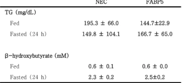
가스 크로마토그래피를 이용하여 간성 지질(hepatic lipids)의 추출(extraction)과 성분 분석(compositional analysis)을 실시하여 본 바 FABP5-OEAV를 처치한 후 급식과 금식을 실시한 그룹에서 간성 TG가 중요하게 증 가하였다(Fig 4A 및 4B 참고). 간성 유리 콜레스테롤 (hepatic free cholesterol)은 급식 상태에서 중요하게 감소하였다(Fig 4A 구글). 이러한 결과에서 FABP5가 정 상 상태에서 간성 TG 생성 증가에 기여할 것으로 추측된 다. FABP5 과발현 상태에서 금식한 혈청 TG와 NEFA 의 농도 변화는 없었다(Table 2 참고).
NEC FABP5
TG (mg/dL)
Fed 195.3 ± 66.0 144.7±22.9
Fasted (24 h) 149.8 ± 104.1 166.7 ± 65.0
β-hydroxybutyrate (mM)
Fed 0.6 ± 0.1 0.6 ± 0.0
Fasted (24 h) 2.3 ± 0.2 2.5±0.2
All serum values were obtained at time of sacrifice. Values are based on the average of four animals in the NEC/fasted group and five animals in all other groups.
Table 2. Serum chemistries in C57BL/6J mice overexpressing FABP5
금식 혈청 β-HBA의 농도는 FABP5-OEAV 처치 상 태에서 중요한 변화가 없었으며, 이는 TG 저장의 증가는 지방산 산화 감소의 결과가 아닌 것으로 추측된다. 결과 적으로, FABP5 과발현은 지방산을 저장하게 하고 그리 고 그 결과 간성 TG 풀(pools)이 증가하게 된다. GK-과 발현 랫트의 결과에서 대부분의 신생 지방산들은 TG 풀 로 저장되고 그리고 FABP5 농도가 높았다[20].
3.3 FABP5의 침묵에 의한 TG 농도의 감소
간에서의 FABP5 발현 억제를 위해서 도움-의존성 벡 터(helper-dependent vectors)를 이용하여 제작하여 사용하였다. 발현 억제에는 shFABP5 벡터와 대조 염기 서열을 포함하고 있는 shSCR을 대조군으로 주사하여 이 용하였다. 쥐를 한 주 동안 보통의 사료를 공급하여 유지 하였으며 그 후에 안락사하여 실험에 이용하였다.
FABP5는 shFABP5를 처치한 그룹에서 대략 75% 정도 감소하였으며 FABP1의 증가는 관찰되지 않았다.
Fig. 3. C57BL/6J mice were injected with 1x10¹¹viral particles of a helper-dependent adenovirus expressing mouse FABP5 or without an expression cassette(NEC).
Western blotting was performed on liver protein extracts with anti-FABP5 and anti-FABP1 antibody. Cyclophilin was used as loading control. Each lane corresponds to an individual animal sacrificed under fed conditions.
FABP5 발현을 억제시킨 그룹에서 간성 TG 양이 상 당히 감소하였으며, Fig 5, 이러한 결과는 FABP5 과발 현에서 TG 와 단일불포화지방산의 증가와는 대조적이다 (Fig 4 참고). FABP5의 결합 능에 따라 이렇게 다양한 영향을 미치는 것으로 추측된다. 오랜 기간의 금식은 간 에서 많은 지방산의 변화를 유도한다. 그 결과, FABP5는 TGs 그리고 PLs의 합성을 위한 특별한 지방산 기질을 분리하는데 있어서 중요한 역할을 하는 것으로 사료된다.
4. 고찰
지방산 순환에 있어서 FABPs의 역할에 대하여 광범 위하게 연구되어 왔다. 여러 개의 FABP 동형들이 있으 며, 이들은 다양한 조직에서 발현이 되고 독특한 기능이 있음을 추측할 수 있다. FABP5는 피부에 많이 발현되는 특성이 있으나, 간장 지방 생성(hepatic lipogenesis)에 서 FABP5가 분명히 어떠한 역할을 하고 있을 것으로 추 측된다. 지방산 합성과 흡수 그리고 콜레스테롤 합성에 관여하는 전사인자인 스테롤 조절 요소-결합 단백질
(sterol regulatory element-bindingprotein, SREBP)-1a 를 과발현 시킨 마우스에서 간성 FABP5가 증가 하였으며 [22], 더 나아가, 서양식을 섭취하거나 또는 높은 포화 지 방을 섭취한 마우스에서 FABP5 발현이 높게 증가하였다 [7,8,15]. 간에서 지방합성과 FABP5와의 관계는 제한되 어 있지 않고 연결되어 있다. FABP5 발현은 각질세포 (keratinocyte) 성장인자-유도 지방생성이 이루어지는 동안에 증가 한다[23,24]. 본 연구에서, 지방생성 경로 (lipogenic pathway)에 대한 FABP5의 연관성을 알아 보고자 한다.
Fig. 4. C57BL/6J mice received NEC (white bars) or FABP5 (black bars), as described in Figure 3.
Lipids were extracted from livers of mice sacrificed under fed (A), or fasted (B), conditions.
Values are based on the average of four animals in the gAd.NEC/fasted group and five animals in all other groups.
PL, phospholipid; TG, triglyceride; CE, cholesterol ester; Chol, cholesterol. * Indicates statistical significance with p < 0.05 compared to gAd.NEC groups.
Fig. 5. C57BL/6J mice were injected with 1x10¹¹viral particles of a helper-dependent adenovirus expressing a scrambled shRNA sequence(shSCR, white bars) or a shRNA to mouse FABP5(shFABP5, black bars).
Lipids were extracted from livers of mice sacrificed under fed (A), or fasted (B) conditions. Values are based on the average of five individual mice per group. PL, phospholipid; TG, triglyceride; CE, cholesterol ester; Chol, cholesterol. * Indicates statistical significance with p < 0.05 compared to gAd.shSCR groups.
전체적인 간성 중성지방의 구성은 음식 그리고/또는 지방조직으로부터 지방산 유입(influx of fatty acids), 베타-산화의정도(degree of β-oxidation), 그리고 초 저밀도지질단백질(very low density lipoproteins, VLDL) 과 같은 중성지방 분비 비율에 의해서 이루어진 다. 흥미롭게도 FABP1, 그리고 FABP5의 결핍은 NEFA 또는 TG의 혈청 농도에 영향이 없어도, 금식 상태를 유 지하는 동안의 간성 중성지방 감소와 연관이 있다[25].
FABP1의 경우,
Fabp1
-/- 쥐에서 TG 분비가 감소하여 TG 감소 그리고 케톤(ketone) β-HBA의 혈청 내 농도 로 볼 때 지방산 산화(fatty acid oxidation)와 연관이 있고, 이러한 각각의 경로 감소는 낮은 농도의 지방산 흡 수 결과이다[25]. 연구 결과에서 볼 때, FABP5는 FABP1보다 다른 자극에 반응하며, 영양소나 호르몬 신 호에 의해 유도되는 FABP5 그리고 FABP1 발현은 다르 게 나타나는 것을 알 수 있었다. 본 연구 결과로 추측컨 대, FABP5는 FABP1과는 다른 경로에 의해 새로운 지질 생성 또는 금식 기간의 증가에 의한 세포내 지방산 농도 증가에 의해 TG 합성을 가능하게 한다.쥐에서, 디아실글리세롤(diacylglycerol) 그리고 아실 (acyl)-CoA로부터 TG 형성에 촉매 작용을 하고 주로 간 에 존재하는 디글리세리드 아실기 전이효소(diglyceride acyltransferase 2, DGAT2)를 과발현 또는 발현을 억 제한 실험에서 간성 TG 저장(hepatic TG storage)을 매개(mediate)하는 것으로 알려졌다[25,26]. FABP5가 DGAT2와 직접적인 상호작용에 의해 TG 합성을 위해 지방산을 배열(sequesters)할 수 있다. SCD1과 직접적인 상호작용은 SCD1에 포화지방산을 전달하거나, 또는 SCD1 에서 DGAT2로 단일불포화 지방산(monounsaturated fatty acids)을 전달해주게 된다. 소포체(endoplasmic reticulum, ER)에 DGAT2와 SCD1이 같이 존재하는 것으 로 알려졌으며[27], 그렇지 않으면, 아마도 FABP5가 지방산 저장고에서 지방산을 막 주변(membranes surrounding)의 DGAT 그리고 SCD1에 전달해 줄 것으로 생각된다. 이러한 가설은 FABP5가 직접적임 상호작용에 의해 막 지방산을 전 달한다는 데서 알 수 있다[28]. 이에 반하여 FABP1은 수 성확산 기전(aqueous diffusion mechanism)에 의해 지방산을 막으로 전달하는 것으로 알려졌다[29].
FABP5 발현 조절에 대한 정확한 기전은 잘 알려지지 않았다. 지방생성(lipogenesis)에 있어서 전사 인자 (transcription factors)가 강하게 관여하고 있다. 이에 대한 직접적인 증거로 SREBP1 그리고 LXR에 의해 양 성조절(positive regulation)이 나타난다는 것이다.
GK-과발현 당뇨병 랫트에서 SREBP-1 그리고 ChREBP 가 증가하였으며[20], 그리고 과당 섭취 랫트 그룹에서 ChREBP mRNA가 증가하는 것을 관찰하였다[30]. 이 전사 인자는 포도당 대사에 의해 활성화 되고 그리고 금 식 상태 동안에는 활성이 저하된다[31,32]. 본 연구 결과에 서 금식 마우스에서 FABP5 mRNA 농도가 낮아짐을 보였으 며, Fig 2, 이는 ChREBP-의존성 발현(ChREBP-dependent expression)이 되는 것으로 추측된다. 다른 연구에서 전사인자 c-myc 또한 FABP5 발현을 양성 조절(positive regulation) 하는 것으로 알려져 있다[33,34]. 이에 대한 확인을 위해서는 FABP5 프로모터 영역(FABP5 promoter region)에 대한 세 부적인 분석이 있어야 될 것이다. 급식 그리고 금식 상태에 서 FABP5 단백질 농도는 전사 기전 조절(transcriptional mechanisms regulating)에 상관없이 비슷하였으며, Fig 2, 이는 FABP5 단백질이 상대적으로 안정적이고 그리고 FABP5 기능은 급식 그리고 금식 상태와 관련이 있을 것으 로 추측된다.
요약하면, 간성 지방생성에 FABP5가 중요한 역할을 하고 있음을 알 수 있었고, 또한 FABP5는 정상 그리고 탄수화물로 유도된 상태에서 간성 TG 저장의 형성에 관 여하고 있음을 알 수 있었다. 앞으로 섭취 물질에 대한 FABP5 발현, 특히 높은 농도의 포화지방(saturated fat), 그리고 어떤 전사인자가 이러한 조절에 관여하는지 연구가 필요하다. FABP5는 간질환인 NASH, 대사증후 군(metabolic syndrome), 그리고 비만(obesity) 등의 치료와 예방에 있어서 중요한 조절 물질로 이용할 수 있 을 것으로 사료된다.
REFERENCES
[1] G. V. Richieri, R. T. Ogata & A. M. Kleinfeld. (1994).
Equilibrium constants for the binding of fatty acids with fatty acid-binding proteins from adipocyte, intestine, heart, and liver measured with the fluorescent probe ADIFAB.
The Journal of Biological Chemistry, 269(39),23918-23930. PMID: 7929039 [2] A. Reese-wagoner, J. Thompson & L. Banaszak. (1999).
Structural properties of the adipocyte lipid binding protein.
Biochimca et Biophysica Acta, 1441(2-3),106-116.
DOI: 10.1016/S1388- 1981(99)00154-7
[3] J. Thompson, J. Ory, A. Reese-wagoner & L. Banaszak.
(1999a). The liver fatty acid binding protein-comparison of cavity properties of intracellular lipid-binding proteins.
Molecular and Cellular Biochemistry, 192(1-2),9-16. PMID: 10331654
[4] J. Thompson, A. Reese-wagoner & L. Banaszak.
(1999b). Liver fatty acid binding protein: species variation and the accommodation of different ligands.
Biochimca et Biophysica Acta 1441(2-3),
117-130.
DOI: 10.1016/s1388-1981(99) 00146-8
[5] M. Furuhashi & G. S. Hotamisligil. (2008). Fatty acid-binding proteins: role in metabolic diseases and potential as drug targets. Nature Reviews Drug Discovery 7(6), 489-503. DOI: 10.1038/ nrd2589 [6] L. Makowski & G. S. Hotamisligil. (2005). The role of
fatty acid binding proteins in metabolic syndrome and atherosclerosis. Current opinion in lipidology 16(5), 543-548. PMID: 16148539
[7] E. P. Newberry, Y. Xie, S. M. Kennedy, J. Luo & N. O.
Davidson. (2006). Protection against Western diet-induced obesity and hepatic steatosis in liver fatty acid-binding protein knockout mice.
Hepatology, 44(5),
1191-1205.
DOI: 10.1002/hep.21369
[8] E. P. Newberry, S. M. Kennedy, Y. Xie, B. T. Sternard, J. Luo & N. O. Davidson. (2008). Diet-induced obesity and hepatic steatosis in L-Fabp / mice is abrogated with SF, but not PUFA, feeding and attenuated after cholesterol supplementation.
American Journal of Physiology Gastrointestinal and Liver Physiology, 294(1),G307-314.
DOI: 10.1152/ajpgi. 00377.2007
[9] G. Siegenthaler, R. Hotz, D. Chatellard-Gruaz, L.
Didierjean, U. Hellman, & J. H. Saurat. (1994).
Purification and characterization of the human epidermal fatty acid-binding protein: localization during epidermal cell differentiation in vivo and in vitro.
Biochemical Journal, 302(Pt 2),363-371. DOI:
10.1042/bj3020363
[10] D. A. Bernlohr, M. A. Simpson, A. V. Hertzel, & L. J.
Banaszak. (1997). Intracellular lipid-binding proteins and their genes.
Annual Review of Nutrition, 17,277-303.
DOI: 10.1146/annurev.nutr.17.1.277
[11] J. amulin, I. Berget, S. Lien, & H. Sundvold. (2008).
Differential gene expression of fatty acid binding proteins during porcine adipogenesis. Comparative Biochemistry and Physiology.
Part B, Biochemical &Molecular Biolology, 151(2),
147-152.
DOI: 10.1016/j.cbpb.2008. 06.010.
[12] N. H. Haunerland & F. Spener. (2004). Fatty acid-binding proteins--insights from genetic manipulations.
Progress in Lipid Research, 43(4),328-349. DOI: 10.1016/j.plipres.2004.05.001
[13] F. Guthmann, C. Schachtrup, A. Tolle, H. Wissel, B.
Binas, H. Kondo, Y. Owada, F. Spener & B. Rustow.
(2004). Phenotype of palmitic acid transport and of
signalling in alveolar type II cells from E/H-FABP
double-knockout mice: contribution of caveolin-1
and PPARgamma.
Biochimca et Biophysica Acta, 1636(2-3),196-204. DOI: 10.1016/j.bbalip.2003.10.015
[14] Y. Owada, H. Takano, H. Yamanaka, H. Kobayashi, Y.
Sugitani, Y. Tomioka, I. Suzuki, R. Suzuki, T. Terui, M.
Mizugaki, H. Tagami, T. Noda & H. KONDO. (2002b).
Altered water barrier function in epidermal-type fatty acid binding protein-deficient mice.
The Journal of Investigative Dermatology, 118(3),430-435. DOI:
10.1046/j.0022-202x.2001.01616.x
[15] M. Hoekstra, M. Stitzinger, E. J. Van Wanrooij, I. N.
Michon, J. K. Kruijt, J. Kamphorst, M. Van Eck, E.
Vreugdenhil, T. J. Van Berkel & J. Kuiper. (2006).
Microarray analysis indicates an important role for FABP5 and putative novel FABPs on a Western-type diet.
Journal of Lipid Research, 47(10),2198-2207.
DOI: 10.1194/ jlr.M600095-JLR200
[16] J. Westerbacka, M. Kolak, T. Kiviluoto, P. Arkkila, J.
Siren, A. Hamsten, R. M. Fisher & H. Yki-Jarvinen.
(2007). Genes involved in fatty acid partitioning and binding, lipolysis, monocyte/macrophage recruitment, and inflammation are overexpressed in the human fatty liver of insulin-resistant subjects.
Diabetes, 56(11),2759-2765. DOI: 10.2337/db07-0156 [17] A. W. Thorburn, L. H. Storlien, A. B. Jenkins, S. Khouri
& E. W. Kraegen. (1989). Fructose-induced in vivo insulin resistance and elevated plasma triglyceride levels in rats.
The American Journal of Clinical Nutrition, 49(6),1155-1163. DOI:
10.1093/ajcn/49.6.1155
[18] S. R. Witting, M. Brown, R. Saxena, S. Nabinger & N.
Morral. (2008). Helper-dependent
Adenovirus-mediated Short Hairpin RNA Expression in the Liver Activates the Interferon Response.
The Journal of Biological Chemistry, 283(4),2120-2128.
DOI: 10.1074/jbc.M704178200
[19] K. A. Lê & L. Tappy. (2006). Metabolic effects of fructose.
Current Opinion in Clinical Nutrition and Metabolic Care, 9(4),469-475. DOI:
10.1097/01.mco.0000232910.61612.4d
[20] N. Morral, H. J. Edenberg, S. R. Witting, J. Altomonte, T. Chu & M. Brown. (2007). Effects of glucose metabolism on the regulation of genes of fatty acid synthesis and triglyceride secretion in the liver.
Journal of Lipid Research ,48(7),
1499-1510. DOI:
10.1194/jlr.M700090- JLR200
[21] K. L. Stanhope & P. J. Havel. (2008). Fructose consumption: potential mechanisms for its effects to increase visceral adiposity and induce dyslipidemia and insulin resistance.
Current opinion in lipidology, 19(1),16-24. DOI: 10.1097/ MOL.0b013e3282f2b24a [22] K. N. Maxwell, R. E. Soccio, E. M. Duncan, E. Sehayek
& J. L. Breslow. (2003). Novel putative SREBP and LXR target genes identified by microarray analysis in liver of cholesterol-fed mice.
Journal of Lipid Research ,44(11),2109-2119. DOI: 10.1194/jlr.M300203- JLR200 [23] R. J. Mason, T. Pan, K. E. Edeen, L. D. Nielsen, F.
Zhang, M. Longphre, M. R. Eckart & S. Neben. (2003).
Keratinocyte growth factor and the transcription
factors C/EBP alpha, C/EBP delta, and SREBP-1c regulate fatty acid synthesis in alveolar type II cells.
Journal of Clinical Investigation, 112(2),
244-255. DOI:
10.1172/JCI16793
[24] Y. Chang, K. Edeen, X. Lu, M. De Leon & R. J. Mason.
(2006). Keratinocyte growth factor induces lipogenesis in alveolar type II cells through a sterol regulatory element binding protein-1c-dependent pathway.
American Journal of Respiratory Cell and Molecular Biology, 35(2),
268-274. DOI: 10.1165/rcmb.2006- 0037OC
[25] J. S. Millar, S. J. Stone, U. J. Tietge, B. Tow, J. T.
Billheimer, J. S. Wong, R. L. Hamilton, R. V. JR. Farese
& D. J. Rader. (2006). Short-term overexpression of DGAT1 or DGAT2 increases hepatic triglyceride but not VLDL triglyceride or apoB production.
J Lipid Res, 47(10),2297-2305. DOI: 10.1194/jlr.M600213 -JLR200 [26] X. X. Yu, S. F. Murray, S. K. Pandey, S. L. Booten, D.
Bao, X. Z. Song, S. Kelly, S. Chen, R. Mckay, B. P.
Monia & S. Bhanot. (2005). Antisense oligonucleotide reduction of DGAT2 expression improves hepatic steatosis and hyperlipidemia in obese mice.
Hepatology, 42(2),
362-371. DOI: 10.1002/hep.20783 [27] W. C. Man, M. Miyazaki, K. Chu & J. Ntambi. (2006).
Colocalization of SCD1 and DGAT2: implying preference for endogenous monounsaturated fatty acids in triglyceride synthesis.
Journal of Lipid Research, 47(9),1928-1939. DOI:
10.1194/jlr.M600172-JLR200
[28] J. Storch & A. E. Thumser. (2000). The fatty acid transport function of fatty acid-binding proteins.
Biochimca et Biophysica Acta, 1486(1),
28-44. DOI:
10.1016/s1388-1981(00)00046-9
[29] K. T. Hsu & J. Storch. (1996). Fatty acid transfer from liver and intestinal fatty acid-binding proteins to membranes occurs by different mechanisms.
The Journal of Biological Chemistry, 271(23),13317-13323. DOI: 10.1074/ jbc.271.23.13317 [30] H. Y. Koo, M. A. Wallig, B. H. Chung, T. Y. Nara, B. H.
Cho & M. T. Nakamura. (2008). Dietary fructose induces a wide range of genes with distinct shift in carbohydrate and lipid metabolism in fed and fasted rat liver.
Biochimica et Biophysica Acta, 1782(5),341-348. DOI: 10.1016/j.bbadis.2008.02.007 [31] H. Yamashita, M. Takenoshita, M. Sakurai, R. K.
Bruick, W. J. Henzel, W. Shillinglaw, D. Arnot & K.
Uyeda. (2001). A glucose-responsive transcription factor that regulates carbohydrate metabolism in the liver.
Proceedings of the National Academy of Science of United State of America, 98(16),9116-9121. DOI:
10.1073/pnas.161284298
[32] L. Ma, L. N. Robinson & H. C. Towle. (2006).
ChREBP*Mlx is the principal mediator of
glucose-induced gene expression in the liver.
The Journal of Biological Chemistry, 281(39),28721-28730. DOI: 10.1074/jbc.M601576200
[33] H. A. Coller, C. Grandori, P. Tamayo, T. Colbert, E. S.
Lander, R. N. Eisenman & T. R. Golub. (2000).
Expression analysis with oligonucleotide microarrays reveals that MYC regulates genes involved in growth, cell cycle, signaling, and adhesion.
Proceedings of the National Academy of Science of United State of America, 97(7),3260-3265. DOI: 10.1073/pnas.97.
7.3260
[34] M. Munz, R. Zeidler & O. Gires. (2005). The tumour-associated antigen EpCAM upregulates the fatty acid binding protein E-FABP.
Cancer Lett, 225(1),151-157. DOI: 10.1016/j.canlet.2004. 11.048
박 재 승(Jae-Seung Park) [정회원]
․ 1993년 2월 : 전주대학교 이공대학 미 생물학과 (이학사)
․ 1996년 2월 : 전북대학교 의과대학 (의학석사)
․ 2000년 2월 : 전북대학교 의과대학 (의학박사)
․ 2000년 ~ 2011년 : 미국 IUPUI- PURDU 의과대학 박사 후 연수 및 연구 교수
․ 2011년 ~ 현재 : 서해대학 임상병리과 교수
․ 관심분야 : 제 2형 당뇨병, 간 대사, 간 종양, 비만
․ E-Mail : [email protected]