Copyrightⓒ 2010, The Korean Academy of Oral Biology
107
Journal of Oral Biology
Characterization of Binding of Treponema denticola to Immobilized Fibrinogen using the Fluorescent Fatty Acid Labeling Method
Jin Hong and Si Young Lee*
Department of Oral Microbiology, College of Dentistry, Research Institute of Oral Science, Gangneung-Wonju National University, Gangneung, 210-702, Korea
(received August 27, 2010 ; revised September 15, 2010 ; accepted September 17, 2010)
Treponema denticola is a gram-negative anaerobe that can cause periodontal disease. The adhesion of this bacterium to host tissues is considered to be the primary event in the colonization and infection of a host. Fibrinogen is generally found in damaged tissues resulting from periodontitis. The binding ability of T. denticola to fibrinogen may therefore be an important virulence factor in inducing periodontal diseases. It has been reported recently that oral spirochetes can be labeled with fluorescent fatty acids and we speculated that this labeling method could be used in an oral spirochete binding assay. The binding of several different strains of T.
denticola to immobilized human fibrinogen was therefore tested using the fluorescent fatty acid labeling method. In the case of immobilized fibrinogen, the T. denticola ATCC 35405 strain showed saturable binding to immobilized fibrinogen. Indeed, all four different T. denticola strains tested in this experiment, T. denticola ATCC 35405, T. denticola ATCC 33520, T. denticola ATCC 35404 and T. denticola OTK showed binding to fibrinogen. The fluorescent fatty acid labeling method thus shows utility in binding assays for T.
denticola, different strains of which can generally bind to immobilized fibrinogen.
Key words: Treponema denticola, fibrinogen, binding, fatty acid, fluorescence
서 론
치주질환은 만성 염증성 질환으로 세균과 이에 대한 인 체의 반응이 주된 원인으로 알려져 있으며, 원인이 되는 세 균 들 중 Porphyromonas gingivalis, Tannerella forsythia 와 Treponema denticola가 이 질환과 밀접한 관련이 있는 것으로 보고되고 있다(Socransky et al., 1998). T. denticola 로 대표되는 구강 spirochetes는 치주낭의 세균 구성 중 중 요한 부분을 차지하고 있으며(Ellen et al., 2005; Yao et al., 1996), 치주질환과 관련된 여러가지 병독력을 지니고 있다. 이 세균은 fibronectin, laminin, collagen 같은 extra- cellular matrix protein에 부착하는 것으로 보고되었으며 (Dawson et al., 1990; Edwards et al., 2005; Haapasalo et al., 1991), 단백질 분해 효소도 지니고 있다(Fenno et al., 1998; Lee et al., 2002).
숙주 조직에 대한 세균의 부착은 숙주에서의 세균 증식 과 감염의 과정에서 첫 번째로 일어나는 일이다. 치주질환 의 발병에 있어서도 첫 번째 과정은 조직 수용기와 세균 부착소의 상호작용에 의한 숙주조직에의 세균 부착일 것 이다(Haapasalo et al., 1991). 지금까지의 연구에 의하면 T. denticola는 상피세포에 부착할 수 있는 능력을 지니고 있으며(Fenno et al., 1996; Grenier et al., 1990) 또한 세 포외 기질성분에 대한 T. denticola의 부착은 세균의 구강 조직 침투에 필수적인 역할을 한다고 알려져 있다(Ellen et al., 1994).
Fibrinogen은 혈청에 존재하는 340-kDa의 단백질로서 조직의 손상시 상처의 회복에 필수적으로 필요하다(Pereira et al., 2002; Rybarczyk et al., 2003). Fibrinogen은 불 용성 중합체의 형태로 혈액응고의 matrix를 형성하는데, Staphylococcus aures, 구강연쇄구균, Candida albicans 그 리고 human platelets를 포함하여 조직의 기질 요소에 특
*Corresponding author: Si Young Lee, Department of Oral Microbiology, College of Dentistry, Research Institute of Oral Science, Gangneung-Wonju National University, Gangneung, 210-702, Korea. Tel: +82-33-640-2455,
Fax: +82-33-642-6410, E-mail: [email protected]
이적으로 부착한다(Lantz et al., 1990; Lee et al., 2001).
치주질환이 있는 경우 조직이 손상을 입고 출혈이 빈번하 게 일어나기 때문에, fibrinogen은 치주조직에 많은 양으 로 존재한다. 그러므로 치주질환 원인 세균들의 fibrinogen 에의 부착은 이들 세균이 가지는 중요한 치주질환 유발 병독 요인으로 작용할 수 있다. Porphyromonas gingivalis 와 Prevotella intermedia의 경우 fibrinogen에의 부착은 이 들 세균에서 중요한 병독인자로서 작용하는 것으로 보고 되고 있다(Lantz et al., 1990).
세균의 부착을 조사하기 위하여 여러가지 연구 방법들 이 사용되어 왔다. 대표적으로, 세균을 방사선 동위원소로 표지하여 단백질에 부착된 세균의 양을 직접 측정하는 방 법(Lee et al., 2001)과 단백질 등에 세균을 부착 후 세 균에 대한 항체를 이용하여 부착된 세균을 측정하는 방법 (Haapasalo et al., 1991)이 이용 되어 왔다. 그러나 항체 를 이용하여 세균 부착을 간접적으로 측정하는 방법은 세 균에 대한 항체를 직접 만들어야 하는 불편 때문에 편리 하게 사용되기 힘들며, 동위원소을 사용하여 세균을 직접 표지하는 방법도 동위원소의 사용 제약으로 인하여 불편 함이 많다.
최근에 구강 spirochetes를 형광표지된 지방산으로 쉽게 표지할 수 있는 방법이 보고 되었으며(Hong et al., 2008), 구강 spirochetes의 부착실험에 응용할 수 있는 방법으로 제시 되었다. 이전에 보고된 T. denticola의 fibrinogen에 대한 연구들은 T. denticola ATCC 35405 한가지 종 만을 실험에 사용하여(Bamford et al., 2007; Haapasalo et al., 1991) 다른 종의 T. denticola도 fibrinogen에의 부착 성질 을 가지는지 알려지지 않고 있다. 본 연구에서는 형광지 방산으로T. denticola를 직접 표지하는 방법을 이 세균의 부착 실험에 최초로 사용하였으며, T. denticola ATCC 35405 뿐만아니라 다른 T. denticola 종들의 fibrinogen 에의 부착 여부도 조사하였다.
재료 및 방법
세균배양
본 실험에서는 구강 spirochetes인 T. denticola ATCC 35405, T. denticola ATCC 33520, T. denticola ATCC 35404, T. denticola OTK를 사용하였다. T. denticola는 0.2% thiamine pyrophosphate, 10% sodium bicarbonate, 1% serum 그리고 0.2% fatty acid 가 첨가된 NOS (new oral spirochete)배지에 접종하였으며 90% N2, 5% CO2 그리고 5%의 H2가 있는 36oC 혐기성 배양기(Sheldon manufacturing, INC, USA)에서 48시간(log phase) 동안 배양하여 실험에 사용하였다.
세균의 형광지방산 표지 방법 및 형광 측정
지방산인 5-octadecanoylaminfluorescein (OAF) (Molecular probe, Eugene, Oregon, USA)를 Dimethyl Sulfoxide (DMSO) (Sigma Chemical Co. St. Louis, Mo, USA)에 1 mg/ml로 녹인 뒤 4oC에서 빛을 차단한 상태로 보관하 였다. 형광 지방산을 상온에서 녹인 뒤 세균의 log-phase 단계에서 세균 배양배지에 1 µg/ml로 첨가하여 한 시간 동안 36oC 혐기성 배양기에서 배양 후0.5% BSA가 포함 된 PBS-BSA buffer로 세균을 두 번 세척한 후 실험에 사용하였다. 형광의 측정은 fluorescent spectrophotometer (F4500, HITACHI, Tokyo, Japan)을 이용하였다. Fluorescent spectrophotometer F4500의 메뉴에서 Time Scan을 선택 하여 30초 지점에서의 형광을 측정하였으며 OAF의 여기 파장은 497 nm, 방출파장은 519 nm였다(Hong et al., 2008).
세균 labeling 효율 (labeling efficiency) (세균 수/ 형광 단위)
세균의 부착실험 후 부착된 세균을lysis solution (Lee et al., 2001)을 이용하여 탈락시킨 후 형광을 측정하기 때문 에 세균 labeling 효율 측정 시 같은 조건을 사용하였다.
형광 수치에 따른 세균 수를 구하기 위하여 OAF로 labeling 된 일정 수의 구강 spirochetes 0.5 ml에 1% SDS, 8M urea 그리고 1M NaCl이 포함된 lysis solution (Lee et al., 2001) 4.5 ml을 더한 후 형광을 측정하였다(OD660= 0.2: 5× 10⁸/ ml). 세균 labeling 효율(labeling efficiency) = (세균 수/
형광단위)로 계산하여 1 형광단위에 해당하는 세균수를 계산하였다.
OAF로 labeling한 T. denticola의 고정된 fibrinogen에 의 부착
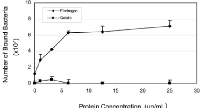
1) Coating된 fibrinogen의 농도에 따른 세균의 부착 세균의 fibrinogen에 대한 부착이 특이적(specific binding) 인지를 조사하기 위하여 coating하는 fibrinogen의 농도 증 가에 따른 세균의 부착 정도를 조사하였다. 세균을 log- phase에서 OAF 1 µg/ml으로 한 시간 동안 혐기성 배양기 에서 labeling한 후 0.5% BSA가 포함된 PBS-BSA buffer 로 두 번 세척 후 실험에 사용하였다. Human fibrinogen (Sigma Chemical Co. St. Louis, Mo, USA)을 0.2%
Sodium Azide (NaN3)가 포함된 0.05M carbonate buffer (pH 9.5)에 녹인 후0 µg/ml에서 25 µg/ml의 농도로 24 well cell culture plate에 0.55 ml 씩 더하여 36oC 일반 배양기에서 18시간 동안 coating하였다. Coating된 각 well 은 0.55 ml의 PBS-BSA buffer로 두 번 세척 후, OAF로 labeling된 25 × 108/ml 농도의 세균 현탁액 0.5 ml을 더한 후 36oC 일반 배양기에서 1시간 동안 배양한 다음 세균 현탁액을 제거하고 각 well은 PBS-BSA buffer로 두 번 세척하였다. 부착된 세균은 1% SDS, 8M urea 그리고 1M
NaCl을 포함한 lysis solution 0.5 ml을 더하여 일반 배양 기 36oC에서 30분 동안 용해시킨 뒤 얻은 용액의 형광 을 측정하였다. 측정된 형광값은 앞의 실험에서 얻은 세 균의 labeling efficiency (세균수/형광단위)를 사용하여 세 균수로 환산하였다. 세균 부착 음성대조군으로 gelatin (Sigma Chemical Co. St. Louis, Mo, USA)에의 T. denticola 부 착을 실험에 포함하였다.
2) 구강 spirochetes종의 차이에 따른 fibrinogen에의 부착 구강 spirochetes의 종의 차이에 따른 fibrinogen의 부착 정도를 조사하기 위하여 T. denticola ATCC 35404, T.
denticola ATCC 33520, T. denticola OTK 그리고 T.
denticola ATCC 35405를 1 µg/ml의 OAF로 36oC 혐기성 배양기에서 한 시간 동안 labeling 한 후0.5% BSA가 포 함된 PBS-BSA buffer로 두 번 세척 후 실험에 사용하였 다. Fibrinogen (100 µg/ml)으로 coating된 24 well cell culture plate에 labeling된 세균0.5 ml (25 × 108/ml)을 각각 넣은 후 위와 같은 방법으로fibrinogen에의 부착정도를 비 교 실험하였다. 세균 간의 부착 정도의 차이는Student t 검사로써 비교하였고, p < 0.05의 값을 유의한 차이를 보 이는 것으로 간주하였다.
결 과
T. denticola의 fluorescent fatty acid에 의한 labeling effi- ciency는 T. denticola ATCC 35404가 relative fluorescent unit당 8.4 × 10⁵ bacteria로 가장 높은 수치를 보였으며T.
denticola ATCC 33520 가4.0 × 10⁵ bcteria로 가장 낮은 수치를 보였다(Table 1). T.denticola의 fibrinogen에의 부 착 특이성을 조사하기 위하여, fibrinogen을 0 µg/ml에서 25µg/ml까지 coating하여 세균의 부착정도를 비교하였다.
그 결과 fibrinogen의 증가되는 농도에 따라 6.25 µg/ml까 지는 세균의 부착이 coating된 fibrinogen의 농도와 비례 하여 계속 증가하였으며 그 이상의 농도에서는 세균의 부 착이 포화되는 양상을 보였다(Fig. 1).
Fibrinogen에의 부착 실험에서는 실험에 사용한 네가지
세균 종 모두 fibrinogen에 부착하는 것으로 관찰되었다. T. denticola ATCC 35405는 T. denticola ATCC 33520 보다 통계적으로 유의하게 더 많이 부착한 것으로 관찰되 었으나, T. denticola ATCC 35404나 T. denticola OTK의 부착 정도와는 통계적으로 유의한 차이가 없었다. 다른 세 균 종들 간에도 통계적으로 유의한 부착 정도의 차이는 관찰되지 않았다(Fig. 2).
고 찰
본 연구에서는 구강 spirochetes의 외막에 형광 지방산을 Table 1. Bacteria count / relative fluorescence unit for labeling effi-
ciency. In order to count the bacterial number according to relative fluorescence units, 0.5 ml of OAF-labeled T. denticola (OD660= 0.2 : 5× 10⁸/ml) was added with lysis buffer and then measured for fluorescence
Bacteria count / relative fluorescence 1 unit T. denticola ATCC 35405 5.8× 10⁵ T. denticola ATCC 35404 8.4× 10⁵ T. denticola ATCC 33520 4.0× 10⁵ T. denticola OTK 5.5× 10⁵
Fig. 1. Adhesion of T. denticola ATCC 35405 to the wells coated with different concentrations (0µg/ml- 25 µg/ml) of fibrinogen. T.
denticola was labeled with 1µg/ml OAF in an anaerobic incubator at 36oC for 1 h. Labeled bacteria was added to fibrinogen coated well and incubated for 1 h. Bound bacteria were detached with lysis buffer and fluorescence was measured with fluorescent spec- trophotometer. Number of bacteria bound was calculated with labeling efficiency. Values indicate means of triplicate determina- tions; standard errors of the mean are indicated by vertical lines.
Fig. 2. Adhesion of strains of T. denticola to immobilized fibrino- gen. T. denticola was labeled with 1µg/ml OAF in an anaerobic incubator at 36oC for 1 h. Labeled bacteria was added to fibrinogen coated well and incubated for 1 h. Bound bacteria were detached with lysis buffer and fluorescence was measured with fluorescent spectrophotometer. Number of bacteria bound was calculated with labeling efficiency of each strain. Values indicate means of tripli- cate determinations; standard errors of the mean are indicated by horizontal lines.
삽입하는 방법을 이용하여 세균을 labeling하였으며, labeling 된 구강 spirochetes을 이용하여 고정된 fibrinogen에의 부 착 실험을 수행하였다. 구강 spirochetes종에 따라 차이를 나타내긴 했지만 비교적 안정적으로 형광지방산이 labeling 됨으로써 fibrinogen 부착 실험에 적용시킬 수 있었다. 또 한 기존의 연구에서 밝혀지지 않았던T. denticola ATCC 35405 이외의 다른 T. denticola 종들도 고정된 fibrinogen 에의 부착 성질을 지니고 있음을 밝혔다.
형광 지방산 labeling을 이용한 구강 spiochetes의 부착 실험은 기존에 사용하던 방사선동위원소를 사용하는 방법 와 항체를 사용하는 ELISA방법에 비하여 간편하게 적용 할 수 있어 앞으로의 구강 spirochetes의 부착실험에 많이 응용될 수 있을 것으로 생각된다. 구강 spirochetes를 형 광 지방산으로 labeling하는 방법을 세균 부착실험에 응용 하는 것은 방사선 동위원소나 항체를 사용하는 방법보다 시간이 단축되고 비용이 적게 들며, 실험 수행에 있어서 과정이 간편하다는 점에서 장점을 지닌다.
세포외 기질단백질에의 세균 부착 능력은 감염의 과정 에서 중요하게 작용한다(Ishihara et al., 1999). Fibrinogen 에 대한 T. denticola의 부착을 매개하는 기작은 fibronectin, laminin, collagen 그리고 kelatin 등과 T. denticola의 부 착을 매개하는 기작과는 명백히 다르다고 보고되고 있다 (Edwards et al., 2003). T. denticola의 외막에 있는 major surface protein (Msp) (Fenno et al., 1998)와 oligopeptide- binding protein (OppA) (Fenno et al., 2000) 그리고 chymotrypsin-like protease (CTLP) (Fenno et al., 1998) 는 조직에 세포병리적인 영향을 미칠 뿐만 아니라 조직에 이 세균이 부착할 때 부착소로서 작용한다. 이들의 연구 에 따르면 T. denticola의 OppA는 용해상태의 plasminogen 과 fibronectin에 부착하는데 관여한다(Fenno et al., 2000).
최근의 보고에 따르면T. denticola ATCC 35405의 chymo- trypsin-like protease (CTLP) complex가 fibrinogen에의 세 균 부착을 매개하는 것으로 알려졌다(Bamford et al., 2007).
본 실험에 사용한 다른 T. denticola 종들의 fibrinogen에 의 부착에도 이들 세균의 CTLP complex가 매개할 것으 로 추정되는데 정확한 사실을 위해서는 이들 세균 종에 대한 추가적인 연구가 필요할 것이다.
Fibrinogen은 조직파괴와 출혈이 동반되는 치주질환이 있는 부위에서 다량으로 발견된다. 치주질환과 관련이 있 는 원인세균들의 fibrinogen과의 상호 반응은 치주질환의 발병 기전을 이해하는데 도움을 줄 것으로 생각된다. 그러 나 치주질환 원인세균 중의 하나로 알려져 있는 T. denticola 의 fibrinogen에 대한 부착에 대한 연구와 이러한 부착이 인체와 T. denticola와의 상호반응에 어떠한 영향을 미쳐 서 치주질환의 발병에 기여하는지 등에 대한 연구는 아직 미흡하다.
본 연구를 통하여 형광지방산을 이용한 T. denticola의 labeling 방법을 이 세균의 부착 실험에 응용하여 사용할
수 있음을 보였고, 기존에 알려져 있는 T. denticola ATCC 35405 종 뿐만아니라 다른 T. denticola 종들도 고정된 fibrinogen에 부착하는 성질을 일반적으로 가지고 있음을 보였다. 앞으로 본 연구 방법 등을 이용하여 T. denticola 의 부착 연구가 좀 더 다양하게 진행되어 숙주 조직과 T.
denticola 간에 특이적인 상호작용을 발견하고 차단한다면 치주질환의 예방과 치료에 도움을 줄 것으로 생각된다.
참 고 문 헌
Bamford CV, Fenno JC, Jenkinson HF, Dymock D. The chymotrypsin-like protease complex of Treponema denticola ATCC 35405 mediates fibrinogen adherence and degradation.
Infect Immun. 2007;75:4364-72.
Dawson JR, Ellen RP. Tip-oriented adherence of Treponema denticola to fibronectin. Infect Immun. 1990;58:3924-8.
Edwards AM, Dymock D, Woodward MJ, Jenkinson HF.
Genetic relatedness and phenotypic characteristics of Treponema associated with human periodontal tissues and ruminant foot disease. Microbiology. 2003;149:1083-93.
Edwards AM, Jenkinson HF, Woodward MJ, Dymock D.
Binding properties and adhesion-mediating regions of the major sheath protein of Treponema denticola ATCC 35405.
Infect Immun. 2005;73:2891-8.
Ellen RP, Dawson JR, Yang PF. Treponema denticola as a model for polar adhesion and cytopathogenicity of spirochetes.
Trends Microbiol. 1994;2:114-9.
Ellen RP, Galimanas VB. Spirochetes at the forefront of periodontal infections. Periodontol 2000. 2005;38:13-32.
Fenno JC, Hannam PM, Leung WK, Tamura M, Uitto VJ, McBride BC. Cytopathic effects of the major surface protein and the chymotrypsinlike protease of Treponema denticola.
Infect Immun. 1998;66:1869-77.
Fenno JC, Muller KH, McBride BC. Sequence analysis, expression, and binding activity of recombinant major outer sheath protein (Msp) of Treponema denticola. J Bacteriol.
1996;178:2489-97.
Fenno JC, Tamura M, Hannam PM, Wong GW, Chan RA, McBride BC. Identification of a Treponema denticola OppA homologue that binds host proteins present in the subgingival environment. Infect Immun. 2000;68:1884-92.
Grenier D, Uitto VJ, McBride BC. Cellular location of a Treponema denticola chymotrypsinlike protease and import- ance of the protease in migration through the basement membrane. Infect Immun. 1990;58:347-51.
Haapasalo M, Singh U, McBride BC, Uitto VJ. Sulfhydryl- dependent attachment of Treponema denticola to laminin and other proteins. Infect Immun. 1991;59:4230-7.
Hong J, Kim KJ, Lee SY. Labeling of oral spirochetes with fluorescent fatty acids. Int J Oral Biol. 2008;33:65-70.
Ishihara K, Okuda K. Molecular pathogenesis of the cell surface proteins and lipids from Treponema denticola. FEMS MicrobiolLett. 1999;181:199-204.
Lantz MS, Allen RD, Bounelis P, Switalski LM, Hook M.
Bacteroides gingivalis and Bacteroides intermedius recognize
different sites on human fibrinogen. J Bacteriol. 1990;172:
716-26.
Lee SY, Bian XL, Wong GW, Hannam PM, McBride BC, Fenno JC. Cleavage of Treponema denticola PrcA polypeptide to yield protease complex-associated proteins Prca1 and Prca2 is dependent on PrtP. J Bacteriol. 2002;184:3864-70.
Lee SY, Kim KK, Choe SJ. Binding of oral streptococci to human fibrinogen. Oral Microbiol Immunol. 2001;16:88-93.
Pereira M, Rybarczyk BJ, Odrljin TM, Hocking DC, Sottile J, Simpson-Haidaris PJ. The incorporation of fibrinogen into extracellular matrix is dependent on active assembly of a
fibronectin matrix. J Cell Sci. 2002;115:609-17.
Rybarczyk BJ, Lawrence SO, Simpson-Haidaris PJ. Matrix- fibrinogen enhances wound closure by increasing both cell proliferation and migration. Blood. 2003;102:4035-43.
Socransky SS, Haffajee AD, Cugini MA, Smith C, Kent RL, Jr. Microbial complexes in subgingival plaque. J Clin Periodontol. 1998;25:134-44.
Yao ES, Lamont RJ, Leu SP, Weinberg A. Interbacterial binding among strains of pathogenic and commensal oral bacterial species. Oral Microbiol Immunol. 1996;11:35-41.